



TPCK
Based on 1 publication(s) in Google Scholar
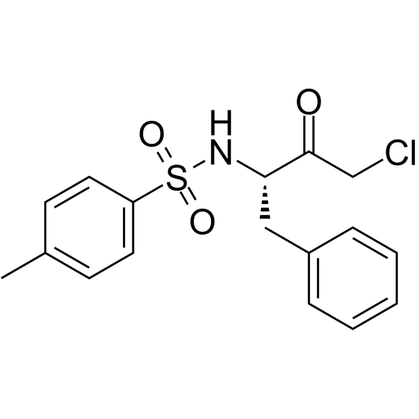
TPCK (L-1-Tosylamido-2-phenylethyl chloromethyl ketone; L-TPCK) is an effective serine protease inhibitor and also a blocker of the PDK1/Akt pathway. TPCK can modify the E7 protein in actively keratinocyte cells. TPCK can induce cellular apoptosis, suppress tumor growth, reduce hypoxic-ischemic brain injury in rat pups, and affect vascular permeability in inflamed rats.
商品は「研究用試薬」です。人や動物の医療用・臨床診断用・食品用の製品ではありません。
研究用途以外に使用した場合、当社は一切の責任を負いかねます。
- 純度 : 98.05%
- CAS 番号: 402-71-1
- 分子式: C17H18ClNO3S
- 分子量:351.85
-
保管条件:Powder -20°C, 3 years , 4°C, 2 years ; In solvent -80°C, 6 months , -20°C, 1 month

To place orders, for customer services and technical support, please contact: MedChemExpress USA
Tel: 609-228-6898 E-mail: [email protected] [email protected]


-
 生物活性
生物活性
-
化学情報
-
溶剤 & 溶解度
- プロトコル
- 純度とドキュメンテーション
- 参考文献
-
 Help & FAQs


-
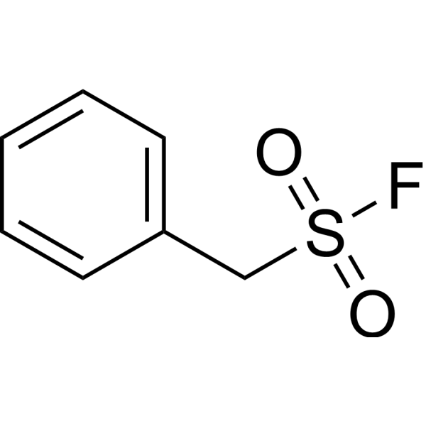
HY-B0496
PMSFCathepsin Inhibitor -
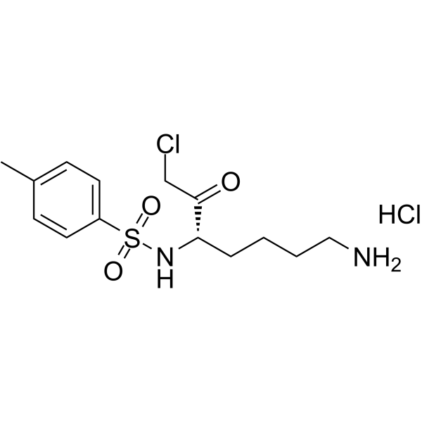
HY-112716
N-alpha-Tosyl-L-lysine chloromethyl ketone hydrochlorideTrypsin Like Protease Inhibitor -
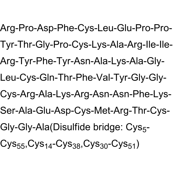
HY-P0017
AprotininBPTI Inhibitor -
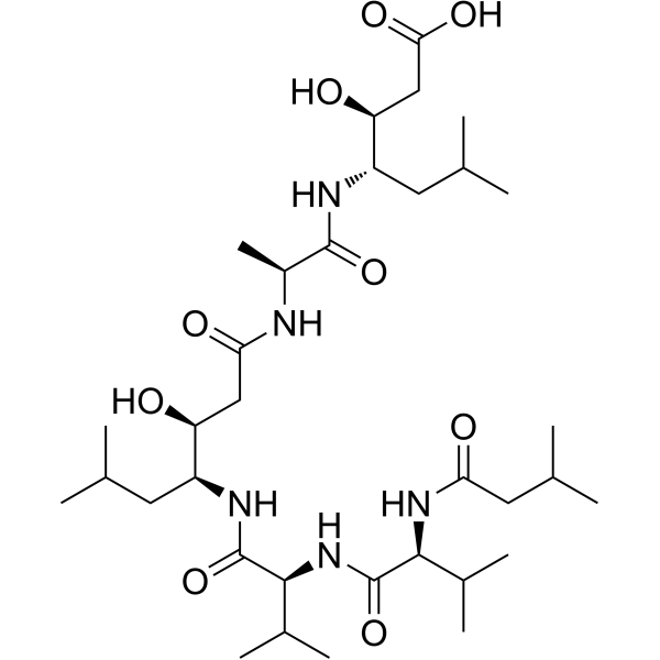
HY-P0018
PepstatinAspartic Protease Inhibitor -
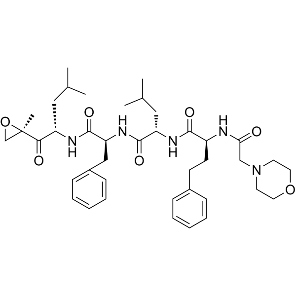
HY-10455
CarfilzomibProteasome Inhibitor -
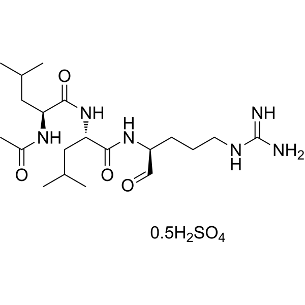
HY-18234A
Leupeptin hemisulfateProtease Inhibitor -
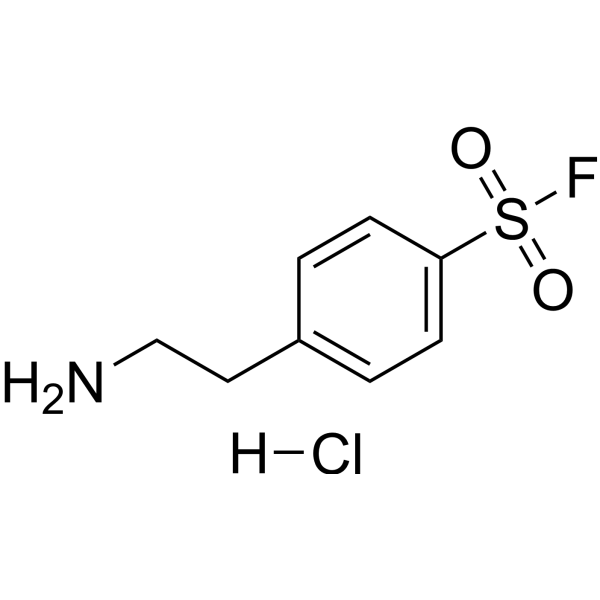
HY-12821
AEBSF hydrochlorideThrombin Inhibitor -
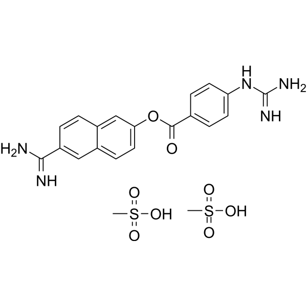
HY-B0190A
Nafamostat mesylateSerine Protease Inhibitor -

HY-10227
BortezomibProteasome Inhibitor -

HY-10455R
Carfilzomib (Standard)Proteasome Inhibitor -
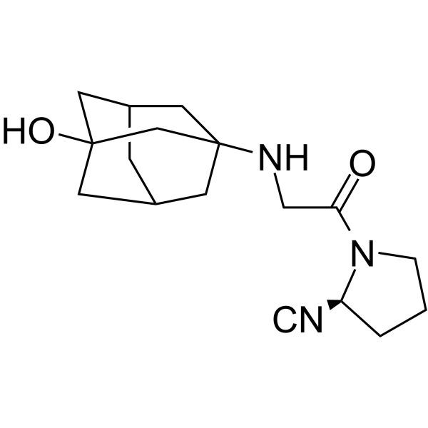
HY-14291
VildagliptinDPP-IV Inhibitor -
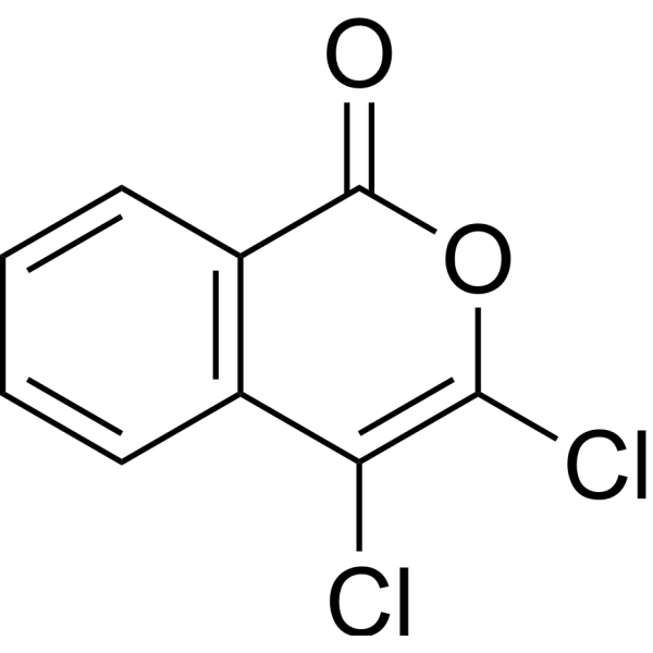
HY-126034
3,4-DichloroisocoumarinSerine-protease Inhibitor
MedChemExpress(MCE)の使用を引用している文献 TPCK
More

生物活性
製品説明
Cellular Effect
|
Cell Line

|
Type | Value | Description | References |
|---|---|---|---|---|
| HEK293 | IC50 |
10.8 μM
Compound: TPCK
|
Inhibition of TNFalpha-induced NFkappaB (unknown origin) activation expressed in HEK293 cells after 6 hrs by luciferase reporter gene assay
Inhibition of TNFalpha-induced NFkappaB (unknown origin) activation expressed in HEK293 cells after 6 hrs by luciferase reporter gene assay
|
[PMID: 23316950] |
| HEK293 | IC50 |
11.1 μM
Compound: TPCK
|
Cytotoxicity against HEK293 cells
Cytotoxicity against HEK293 cells
|
[PMID: 24533857] |
| HEK293 | IC50 |
3.76 μM
Compound: TPCK
|
Inhibition of TNF-alpha-induced NFkappaB activation in human HEK293 cells after 6 hrs by luciferase reporter gene assay
Inhibition of TNF-alpha-induced NFkappaB activation in human HEK293 cells after 6 hrs by luciferase reporter gene assay
|
[PMID: 21261296] |
| HEK293 | IC50 |
3.8 μM
Compound: TPCK
|
Inhibition of TNFalpha-induced NF-kappaB activity expressed in human HEK 293 cells after 6 hrs by luciferase reporter gene assay
Inhibition of TNFalpha-induced NF-kappaB activity expressed in human HEK 293 cells after 6 hrs by luciferase reporter gene assay
|
[PMID: 22850207] |
| HEK293 | IC50 |
3.8 μM
Compound: TPCK
|
Cytotoxicity against human HEK293 cells assessed as cell survival by SRB assay
Cytotoxicity against human HEK293 cells assessed as cell survival by SRB assay
|
[PMID: 23316950] |
| HEK293 | IC50 |
3.8 μM
Compound: TPCK
|
Inhibition of TNFalpha-induced nuclear factor-kappa-B activation in human HEK293 cells at by luciferase reporter gene assay
Inhibition of TNFalpha-induced nuclear factor-kappa-B activation in human HEK293 cells at by luciferase reporter gene assay
|
[PMID: 23994869] |
| HEK293 | IC50 |
3.8 μM
Compound: TPCK
|
Inhibition of TNF-alpha-induced NF-kappaB activity in HEK293 cells after 6 hrs by luciferase reporter gene assay
Inhibition of TNF-alpha-induced NF-kappaB activity in HEK293 cells after 6 hrs by luciferase reporter gene assay
|
[PMID: 24533857] |
| HEK293 | IC50 |
3.8 μM
Compound: TPCK
|
Inhibition of TNF-alpha-induced NF-kappaB activity in HEK293 cells after 6 hrs by luciferase reporter gene assay
Inhibition of TNF-alpha-induced NF-kappaB activity in HEK293 cells after 6 hrs by luciferase reporter gene assay
|
[PMID: 24992702] |
| HEK293 | IC50 |
3.8 μM
Compound: TPCK
|
Inhibition of TNFalpha-induced NF-kappaB activity (unknown origin) transfected in HEK293 cells after 6 hrs by luciferase reporter gene assay
Inhibition of TNFalpha-induced NF-kappaB activity (unknown origin) transfected in HEK293 cells after 6 hrs by luciferase reporter gene assay
|
[PMID: 26343828] |
| HEK293 | IC50 |
3.8 μM
Compound: TPCK
|
Inhibition of TNF-alpha-activated NFkappaB (unknown origin) transfected in HEK293 cells after 6 hrs by luciferase reporter gene assay
Inhibition of TNF-alpha-activated NFkappaB (unknown origin) transfected in HEK293 cells after 6 hrs by luciferase reporter gene assay
|
[PMID: 27196335] |
| HEK293 | IC50 |
4.9 μM
Compound: N-tosyl-L-phenylalanine-chloromethyl ketone
|
Inhibition of TNF-alpha-induced NFkappaB activation in human HEK293 cells after 6 hrs by luciferase assay
Inhibition of TNF-alpha-induced NFkappaB activation in human HEK293 cells after 6 hrs by luciferase assay
|
[PMID: 21978950] |
| HEK293 | IC50 |
5.05 μM
Compound: TPCK
|
Inhibition of TNF-alpha activated nuclear factor-kappa B in HEK293 cells after 6 hrs by luciferase reporter gene assay
Inhibition of TNF-alpha activated nuclear factor-kappa B in HEK293 cells after 6 hrs by luciferase reporter gene assay
|
[PMID: 26048799] |
| HEK293 | IC50 |
5.09 μM
Compound: TPCK
|
Inhibition of NF-kappaB expressed in human HEK293 cells by luciferase reporter gene based luminometric analysis
Inhibition of NF-kappaB expressed in human HEK293 cells by luciferase reporter gene based luminometric analysis
|
[PMID: 21105712] |
| HEK293 | IC50 |
5.09 μM
Compound: TPCK
|
Inhibition of TNFalpha-activated NF-kappaB expressed in HEK293 cells by luciferase reporter gene assay
Inhibition of TNFalpha-activated NF-kappaB expressed in HEK293 cells by luciferase reporter gene assay
|
[PMID: 22115839] |
| HEK293 | IC50 |
5.09 μM
Compound: TPCK
|
Inhibition of TNF-alpha activated NF-kappaB expressed in HEK293 cells by luciferase reporter gene assay
Inhibition of TNF-alpha activated NF-kappaB expressed in HEK293 cells by luciferase reporter gene assay
|
[PMID: 22386564] |
| HEK293 | IC50 |
5.09 μM
Compound: TPCK
|
Inhibition of TNFalpha-induced NF-kappaB activity expressed in HEK293 cells by luciferase reporter gene assay
Inhibition of TNFalpha-induced NF-kappaB activity expressed in HEK293 cells by luciferase reporter gene assay
|
[PMID: 23142320] |
| HEK293 | IC50 |
5.09 μM
Compound: TPCK
|
Inhibition of TNF-alpha induced NF-kappaB transcriptional activity in HEK293 cells expressing NF-kappaB-Luc measured after 48 hrs by luciferase assay
Inhibition of TNF-alpha induced NF-kappaB transcriptional activity in HEK293 cells expressing NF-kappaB-Luc measured after 48 hrs by luciferase assay
|
[PMID: 27155469] |
| HEK293 | IC50 |
5.09 μM
Compound: TPCK
|
Inhibition of TNFalpha-activated nuclear factor-kappa B activity (unknown origin) expressed in HEK293 cells incubated for 6 hrs by luciferase reporter gene based assay
Inhibition of TNFalpha-activated nuclear factor-kappa B activity (unknown origin) expressed in HEK293 cells incubated for 6 hrs by luciferase reporter gene based assay
|
[PMID: 27210437] |
| HEK293 | IC50 |
5.1 μM
Compound: TPCK
|
Inhibition of TNFalpha-induced human NFkappaB activity in HEK293 cells incubated for 6 hrs followed by compound wash out measured after 5 mins by by luciferase assay
Inhibition of TNFalpha-induced human NFkappaB activity in HEK293 cells incubated for 6 hrs followed by compound wash out measured after 5 mins by by luciferase assay
|
[PMID: 22712432] |
| HEK293 | IC50 |
5.3 μM
Compound: TPCK
|
Inhibition of TNFalpha-induced NFkappaB activation in human HEK293 cells after 6 hrs by luciferase assay
Inhibition of TNFalpha-induced NFkappaB activation in human HEK293 cells after 6 hrs by luciferase assay
|
[PMID: 32163285] |
体外実験
TPCK shows slight toxicity to mammalian cells (CC50 = 138.8 µM), but it significantly inhibits the pre-flagellate morphology of L. amazonensis PH8 and Josefa strains (IC50 values of 14.6 µM and 31.7 µM, respectively), with an IC50 value of 11.3 µM for the pre-flagellate morphology of L. infantum. Additionally, TPCK is also effective against the intracellular amastigote form, showing IC50 values of 14.2 µM and 16.6 µM for L. amazonensis PH8 and Josefa strains, and 21.7 µM for the amastigote form of L. infantum[2].
TPCK (40 μM, 1 h) enhances wortmannin-dependent caspase activity in LNCaP cells but inhibits TRAIL-dependent caspase activity[3].
TPCK (40 μM, 1 h) inhibits the formation of TRAIL-DISC in PC3 cells but does not inhibit the formation of Fas-DISC and reduces caspase2 levels[3].
TPCK (0-40 μM, 24 h) kills PC3 cells in a dose-dependent manner, inducing apoptosis[3].
TPCK (40 μM, 3 h) reduces the level of BAD-ser136 in LNCaP cells and eliminates AR in the cytoplasm and granules[3].
MedChemExpress (MCE) has not independently confirmed the accuracy of these methods. They are for reference only.
-
Cell Line:PC3; LNCaP
-
Concentration:40 μM
-
Incubation Time:1 h; 3 h
-
Result:Reduced the level of caspase-2 and inhibited TRAIL-induced activation of caspase-3, -7, -8, and -9. Did not reduce BAD levels, but reduced the level of BAD-ser136 and eliminated AR in the cytosol and granules.
-
Cell Line:PC3
-
Concentration:0, 2.5, 5, 10, 20, 40 μM
-
Incubation Time:24 h
-
Result:Killed PC3 cells in a dose-dependent manner.
体内実験
TPCK (5-100 mg/kg, intraperitoneal injection, single dose) diminishes DNA fragmentation, nitric oxide production, and brain damage in a neonatal mouse model of hypoxic-ischemic brain injury[4].
TPCK (0.5-20 mg/kg, injected into the air sac, single dose) increases vascular permeability in inflamed rats, which then decline[5].
MedChemExpress (MCE) has not independently confirmed the accuracy of these methods. They are for reference only.
-
Animal Model:BALB/c mice infected with L. amazonensis [2]
-
Dosage:15, 30, 45, 60 mg/kg; three times a week; 10 times
-
Administration:Intraperitoneal injection (i.p.)
-
Result:Reduced lesion size and parasite burden in foot pads and spleen.
-
Animal Model:7-day-old rats with right carotid artery ligation[4]
-
Dosage:5, 10, 20, 50, 100 mg/kg; single dose
-
Administration:Intraperitoneal injection (i.p.)
-
Result:Reduced the weight of the right hemisphere of the brain, reduced the number of DNA breaks in cells, abolished the increase in nitrate/nitrite metabolites within 6 hours after injury, and reduced DNA fragmentation, nitric oxide production, and brain damage.
-
Animal Model:Carrageenan-air sac inflammation rats[5]
-
Dosage:0.5, 4, 20 mg/kg; single dose
-
Administration:Injection into the air sac
-
Result:Showed that vascular permeability increased rapidly and then gradually decreased.
- Detection of Apoptosis
- TUNEL staining for apoptotic DNA fragmentation
- Annexin V plus membrane-impermeant dye apoptosis staining
- Patient-Derived Xenograft (PDX)
- Apoptosis Solutions
- Patient-Derived Orthotopic Xenograft (PDOX)
- Transepithelial/transendothelial electrical resistance assay
- Subcutaneous Cell-Line-Derived Xenograft
- Orthotopic Cell-Line Xenograft


化学情報
-
CAS 番号 402-71-1
-
性状 Solid
-
分子量 351.85
-
分子式 C17H18ClNO3S
-
Color White to off-white
-
SMILES
CC1=CC=C(C=C1)S(N[C@@H](CC2=CC=CC=C2)C(CCl)=O)(=O)=O
-
別名
L-1-Tosylamido-2-phenylethyl chloromethyl ketone; L-TPCK
-
輸送条件
Room temperature in continental US; may vary elsewhere.
-
保管条件
Powder -20°C 3 years 4°C 2 years In solvent -80°C 6 months -20°C 1 month


Publications (1)
-
Journal Impact Factor
-
Most Recent
-
Int J Biol Macromol
Evaluation and mechanism of immune enhancement effects of Pleurotus ferulae polysaccharides-gold nanoparticles. [Abstract]2023 Feb 1:227:1015-1026. PMID: 36460244


溶剤 & 溶解度
体外:
DMSO : 100 mg/mL (284.21 mM; Need ultrasonic; Hygroscopic DMSO has a significant impact on the solubility of product, please use newly opened DMSO)
Please refer to the solubility information to select the appropriate solvent. Once prepared, please aliquot and store the solution to prevent product inactivation from repeated freeze-thaw cycles.
Storage method and period of stock solution: -80°C, 6 months; -20°C, 1 month. When stored at -80°C, please use it within 6 months. When stored at -20°C, please use it within 1 month.
Please refer to the solubility information to select the appropriate solvent. Once prepared, please aliquot and store the solution to prevent product inactivation from repeated freeze-thaw cycles.
Storage method and period of stock solution: -80°C, 6 months; -20°C, 1 month. When stored at -80°C, please use it within 6 months. When stored at -20°C, please use it within 1 month.
濃度 (開始) × 体積 (開始) = 濃度 (終了) × 体積 (終了)
体内:
Select the appropriate dissolution method based on your experimental animal and administration route.
- For the following dissolution methods, please ensure to first prepare a clear stock solution using an In Vitro approach and then sequentially add co-solvents:
- To ensure reliable experimental results, the clarified stock solution can be appropriately stored based on storage conditions. As for the working solution for In Vivo experiments, it is recommended to prepare freshly and use it on the same day.
- The percentages shown for the solvents indicate their volumetric ratio in the final prepared solution. If precipitation or phase separation occurs during preparation, heat and/or sonication can be used to aid dissolution.
Add each solvent one by one: 10% DMSO 40% PEG300 5% Tween-80 45% Saline
Solubility: ≥ 2.5 mg/mL (7.11 mM); Clear solution
This protocol yields a clear solution of ≥ 2.5 mg/mL (saturation unknown).
Taking 1 mL working solution as an example, add 100 μL DMSO stock solution (25.0 mg/mL) to 400 μL PEG300, and mix evenly; then add 50 μL Tween-80 and mix evenly; then add 450 μL Saline to adjust the volume to 1 mL.
Preparation of Saline: Dissolve 0.9 g sodium chloride in ddH₂O and dilute to 100 mL to obtain a clear Saline solution.
In Vivo Dissolution Calculator
Please enter the basic information of animal experiments:
-
-
-
-
Recommended: Prepare an additional quantity of animals to account for potential losses during experiments.
Please enter your animal formula composition:
-
%DMSO +
Recommended: Keep the proportion of DMSO in working solution below 2% if your animal is weak.
-
%+
-
+%Tween-80 + +
-
%Saline +
The co-solvents required include: DMSO, . All of co-solvents are available by MedChemExpress (MCE). , Tween 80. All of co-solvents are available by MedChemExpress (MCE).
Working solution concentration: 0.22 mg/mL
Method for preparing stock solution: mg drug dissolved in μL DMSO. Stock solution concentration: mg/mL.
1. Take μL DMSO stock solution;
2. Add μL .
μL , mix evenly;
3. Then add μL Tween 80, mix evenly;
4. Then add μL
Please ensure that the stock solution in the first step is dissolved to a clear state, and add co-solvents in sequence. You can use ultrasonic heating (ultrasonic cleaner, recommended frequency 20-40 kHz), vortexing, etc. to assist dissolution.


純度とドキュメンテーション
-
データシート (281 KB)
-
SDS (615 KB)
- English - EN (615 KB)
- Français - FR (615 KB)
- Deutsch - DE (615 KB)
- Norwegian - NO (615 KB)
- Español - ES (615 KB)
- Swedish - SV (615 KB)
- Italian - IT (615 KB)
- Korean - KR (615 KB)
- Portuguese - PT (615 KB)
-
取扱説明書 (2659 KB)
参考文献
[1]. H Stöppler, et al. The serine protease inhibitors TLCK and TPCK react with the RB-binding core of HPV-18 E7 protein and abolish its RB-binding capability. Virology. 1996 Mar 15;217(2):542-53. [Content Brief]
[2]. Patrícia de A Machado, et al. Effects of a Serine Protease Inhibitor N- p-Tosyl-L-phenylalanine Chloromethyl Ketone (TPCK) on Leishmania amazonensis and Leishmania infantum. Pharmaceutics.2022 Jun 29;14(7):1373. [Content Brief]
[3]. M H LeBlanc, et al. N-tosyl-L-phenylalanyl-chloromethylketone reduces hypoxic-ischemic brain injury in rat pups. Eur J Pharmacol. 2000 Mar 3;390(3):249-56 [Content Brief]
[4]. K Watanabe, et al. Vascular permeability changes by proteinase inhibitors in carrageenin-induced inflammation in rats. Agents Actions. 1986 Mar;17(5-6):472-7. [Content Brief]


Complete Stock Solution Preparation Table
Please refer to the solubility information to select the appropriate solvent. Once prepared, please aliquot and store the solution to prevent product inactivation from repeated freeze-thaw cycles.
Storage method and period of stock solution: -80°C, 6 months; -20°C, 1 month. When stored at -80°C, please use it within 6 months. When stored at -20°C, please use it within 1 month.
| Optional Solvent | Concentration Solvent Mass | 1 mg | 5 mg | 10 mg | 25 mg |
|---|---|---|---|---|---|
| DMSO | 1 mM | 2.8421 mL | 14.2106 mL | 28.4212 mL | 71.0530 mL |
| 5 mM | 0.5684 mL | 2.8421 mL | 5.6842 mL | 14.2106 mL | |
| 10 mM | 0.2842 mL | 1.4211 mL | 2.8421 mL | 7.1053 mL | |
| 15 mM | 0.1895 mL | 0.9474 mL | 1.8947 mL | 4.7369 mL | |
| 20 mM | 0.1421 mL | 0.7105 mL | 1.4211 mL | 3.5527 mL | |
| 25 mM | 0.1137 mL | 0.5684 mL | 1.1368 mL | 2.8421 mL | |
| 30 mM | 0.0947 mL | 0.4737 mL | 0.9474 mL | 2.3684 mL | |
| 40 mM | 0.0711 mL | 0.3553 mL | 0.7105 mL | 1.7763 mL | |
| 50 mM | 0.0568 mL | 0.2842 mL | 0.5684 mL | 1.4211 mL | |
| 60 mM | 0.0474 mL | 0.2368 mL | 0.4737 mL | 1.1842 mL | |
| 80 mM | 0.0355 mL | 0.1776 mL | 0.3553 mL | 0.8882 mL | |
| 100 mM | 0.0284 mL | 0.1421 mL | 0.2842 mL | 0.7105 mL |


TPCK 関連分類
HY-124379 関連分類
 Powered by Bioz