



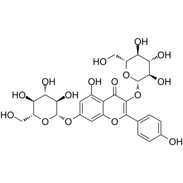
Kaempferol-3,7-di-O-β-glucoside

Based on 1 Customer Validation
Kaempferol-3,7-di-O-β-glucoside (Kaempferol 3,7-diglucoside), a flavonol, possesses enzyme inhibition property towards α-amylase, α-glucosidase and Acetylcholinesterase. Kaempferol-3,7-di-O-β-glucoside protects differentiating neuronal cells, SH-SY5Y from Amyloid β peptide-induced injury. Kaempferol-3,7-di-O-β-glucoside has the potential for Alzheimer's research.
For research use only. We do not sell to patients.
- Purity : 99.59%
- CAS No.: 25615-14-9
- Formula: C27H30O16
- Molecular Weight:610.52
-
Storage:
4°C, sealed storage, away from moisture and light
* In solvent : -80°C, 6 months; -20°C, 1 month (sealed storage, away from moisture and light)

To place orders, for customer services and technical support, please contact: MedChemExpress USA
Tel: 609-228-6898 E-mail: [email protected] [email protected]


-
 Biological Activity
-
Chemical Information
- Protocols
- Purity & Documentation
- References
-
 Help & FAQs


-
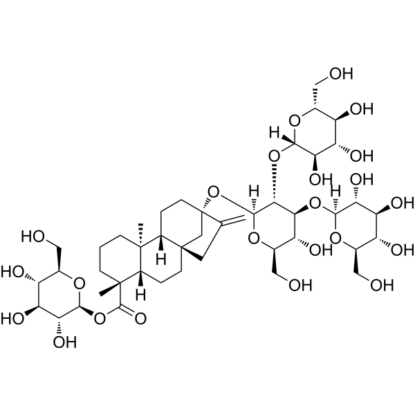
HY-N0466
Rebaudioside Aα-Glucosidase Inhibitor -
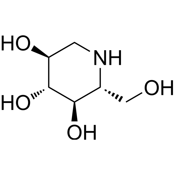
HY-14860
1-Deoxynojirimycinα-glucosidase Inhibitor -
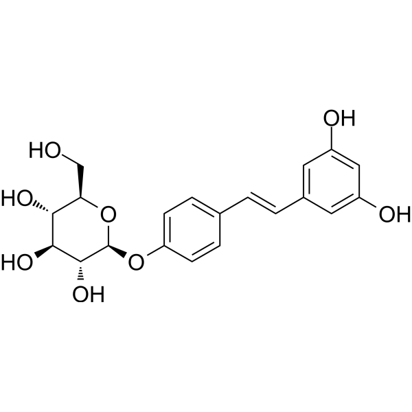
HY-N4195
Resveratrolosideα-Glucosidase Inhibitior -

HY-14860R
1-Deoxynojirimycin (Standard)α-glucosidase Inhibitor -
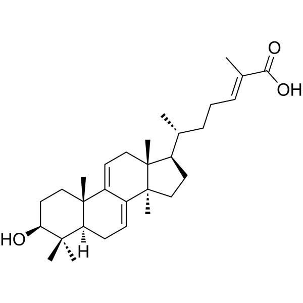
HY-125713
Ganoderic acid YGlucosidase Inhibitor -
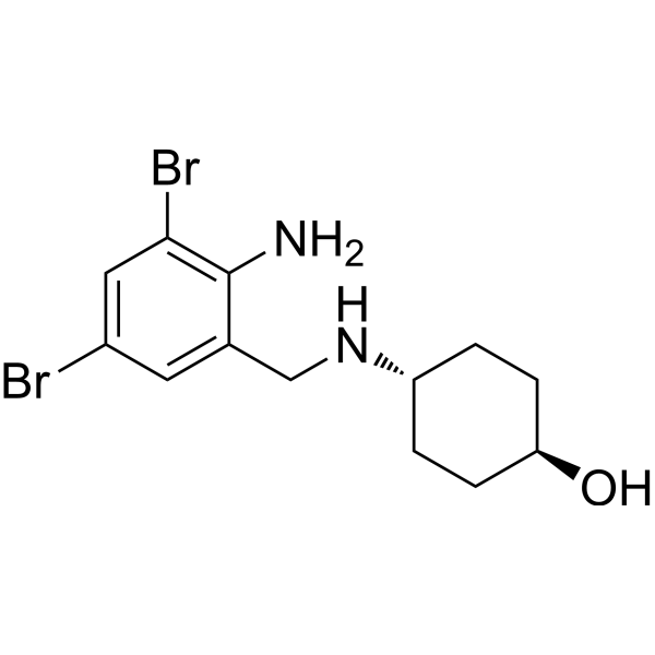
HY-B1039
AmbroxolBromhexine Metabolite -

HY-N0466R
Rebaudioside A (Standard)Standard of Rebaudioside A -
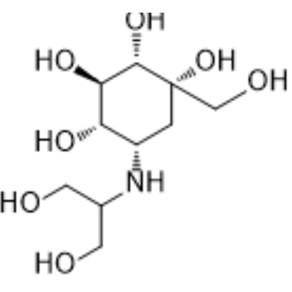
HY-B0025
VogliboseGlucosidase Inhibitor -
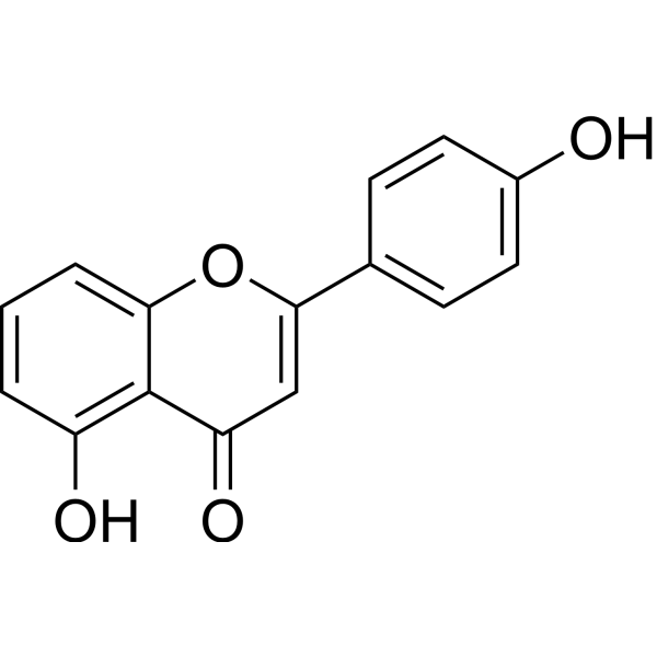
HY-N1881
4',5-DihydroxyflavoneColA Inhibitor -
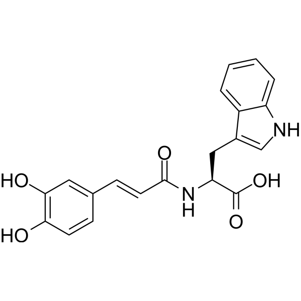
HY-N10002
CaffeoyltryptophanPTP1B Inhibitor -
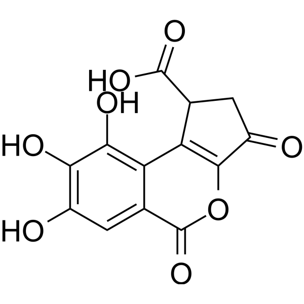
HY-N4095
Brevifolincarboxylic acidPhenolic Compound -
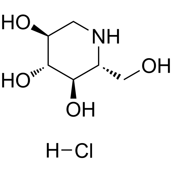
HY-14860A
1-Deoxynojirimycin hydrochlorideα-glucosidase Inhibitor


Biological Activity
Description
Cellular Effect
|
Cell Line

|
Type | Value | Description | References |
|---|---|---|---|---|
| BV-2 | IC50 |
28.89 μM
Compound: 16
|
Antineuroinflammatory activity against mouse BV2 cells assessed as inhibition of LPS-induced nitric oxide production after 24 hrs by Griess method
Antineuroinflammatory activity against mouse BV2 cells assessed as inhibition of LPS-induced nitric oxide production after 24 hrs by Griess method
|
[PMID: 23149227] |
In Vitro
Kaempferol-3,7-di-O-β-glucoside (Kaempferol 3,7-diglucoside; 0.1, 100 μM) has effective inhibition in Aβ1-42 fibril formation. Kaempferol-3,7-di-O-β-glucoside has less neurotoxicity on the cultured SH-SY5Y cells[1].
MedChemExpress (MCE) has not independently confirmed the accuracy of these methods. They are for reference only. Further protocols information, click here.


Chemical Information
-
CAS No. 25615-14-9
-
Appearance Solid
-
Molecular Weight 610.52
-
Formula C27H30O16
-
Color Off-white to light yellow
-
SMILES
O=C1C(O[C@@H]([C@@H]([C@H]2O)O)O[C@@H]([C@H]2O)CO)=C(C(C=C3)=CC=C3O)OC4=CC(O[C@@H]([C@@H]([C@H]5O)O)O[C@@H]([C@H]5O)CO)=CC(O)=C14
-
Synonyms
Kaempferol 3,7-diglucoside
-
Structure Classification
-
Initial Source
-
Shipping
Room temperature in continental US; may vary elsewhere.
-
Storage
4°C, sealed storage, away from moisture and light
* In solvent : -80°C, 6 months; -20°C, 1 month (sealed storage, away from moisture and light)

Protocols
-
Amyloid: Congo Red Amyloid Staining
Congo red amyloid staining is a histochemical method used to detect extracellular amyloid deposits in tissue sections based on the affinity of Congo red dye for β-pleated sheet-rich protein aggregates. When bound to amyloid, Congo red produces characteristic apple-green birefringence under polarized light microscopy, which is widely regarded as a diagnostic feature of amyloid deposition in histopathology. The diagnostic principle relies on the combination of dye binding (congophilia) and optical anisotropy under polarized illumination, which distinguishes amyloid from most non-amyloid eosinophilic extracellular deposits in routine histological evaluation. Amyloid identification by Congo red staining remains a cornerstone in diagnostic pathology despite the availability of adjunct methods such as immunohistochemistry and mass spectrometry, particularly because of its ability to localize deposits directly within tissue architecture. The specificity of Congo red-positive deposits is incre


Purity & Documentation
-
Data Sheet (272 KB)
-
SDS (252 KB)
- English - EN (252 KB)
- Français - FR (252 KB)
- Deutsch - DE (252 KB)
- Norwegian - NO (252 KB)
- Español - ES (252 KB)
- Swedish - SV (252 KB)
- Italian - IT (252 KB)
- Korean - KR (252 KB)
- Portuguese - PT (252 KB)
-
Handling Instructions (2659 KB)
References

Calculators
Concentration (start) × Volume (start) = Concentration (final) × Volume (final)


Kaempferol-3,7-di-O-β-glucoside Related Classifications
HY-N8161 Related Classifications
 Powered by Bioz