



SJFδ
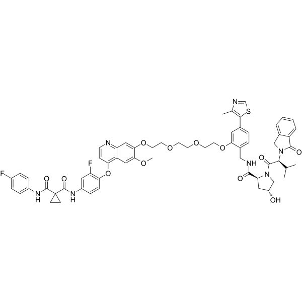
SJFδ is a p38δ PROTAC degrader with a DC50 of 46.17 nM. SJFδ selectively induces ubiquitin-proteasome-dependent degradation of p38δ by forming a ternary complex with p38δ and the VHL E3 ligase, with no effect on other p38 isoforms or members of the MAPK family. SJFδ can be used for breast cancer research.
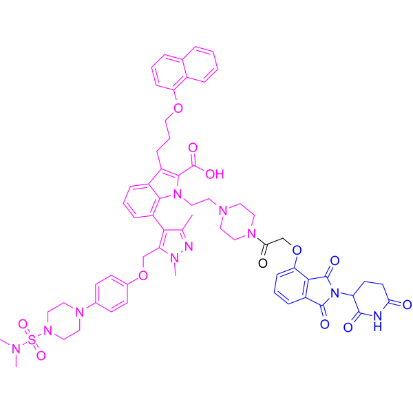
(Pink: p38δ ligand (HY-45124); Blue: VHL ligand (HY-151227); Black: linker (HY-133143)).
연구목적의 판매만을 진행합니다. 환자를 대상으로 한 판매는 하지 않습니다.
- CAS No.: 2254609-23-7
- 화학식: C62H63F2N7O12S
- 분자량:1168.27
-
보관:
Please store the product under the recommended conditions in the Certificate of Analysis.

To place orders, for customer services and technical support, please contact: MedChemExpress USA
Tel: 609-228-6898 E-mail: [email protected] [email protected]


-
 Biological Activity
-
Chemical Information
- Protocol
- 순도&문서
- References
-
 Help & FAQs


-
HY-114404
SJFαPROTAC p38α Degrader -
HY-128360
dMCL1-2MCL1 PROTAC Degrader -
HY-100972
ARV-771BET PROTAC Degrader -
HY-123921
Gefitinib-based PROTAC 3EGFR PROTAC Degrader -
HY-101519
BETd-260BET PROTAC Degrader -
HY-128600
ERD-308ER PROTAC Degrader -
HY-111846
PROTAC ERα Degrader-2PROTAC ERα Degrader -
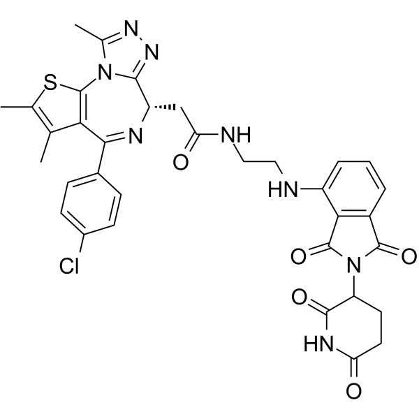
HY-123844
dBET57BRD4 PROTAC Degrader -
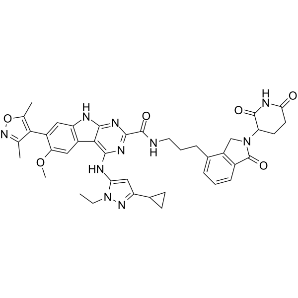
HY-114228
PROTAC BET degrader-2BRD2/BRD3/BRD4 PROTAC Degrader -
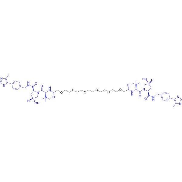
HY-111593
Homo-PROTAC pVHL30 degrader 1pVHL30/19 PROTAC Degrader -
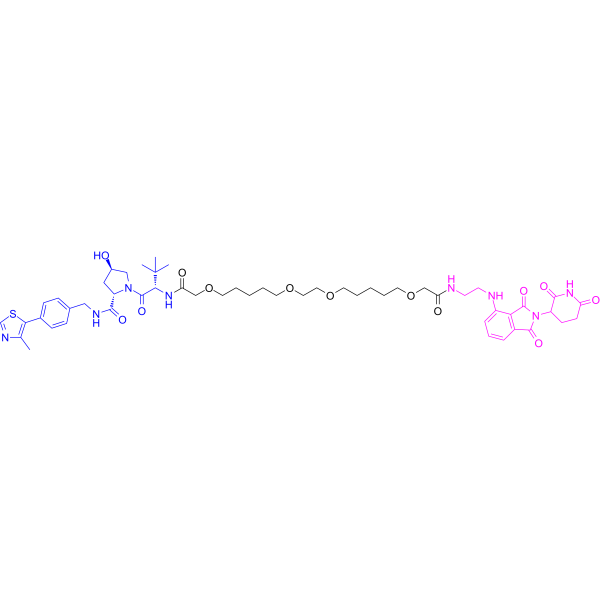
HY-128845
PROTAC CRBN Degrader-1PROTAC CRBN Degrader -

HY-100972R
ARV-771 (Standard)Reference Standard
All PROTACs Isoforms
More

Biological Activity
제품 설명
IC50 & Target
[4]|
p38δ 46.17 nM (DC50) |
Cellular Effect
|
Cell Line

|
Type | Value | Description | References |
|---|---|---|---|---|
| MDA-MB-231 | DC50 |
46.17 nM
|
Selective degradation of p38δ in human MDA-MB-231 breast cancer cells assessed via immunoblotting.
Selective degradation of p38δ in human MDA-MB-231 breast cancer cells assessed via immunoblotting.
|
30631068 |
In Vitro
PROTAC SJFδ forms a stable ternary complex with p38δ and VHL, with a ternary Kd of 436 nM and a dissociation half-life of 38 s, which is more stable than the ternary complex formed by the related PROTAC SJFα[1].
PROTAC SJFδ does not induce substantial ubiquitination of p38α in transfected HeLa cells[1].
SJFδ preferentially degrades p38δ kinase in cells with a DC50 of 46 nM and a maximal degradation of 99%[3].
SJFδ (0.025-2.5 μM) forms stable, dose-dependent ternary complexes with endogenous p38δ and VBC in MDA-MB-231 whole-cell lysate[4].
SJFδ (up to 5 μM; 60 s injection, 300 s dissociation monitoring) forms a high-affinity p38δ:SJFδ:VHL ternary complex with a Kd of 436 nM, which has a longer half-life and stronger affinity than the p38δ:SJFα:VHL ternary complex[4].
SJFδ (1 μM; 2 h) induces significant cellular ubiquitination of FLAG-p38δ in transfected HeLa cells[4].
SJFδ (0.025-2.5 μM; 6 h-24 h) potently and selectively degrades p38δ with a DC50 of 46.17 nM in MDA-MB-231 human breast cancer cells, via a rapid, sustained, proteasome-dependent mechanism, and also degrades mutant p38δK220E/T221E in HeLa cells[4].
MedChemExpress (MCE) has not independently confirmed the accuracy of these methods. They are for reference only. Further protocols information, click here.
-
Cell Line:MDA-MB-231 human breast cancer cells, HeLa cells
-
Concentration:0.025, 0.25, 2.5 μM (dose-response); 250 nM (6 h incubation, 24 h incubation, washout assays); 100 nM, 500 nM, 1 μM, 2 μM (mutant p38δ degradation); 1 μM (proteasome inhibitor/NAE inhibitor pre-treatment)
-
Incubation Time:6 h, 24 h, 30, 90, 180, 360 min (cycloheximide chase); 30 min (proteasome inhibitor/NAE inhibitor pre-treatment); 1 h (cycloheximide pre-treatment)
-
Result:Selectively degraded p38δ with a DC50 of 46.17 nM and a Dmax of 99.41%.
Did not degrade p38α, p38β, p38γ, ERK1, ERK2, JNK1, or JNK2 at concentrations up to 2.5 μM.
Pre-treatment with 1 μM proteasome inhibitor or 1 μM NAE inhibitor completely rescued p38δ degradation.
Showed no significant changes in p38δ mRNA levels after 24 h treatment.
Decreased p38δ abundance over 30-360 min in cycloheximide chase assays.
Maintained sustained p38δ degradation for 24 h post-washout.
Degraded wild-type p38δ and mutant p38δ (K220E/T221E) with similar efficacy in transfected HeLa cells.
-
Cell Line:HeLa cells co-expressing FLAG-p38δ and HA-ubiquitin
-
Concentration:1 μM
-
Incubation Time:2 h
-
Result:Induced substantial poly-ubiquitination of FLAG-p38δ, with prominent high-molecular-weight HA-ubiquitin smears far more abundant than in DMSO- or SJFα-treated cells.
Confirmed increased poly-ubiquitinated p38δ via TUBE1 immunoprecipitation.


Chemical Information
-
CAS No. 2254609-23-7
-
분자량 1168.27
-
화학식 C62H63F2N7O12S
-
SMILES
FC1=CC=C(NC(C2(CC2)C(NC3=CC(F)=C(OC4=C(C=C(OC)C(OCCOCCOCCOC5=CC(C6=C(C)N=CS6)=CC=C5CNC([C@@H]7C[C@@H](O)CN7C([C@@H](N8C(C9=CC=CC=C9C8)=O)C(C)C)=O)=O)=C%10)C%10=NC=C4)C=C3)=O)=O)C=C1
-
선적
Room temperature in continental US; may vary elsewhere.
-
보관
Please store the product under the recommended conditions in the Certificate of Analysis.

Protocol
-
Breast Cancer Modeling
Breast cancer is a heterogeneous cancer, and it has been distinguished into four subtypes: luminal A, luminal B, HER2-positive and basal-like. Molecular mutations, epigenetic alterations, hormone exposure and immune microenvironment are related to the progression of breast cancer.


순도&문서
References
[2]. Sun X, et al. PROTACs: great opportunities for academia and industry. Signal transduction and targeted therapy. 2019;4:64. [Content Brief]
[3]. Luo H, et al. Advancing Design Strategy of PROTACs for Cancer Therapy. MedComm. 2025 Jul;6(7):e70258. [Content Brief]
[4]. Smith BE, et al. Differential PROTAC substrate specificity dictated by orientation of recruited E3 ligase. Nature communications. 2019 Jan 10;10(1):131. [Content Brief]

Calculators
Concentration (start) × Volume (start) = Concentration (final) × Volume (final)


SJFδ Related Classifications
HY-114405 Related Classifications
 Powered by Bioz