



MYC degrader 1
Based on 2 publication(s) in Google Scholar
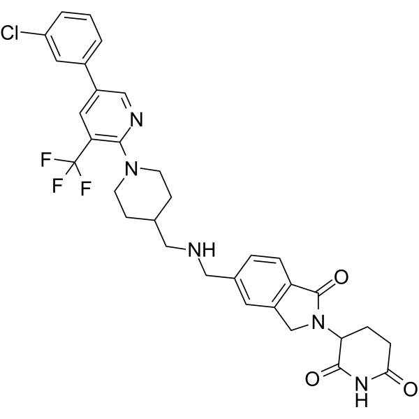
MYC degrader 1 is a MYC degrader that recruits Cereblon (CRBN), with a Kd value of 145 nM and oral activity. MYC degrader 1 specifically binds MYC and GSPT1, recruits the CRBN E3 ubiquitin ligase complex, and induces MYC degradation via the ubiquitin-proteasome pathway. MYC degrader 1 downregulates KLHL42 expression, restores pRB1 protein levels, and re-establishes the sensitivity of MYC-overexpressing cancer cells to CDK4/6 inhibitors. MYC degrader 1 combined with Palbociclib (HY-50767) shows significant inhibition of tumor growth. MYC degrader 1 can be used in studies related to bladder cancer, prostate cancer, and breast cancer.
연구목적의 판매만을 진행합니다. 환자를 대상으로 한 판매는 하지 않습니다.
- Purity : 99.61%
- CAS No.: 2946670-96-6
- 화학식: C32H31ClF3N5O3
- 분자량:626.07
-
보관:
-20°C, sealed storage, away from moisture and light
* In solvent : -80°C, 6 months; -20°C, 1 month (sealed storage, away from moisture and light)

To place orders, for customer services and technical support, please contact: MedChemExpress USA
Tel: 609-228-6898 E-mail: [email protected] [email protected]


-
 Biological Activity
-
Chemical Information
-
용액&용해도
- Protocol
- 순도&문서
- References
-
 Help & FAQs


-
HY-145898
WBC100c-Myc Inhibitor -
HY-129600
MYCi361c-Myc Inhibitor -
HY-144878
VPC-70619N-Myc Inhibitor -
HY-122683
sAJM589Myc Inhibitor -
HY-12702
10058-F4c-Myc Inhibitor -

HY-10071
Y-27632ROCK Inhibitor -
HY-Y0873A
PEG400 -
HY-Y1893
Solutol HS-15 -
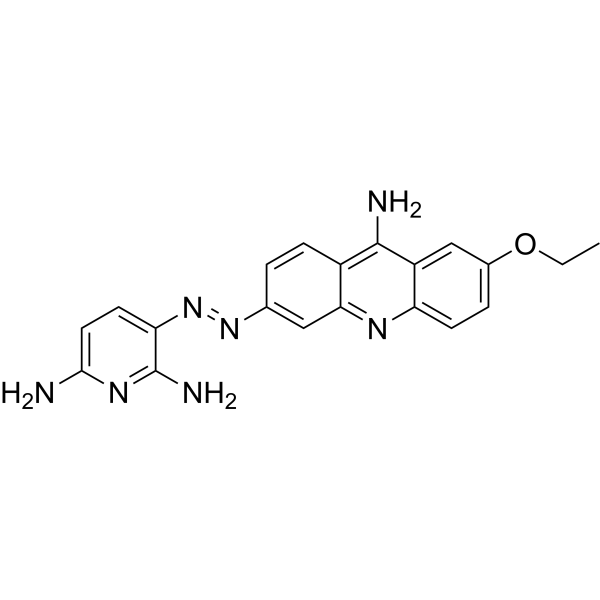
HY-124675
MYCMI-6MYC:MAX Interaction Inhibitor -
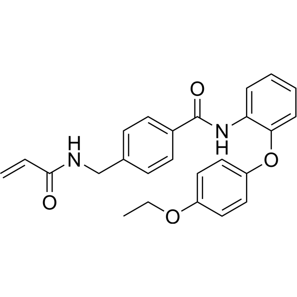
HY-134761
EN4Covalent Ligand of c-MYC -
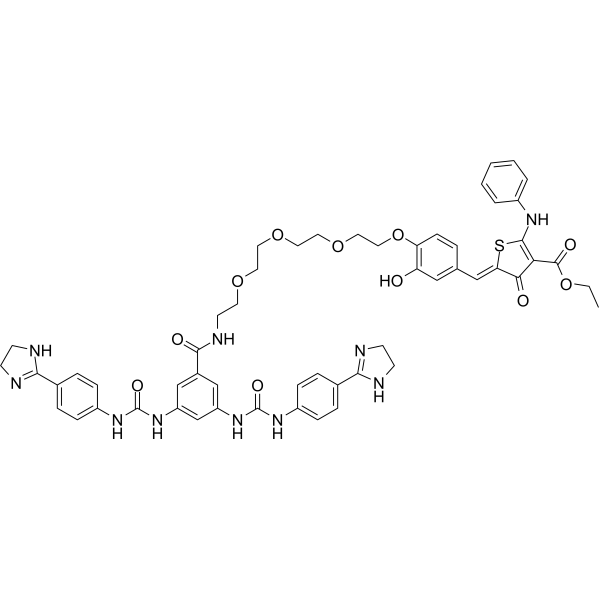
HY-153713
MYC-RIBOTACMYC-RIBOTAC -
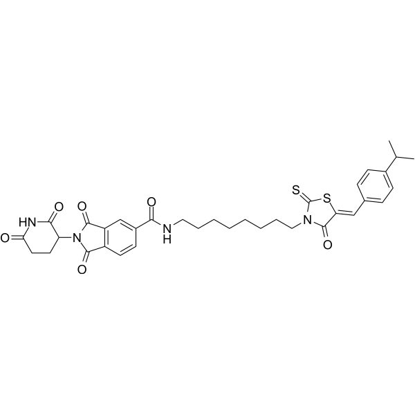
HY-150259
MDEG-541MYC GSPT1 GSPT2 PLK1 Degrader
Publications Citing Use of MedChemExpress (MCE) MYC degrader 1
More

Biological Activity
제품 설명
Cellular Effect
|
Cell Line

|
Type | Value | Description | References |
|---|---|---|---|---|
| T-24 | IC50 |
12-78 nM
|
Inhibition of cell viability in human T24 bladder cancer cells.
Inhibition of cell viability in human T24 bladder cancer cells.
|
38424044 |
| LNCaP C4-2 | IC50 |
12-78 nM
|
Inhibition of cell viability in human C4-2 prostate cancer cells.
Inhibition of cell viability in human C4-2 prostate cancer cells.
|
38424044 |
| MDA-MB-231 | IC50 |
12-78 nM
|
Inhibition of cell viability in human MDA-MB-231 breast cancer cells.
Inhibition of cell viability in human MDA-MB-231 breast cancer cells.
|
38424044 |
In Vitro
MYC degrader 1 (compound A80.2HCl) binds to purified MYC protein with a dissociation constant of 145 nM[1].
MYC degrader 1 directly binds to the CRBN protein[1].
MYC degrader 1 promotes the interaction between endogenous CRBN and MYC in T24 bladder cancer cells[1].
MYC degrader 1 mediates MYC degradation in T24 bladder cancer cells in a CRBN-dependent manner[1].
MYC degrader 1 inhibits colony formation of T24, UMUC14 bladder cancer cells, C4-2 prostate cancer cells, and MDA-MB-231 breast cancer cells, and its activity depends on MYC expression[1].
MYC degrader 1 inhibits cell viability, with an IC50 range of 12 nM to 78 nM in T24 and UMUC14 bladder cancer cells, C4-2 prostate cancer cells, and MDA-MB-231 breast cancer cells, and its activity is dependent on MYC expression[1].
MYC degrader 1 enhances the efficacy of CDK4/6 inhibitors (Palbociclib, Abemaciclib (HY-16297A), Ribociclib (HY-15777)) in T24 and UMUC14 bladder cancer cells, reduces the IC50 of Palbociclib, and induces synergistic cytotoxicity[1].
MYC degrader 1 enhances the ability of CDK4/6 inhibitors (Palbociclib, Abemaciclib, Ribociclib) to suppress colony formation of T24 and UMUC14 bladder cancer cells[1].
MYC degrader 1 (2-50 nM) induces proteasomal degradation of MYC, reduces KLHL42 expression, and increases the stability of pRB1 protein in T24 bladder cancer cells, C4-2 prostate cancer cells, and MDA-MB-231 breast cancer cells[1].
MYC degrader 1 (10 nM; 24 h) inhibits the transcriptional activity of MYC and downregulates the target genes of MYC and E2F in T24 bladder cancer cells[1].
MYC degrader 1 (10 nM; 24 h) combined with Palbociclib (HY-50767) (1 μM) shows stronger inhibition of MYC and E2F target genes in T24 bladder cancer cells than either agent alone[1].
MYC degrader 1 overcomes the acquired resistance of cells to CDK4/6 inhibitors by resensitizing T24 bladder cancer cells to Palbociclib[1].
MedChemExpress (MCE) has not independently confirmed the accuracy of these methods. They are for reference only.
-
Cell Line:T24, C4-2, MDA-MB-231, 22RV1, T47D and UMUC14 cell lines
-
Concentration:0, 10, 25, 50, 100, 500 and 1000 nM
-
Incubation Time:24 h
-
Result:Reduced KLHL42 levels and increased RB1 levels.
In Vivo
MYC degrader 1 (6 mg/kg; p.o.; once daily; study terminated on day 30) enhances the antitumor efficacy of Palbociclib in UMUC14 bladder cancer xenografts[1].
MYC degrader 1 inhibits the growth of T24 bladder cancer xenografts in a MYC-dependent manner and reduces MYC protein levels in tumor tissues[1].
MYC degrader 1 (6 mg/kg; p.o.; once daily; for 7 consecutive days) enhances the antitumor efficacy of Palbociclib in MYC-amplified breast cancer mini-PDX models[1].
MedChemExpress (MCE) has not independently confirmed the accuracy of these methods. They are for reference only.
-
Animal Model:Male nude mice were subcutaneously inoculated with T24 cells (1×10⁶ suspended in 100 μL of serum-free DMEM, mixed with Matrigel at a 1:1 ratio) on the right side of the abdomen to induce tumor formation. Subsequent experiments were started when the tumor volume reached 100-150 mm3[1]
-
Dosage:6 mg/kg
-
Administration:p.o.; daily; harvesting tumors on the 30th day after cancer cell inoculation
-
Result:Significantly inhibited T24 xenograft tumor growth.
Produced more profound reduction in tumor volume when combined with palbociclib compared to either single agent.\nProduced more profound reduction in UMUC14 xenograft tumor volume when combined with palbociclib compared to either single agent.
-
Animal Model:Male NSG mice[1]
-
Dosage:6 mg/kg (in combination with palbociclib)
-
Administration:p.o.; daily (in combination with Palbociclib); harvesting tumors on the 30th day after cancer cell inoculation
-
Result:Significantly inhibited T24 xenograft tumor growth.
Had little effect on MYC-knockdown xenograft tumors.
Markedly decreased MYC protein levels in treated tumors.
-
Animal Model:5-week-old female nu/nu mice[1]
-
Dosage:6 mg/kg
-
Administration:p.o.; daily; 7 days
-
Result:Showed no significant effect on tumor cell growth in MYC-amplified breast cancer mini-PDX models when administered alone.
Markedly enhanced the growth-inhibitory efficacy of palbociclib when combined.


Chemical Information
-
CAS No. 2946670-96-6
-
Appearance Solid
-
분자량 626.07
-
화학식 C32H31ClF3N5O3
-
Color White to off-white
-
SMILES
ClC1=CC(C2=CC(C(F)(F)F)=C(N3CCC(CNCC4=CC(CN(C5C(NC(CC5)=O)=O)C6=O)=C6C=C4)CC3)N=C2)=CC=C1
-
선적
Room temperature in continental US; may vary elsewhere.
-
보관
-20°C, sealed storage, away from moisture and light
* In solvent : -80°C, 6 months; -20°C, 1 month (sealed storage, away from moisture and light)


Publications (2)
-
Journal Impact Factor
-
Most Recent
-
Adv Sci (Weinh)
BCR::ABL1-Induced Enhancer Reprogramming Uncovers Hypersensitivity of Ph+B-ALL Cells to Enhancer-Targeting Drugs. [Abstract]2026 Mar 1:e17231. PMID: 41764406 -


용액&용해도
In Vitro:
DMSO : 100 mg/mL (159.73 mM; Need ultrasonic; Hygroscopic DMSO has a significant impact on the solubility of product, please use newly opened DMSO)
Please refer to the solubility information to select the appropriate solvent. Once prepared, please aliquot and store the solution to prevent product inactivation from repeated freeze-thaw cycles.
Storage method and period of stock solution: -80°C, 6 months; -20°C, 1 month (sealed storage, away from moisture and light). When stored at -80°C, please use it within 6 months. When stored at -20°C, please use it within 1 month.
Please refer to the solubility information to select the appropriate solvent. Once prepared, please aliquot and store the solution to prevent product inactivation from repeated freeze-thaw cycles.
Storage method and period of stock solution: -80°C, 6 months; -20°C, 1 month (sealed storage, away from moisture and light). When stored at -80°C, please use it within 6 months. When stored at -20°C, please use it within 1 month.
Concentration (start) × Volume (start) = Concentration (final) × Volume (final)
In Vivo:
Select the appropriate dissolution method based on your experimental animal and administration route.
- For the following dissolution methods, please ensure to first prepare a clear stock solution using an In Vitro approach and then sequentially add co-solvents:
- To ensure reliable experimental results, the clarified stock solution can be appropriately stored based on storage conditions. As for the working solution for In Vivo experiments, it is recommended to prepare freshly and use it on the same day.
- The percentages shown for the solvents indicate their volumetric ratio in the final prepared solution. If precipitation or phase separation occurs during preparation, heat and/or sonication can be used to aid dissolution.
Add each solvent one by one: 10% DMSO 90% (20% SBE-β-CD in Saline)
Solubility: ≥ 2.5 mg/mL (3.99 mM); Clear solution
This protocol yields a clear solution of ≥ 2.5 mg/mL (saturation unknown).
Taking 1 mL working solution as an example, add 100 μL DMSO stock solution (25.0 mg/mL) to 900 μL 20% SBE-β-CD in Saline, and mix evenly.
Preparation of 20% SBE-β-CD in Saline (4°C, storage for one week): 2 g SBE-β-CD powder is dissolved in 10 mL Saline, completely dissolve until clear.
In Vivo Dissolution Calculator
Please enter the basic information of animal experiments:
-
-
-
-
Recommended: Prepare an additional quantity of animals to account for potential losses during experiments.
Please enter your animal formula composition:
-
%DMSO +
Recommended: Keep the proportion of DMSO in working solution below 2% if your animal is weak.
-
%+
-
+%Tween-80 + +
-
%Saline +
The co-solvents required include: DMSO, . All of co-solvents are available by MedChemExpress (MCE). , Tween 80. All of co-solvents are available by MedChemExpress (MCE).
Working solution concentration: 0.22 mg/mL
Method for preparing stock solution: mg drug dissolved in μL DMSO. Stock solution concentration: mg/mL. * In solvent : -80°C, 6 months; -20°C, 1 month (sealed storage, away from moisture and light)
1. Take μL DMSO stock solution;
2. Add μL .
μL , mix evenly;
3. Then add μL Tween 80, mix evenly;
4. Then add μL
Please ensure that the stock solution in the first step is dissolved to a clear state, and add co-solvents in sequence. You can use ultrasonic heating (ultrasonic cleaner, recommended frequency 20-40 kHz), vortexing, etc. to assist dissolution.


순도&문서
-
Data Sheet (295 KB)
-
SDS (252 KB)
- English - EN (252 KB)
- Français - FR (252 KB)
- Deutsch - DE (252 KB)
- Norwegian - NO (252 KB)
- Español - ES (252 KB)
- Swedish - SV (252 KB)
- Italian - IT (252 KB)
- Korean - KR (252 KB)
- Portuguese - PT (252 KB)
-
Handling Instructions (2659 KB)
References


Complete Stock Solution Preparation Table
Please refer to the solubility information to select the appropriate solvent. Once prepared, please aliquot and store the solution to prevent product inactivation from repeated freeze-thaw cycles.
Storage method and period of stock solution: -80°C, 6 months; -20°C, 1 month (sealed storage, away from moisture and light). When stored at -80°C, please use it within 6 months. When stored at -20°C, please use it within 1 month.
| Optional Solvent | Concentration Solvent Mass | 1 mg | 5 mg | 10 mg | 25 mg |
|---|---|---|---|---|---|
| DMSO | 1 mM | 1.5973 mL | 7.9863 mL | 15.9727 mL | 39.9316 mL |
| 5 mM | 0.3195 mL | 1.5973 mL | 3.1945 mL | 7.9863 mL | |
| 10 mM | 0.1597 mL | 0.7986 mL | 1.5973 mL | 3.9932 mL | |
| 15 mM | 0.1065 mL | 0.5324 mL | 1.0648 mL | 2.6621 mL | |
| 20 mM | 0.0799 mL | 0.3993 mL | 0.7986 mL | 1.9966 mL | |
| 25 mM | 0.0639 mL | 0.3195 mL | 0.6389 mL | 1.5973 mL | |
| 30 mM | 0.0532 mL | 0.2662 mL | 0.5324 mL | 1.3311 mL | |
| 40 mM | 0.0399 mL | 0.1997 mL | 0.3993 mL | 0.9983 mL | |
| 50 mM | 0.0319 mL | 0.1597 mL | 0.3195 mL | 0.7986 mL | |
| 60 mM | 0.0266 mL | 0.1331 mL | 0.2662 mL | 0.6655 mL | |
| 80 mM | 0.0200 mL | 0.0998 mL | 0.1997 mL | 0.4991 mL | |
| 100 mM | 0.0160 mL | 0.0799 mL | 0.1597 mL | 0.3993 mL |


MYC degrader 1 Related Classifications
HY-162318 Related Classifications
 Powered by Bioz