



BCP-T.A

Based on 1 Customer Validation
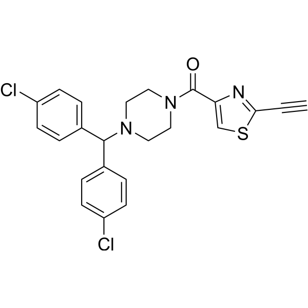
BCP-T.A, a tunable heterocyclic electrophile, is a potent ferroptosis inducer by binding to GPX4. BCP-T.A is a click chemistry reagent, it contains an Alkyne group and can undergo copper-catalyzed azide-alkyne cycloaddition (CuAAc) with molecules containing Azide groups.
연구목적의 판매만을 진행합니다. 환자를 대상으로 한 판매는 하지 않습니다.
- Purity : ≥99.0%
- CAS No.: 2786829-70-5
- 화학식: C23H19Cl2N3OS
- 분자량:456.39
-
보관:Powder -20°C, 3 years ; In solvent -80°C, 6 months , -20°C, 1 month

To place orders, for customer services and technical support, please contact: MedChemExpress USA
Tel: 609-228-6898 E-mail: [email protected] [email protected]


-
 Biological Activity
-
Chemical Information
- Protocol
- 순도&문서
- References
-
 Help & FAQs


-
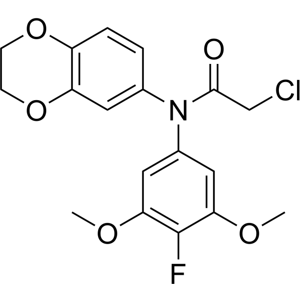
HY-155663
GPX4-IN-5GPX4 Inhibitor -
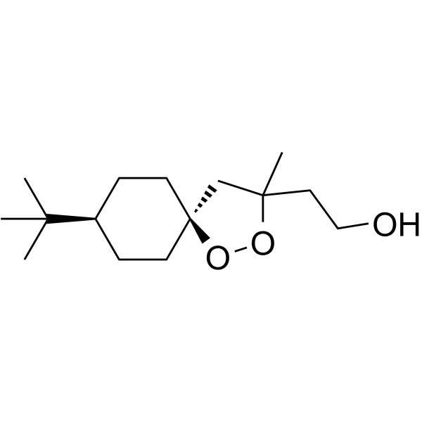
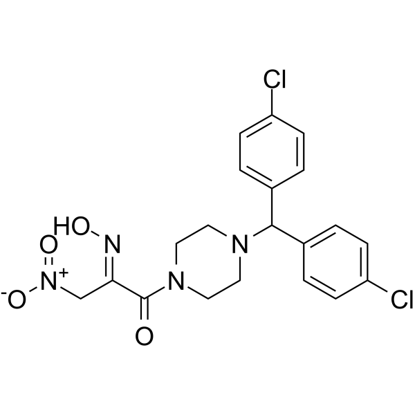
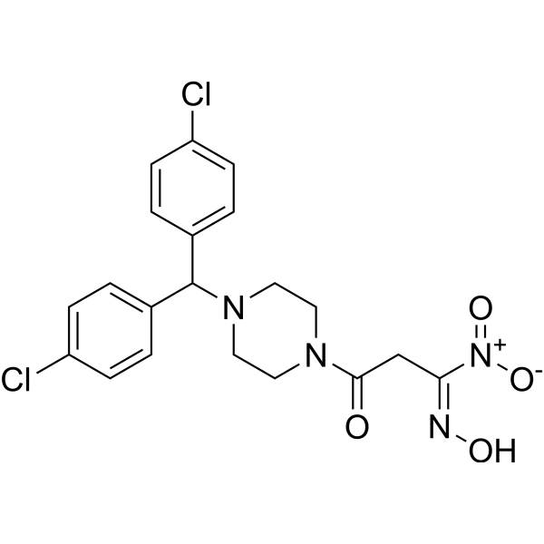
HY-129457
FINO2Ferroptosis Inducer -
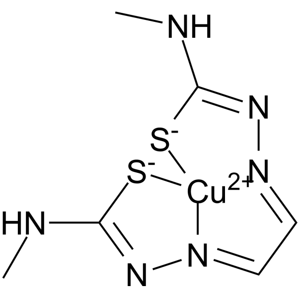
HY-139324
Cu(II)GTSMGSK3β/AβOs Inhibitor -
HY-138153
JKE-1674GPX4 Inhibitor -
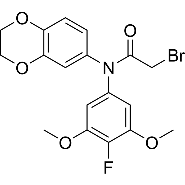
HY-155664
GPX4-IN-6GPX4 Inhibitor -
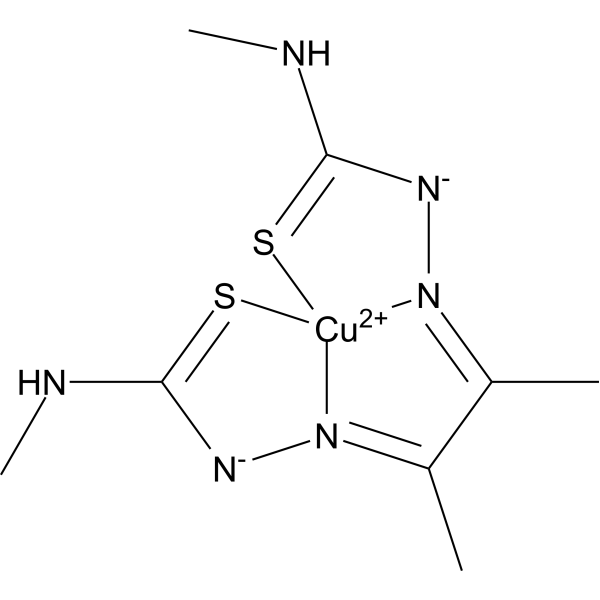
HY-139827
CuATSMFerroptosis Inhibitor -
HY-139001
JKE-1716GPX4 Inhibitor -

HY-163995
GPX4-IN-13GPX4 Inhibitor -

HY-158740
1(R)-(Trifluoromethyl)oleyl alcoholFerroptosis Inhibitor


Biological Activity
제품 설명
IC50 & Target
|
GPX4 |
Cellular Effect
|
Cell Line

|
Type | Value | Description | References |
|---|---|---|---|---|
| HCT-116 | IC50 |
>1 μM
Compound: BCP-TA
|
Cytotoxicity against human HCT-116 cells assessed as reduction in cell viability incubated for 3 days by methylene blue staining based analysis
Cytotoxicity against human HCT-116 cells assessed as reduction in cell viability incubated for 3 days by methylene blue staining based analysis
|
[PMID: 35984756] |
| HeLa | IC50 |
0.242 μM
Compound: BCP-TA
|
Cytotoxicity against human HeLa cells assessed as reduction in cell viability incubated for 3 days by methylene blue staining based analysis
Cytotoxicity against human HeLa cells assessed as reduction in cell viability incubated for 3 days by methylene blue staining based analysis
|
[PMID: 35984756] |
| HT-1080 | IC50 |
0.019 μM
Compound: BCP-TA
|
Cytotoxicity against human HT-1080 cells assessed as reduction in cell viability incubated for 3 days by methylene blue staining based analysis
Cytotoxicity against human HT-1080 cells assessed as reduction in cell viability incubated for 3 days by methylene blue staining based analysis
|
[PMID: 35984756] |
| MDA-MB-231 | IC50 |
0.021 μM
Compound: BCP-TA
|
Cytotoxicity against human MDA-MB-231 cells assessed as reduction in cell viability incubated for 3 days by methylene blue staining based analysis
Cytotoxicity against human MDA-MB-231 cells assessed as reduction in cell viability incubated for 3 days by methylene blue staining based analysis
|
[PMID: 35984756] |
| MDA-MB-468 | IC50 |
0.084 μM
Compound: BCP-TA
|
Cytotoxicity against human MDA-MB-468 cells assessed as reduction in cell viability incubated for 3 days by methylene blue staining based analysis
Cytotoxicity against human MDA-MB-468 cells assessed as reduction in cell viability incubated for 3 days by methylene blue staining based analysis
|
[PMID: 35984756] |
| MEF | IC50 |
0.01 μM
Compound: BCP-TA
|
Cytotoxicity against mouse MEF cells assessed as reduction in cell viability incubated for 3 days by methylene blue staining based analysis
Cytotoxicity against mouse MEF cells assessed as reduction in cell viability incubated for 3 days by methylene blue staining based analysis
|
[PMID: 35984756] |
| NCI-H522 | IC50 |
0.017 μM
Compound: BCP-TA
|
Cytotoxicity against human NCI-H522 cells assessed as reduction in cell viability incubated for 3 days by methylene blue staining based analysis
Cytotoxicity against human NCI-H522 cells assessed as reduction in cell viability incubated for 3 days by methylene blue staining based analysis
|
[PMID: 35984756] |
| NCI-H522 | IC50 |
7.894 μM
Compound: BCP-TA
|
Cytotoxicity against human NCI-H522 cells assessed as reduction in cell viability incubated for 3 days in presence of ferroptosis inhibitor, liproxstatin-1 by methylene blue staining based analysis
Cytotoxicity against human NCI-H522 cells assessed as reduction in cell viability incubated for 3 days in presence of ferroptosis inhibitor, liproxstatin-1 by methylene blue staining based analysis
|
[PMID: 35984756] |
| U2OS | IC50 |
0.367 μM
Compound: BCP-TA
|
Cytotoxicity against human U2OS cells assessed as reduction in cell viability incubated for 3 days by methylene blue staining based analysis
Cytotoxicity against human U2OS cells assessed as reduction in cell viability incubated for 3 days by methylene blue staining based analysis
|
[PMID: 35984756] |
| WI-38 | IC50 |
0.022 μM
Compound: BCP-TA
|
Cytotoxicity against human WI-38 cells assessed as reduction in cell viability incubated for 3 days by methylene blue staining based analysis
Cytotoxicity against human WI-38 cells assessed as reduction in cell viability incubated for 3 days by methylene blue staining based analysis
|
[PMID: 35984756] |
In Vitro
BCP-T.A induces ferroptosis in various cell lines (NCI-H522, T-1080, MDA-MB-468, MDA-MB-231, HeLa, HCT-116, U2OS, WI-38, and MEFS) with IC50 values of 10 nM-367 nM[1].
BCP-T.A (0.5 μM, 3 h) binds to GPX4 and increases lipid peroxides, indicated by cellular thermal shift assay (CETSA)[1].
MedChemExpress (MCE) has not independently confirmed the accuracy of these methods. They are for reference only. Further protocols information, click here.


Chemical Information
-
CAS No. 2786829-70-5
-
Appearance Solid
-
분자량 456.39
-
화학식 C23H19Cl2N3OS
-
Color White to light yellow
-
SMILES
O=C(N1CCN(C(C2=CC=C(Cl)C=C2)C3=CC=C(Cl)C=C3)CC1)C4=CSC(C#C)=N4
-
선적
Room temperature in continental US; may vary elsewhere.
-
보관
Powder -20°C 3 years In solvent -80°C 6 months -20°C 1 month

Protocol
-
EdU Incorporation Assay (Click Chemistry-Based DNA Synthesis Measurement)
The EdU incorporation assay measures DNA synthesis by adding the thymidine analog 5-ethynyl-2′-deoxyuridine to cells or tissues, where it is incorporated into newly synthesized DNA during S phase. Incorporated EdU is detected by copper-catalyzed azide-alkyne cycloaddition, in which a fluorescent azide covalently reacts with the ethynyl group on EdU, allowing S-phase cells to be detected by fluorescence microscopy, flow cytometry, or high-content imaging. EdU detection does not require DNA denaturation or anti-BrdU antibody access, which preserves sample structure and improves compatibility with immunostaining and multiparameter cytometry compared with BrdU-based detection. EdU can be cytotoxic in a cell-type- and exposure-dependent manner, so pulse duration, concentration, and continuous-labeling designs should be validated for each cell type.
-
Ferroptosis Solutions
Ferroptosis is an iron-dependent, non-apoptotic form of regulated cell death characterized by lethal lipid peroxidation and sensitivity to suppression by iron chelators or lipophilic radical-trapping antioxidants. The core pathway links cystine uptake through system Xc−, glutathione availability, GPX4-dependent detoxification of phospholipid hydroperoxides, iron-dependent oxidative reactions, and polyunsaturated-phospholipid metabolism into a cell-death program that is biochemically and morphologically distinct from apoptosis, necrosis, and autophagy. The ferroptosis pathway is experimentally linked to phenotype through chemical and genetic perturbation. Erastin induces ferroptosis by inhibiting cystine uptake through system Xc− and weakening antioxidant defenses, while GPX4 inhibition or depletion causes lipid peroxide accumulation and ferroptotic cancer-cell death. ACSL4 and oxidizable arachidonoyl- or adrenoyl-containing phosphatidylethanolamines shape ferroptosis sensitivity by con


순도&문서
-
Data Sheet (267 KB)
-
SDS (251 KB)
- English - EN (251 KB)
- Français - FR (251 KB)
- Deutsch - DE (251 KB)
- Norwegian - NO (251 KB)
- Español - ES (251 KB)
- Swedish - SV (251 KB)
- Italian - IT (251 KB)
- Korean - KR (251 KB)
- Portuguese - PT (251 KB)
-
Handling Instructions (2659 KB)
References

Calculators
Concentration (start) × Volume (start) = Concentration (final) × Volume (final)


BCP-T.A Related Classifications
HY-151212 Related Classifications
 Powered by Bioz