



Hexoprenaline

Based on 1 Customer Validation
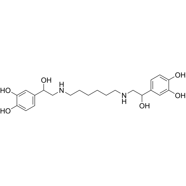
Hexoprenaline is an orally active and selective β-adrenergic receptor agonist. Hexoprenaline can dilate the bronchi. Hexoprenaline can activate adenylate cyclase and increases 14C-Aminopyrine uptake. Hexoprenaline exhibits anti-infection and anti-inflammmation effect. Hexoprenaline can increase placental weight and blood flow. Hexoprenaline can be used for the researches of inflammation, immunology, infection, endocrinology and neurological disease, such as asthma, chronic bronchitis, sepsis and intoxication of organophosphorus compounds.
연구목적의 판매만을 진행합니다. 환자를 대상으로 한 판매는 하지 않습니다.
- Purity : 96.0%
- CAS No.: 3215-70-1
- 화학식: C22H32N2O6
- 분자량:420.50
-
보관:Powder -20°C, 3 years , 4°C, 2 years ; In solvent -80°C, 6 months , -20°C, 1 month

To place orders, for customer services and technical support, please contact: MedChemExpress USA
Tel: 609-228-6898 E-mail: [email protected] [email protected]


-
 Biological Activity
-
Chemical Information
- Protocol
- 순도&문서
- References
-
 Help & FAQs


All Adrenergic Receptor Isoforms
More

Biological Activity
제품 설명
IC50 & Target
[1]|
β-adrenoceptor |
In Vitro
Hexoprenaline (10 nM-1 mM) activates adenylate cyclase in isolated rat gastric mucosal cells[2].
Hexoprenaline (10-10000 nM) increasingly stimulate 14C-Aminopyrine uptake in isolated rat gastric mucosal cells[2].
MedChemExpress (MCE) has not independently confirmed the accuracy of these methods. They are for reference only. Further protocols information, click here.
In Vivo
Hexoprenaline (0.5 mg diet, p.o., daily) increases placental weight and blood flow in blood flow[5].
MedChemExpress (MCE) has not independently confirmed the accuracy of these methods. They are for reference only.
-
Animal Model:Mice with E. coli O157:H7-induced sepsis[3]
-
Dosage:1.5 mg/kg
-
Administration:Subcutaneous injection
-
Result:Reduced the mortality by 1.98-fold at 4 h and by 1.56-fold at 24 h.
Decreased plasma TNF-α, IL-1β, and IL-6 concentrations.


Chemical Information
-
CAS No. 3215-70-1
-
Appearance Solid
-
분자량 420.50
-
화학식 C22H32N2O6
-
Color Light brown to brown
-
SMILES
OC(CNCCCCCCNCC(O)C1=CC=C(C(O)=C1)O)C2=CC=C(C(O)=C2)O
-
선적
Room temperature in continental US; may vary elsewhere.
-
보관
Powder -20°C 3 years 4°C 2 years In solvent -80°C 6 months -20°C 1 month

Protocol
-
Research Protocol for Infectious Diseases
Infectious-disease experiments test how pathogens interact with host barriers, innate immune receptors, inflammatory signaling, pathogen replication, and tissue injury; pattern-recognition receptors such as TLRs, RIG-I-like receptors, NOD-like receptors, and inflammasomes detect microbial molecules and activate NF-κB, interferon, and cytokine responses. The central hypothesis is that infection severity reflects the balance between pathogen burden and host response: protective inflammation restricts pathogen growth, whereas excessive or mislocalized inflammation contributes to tissue damage and disease phenotype. Unresolved questions include which host pathways are protective versus pathogenic, why some infection models fail to translate to human disease, and which combined readouts best predict clinically relevant infection outcomes.
-
LPS-Induced Endotoxemia/Systemic Inflammation
Lipopolysaccharide (LPS)-induced endotoxemia is a widely used in vivo model of acute systemic inflammation in which LPS, a Gram-negative bacterial endotoxin, activates innate immune signaling primarily through TLR4, leading to rapid and transient induction of pro-inflammatory cytokines such as TNF-α, IL-6, and IL-1β in circulation and tissues. This cytokine surge is commonly used as a measurable readout of systemic inflammatory activation and immune dysregulation, and is typically assessed within hours after intraperitoneal LPS administration in mouse models of endotoxemia. The model captures key features of systemic inflammatory response syndrome, including cytokine release, immune cell activation, and downstream tissue responses, and has been used to evaluate anti-inflammatory interventions such as cytokine modulation, lipid mediators, and immune cell-targeting therapies.
-
Research Protocol for Inflammation-related Diseases
The NLRP3 inflammasome is a cytosolic innate immune signaling platform that integrates priming signals and danger-signal activation to promote caspase-1 activation, maturation of IL-1β and IL-18, and gasdermin D-mediated pyroptotic cell death. The core experimental logic is to determine whether inflammatory disease phenotypes are driven by increased NLRP3 expression, ASC-containing inflammasome assembly, caspase-1 cleavage, GSDMD cleavage, and extracellular release of IL-1β/IL-18 rather than by nonspecific cell injury alone. The pathway is strongly linked to inflammation-related disease phenotypes because monosodium urate crystals activate NALP3/NLRP3 inflammasome signaling in gout-like crystal inflammation, cholesterol crystals activate NLRP3 inflammasomes in atherogenesis models, and DSS-induced intestinal inflammation has been reported to involve NLRP3 inflammasome activity. However, experimental colitis studies also show context-dependent protective effects of NLRP3 inflammasome co
-
Research Protocol for Neurological Diseases
PINK1/Parkin-mediated mitophagy pathway is a mitochondrial quality-control signaling axis in which mitochondrial depolarization stabilizes PINK1 on damaged mitochondria, activates Parkin recruitment and E3 ubiquitin ligase activity, promotes ubiquitination of outer mitochondrial membrane proteins, recruits selective autophagy adaptors, and drives lysosomal degradation of damaged mitochondria. In neurological disease research, this pathway is experimentally important because neurons, especially dopaminergic neurons, are highly dependent on mitochondrial integrity, and defective mitochondrial turnover can lead to mitochondrial dysfunction, oxidative stress, impaired neuronal survival, α-synuclein accumulation, and neuroinflammatory damage-associated signals. The genetic disease link is strongest in Parkinson’s disease because mutations in PRKN/parkin cause autosomal recessive juvenile parkinsonism, mutations in PINK1 cause hereditary early-onset Parkinson’s disease, and Drosophila studie


순도&문서
-
Data Sheet (269 KB)
-
SDS (254 KB)
- English - EN (254 KB)
- Français - FR (254 KB)
- Deutsch - DE (254 KB)
- Norwegian - NO (254 KB)
- Español - ES (254 KB)
- Swedish - SV (254 KB)
- Italian - IT (254 KB)
- Korean - KR (254 KB)
- Portuguese - PT (254 KB)
-
Handling Instructions (2659 KB)
References
[1]. Schonell M, et al. Bronchodilator effect of hexoprenaline aerosol in bronchial asthma and chronic bronchitis. Med J Aust. 1977 Dec 17;2(25):828-30. [Content Brief]
[2]. Ruoff HJ, et al. Adrenergic stimulation of isolated rat gastric mucosal cells. Effect on adenylate cyclase and 14C-aminopyrine uptake. Naunyn Schmiedebergs Arch Pharmacol. 1982 Aug;320(2):175-81. [Content Brief]
[5]. Lipshitz J, et al. Effect of long-term administration of beta 2-sympathomimetic drug in the diet-restricted pregnant rat model. Am J Obstet Gynecol. 1986 Feb;154(2):314-7. [Content Brief]

Calculators
Concentration (start) × Volume (start) = Concentration (final) × Volume (final)


Hexoprenaline Related Classifications
HY-121185 Related Classifications
 Powered by Bioz