



L-Citrulline (GMP)
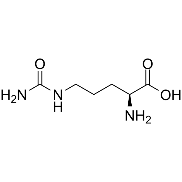
L-Citrulline GMP is an orally active, blood-brain barrier permeable neutral α-amino acid. L-Citrulline GMP inhibits the mitochondrial translocation of Drp1 and maintains mitochondrial homeostasis via a NO-dependent pathway, thereby reducing excessive ROS production. Consequently, L-Citrulline GMP protects sperm DNA integrity, stabilizes the blood-testis barrier and improves semen quality. L-Citrulline GMP can be used in research related to male infertility, mycobacterial infection (tuberculosis) and radiation combined injury.
For research use only. We do not sell to patients.
- CAS No.: 372-75-8
- Formula: C6H13N3O3
- Molecular Weight:175.19
-
Storage:
Please store the product under the recommended conditions in the Certificate of Analysis.


To place orders, for customer services and technical support, please contact: MedChemExpress USA
Tel: 609-228-6898 E-mail: [email protected] [email protected]


-
 Biological Activity
-
Chemical Information
- Protocols
- Purity & Documentation
- References
-
 Help & FAQs


-
HY-N0391
L-CitrullineEndogenous Metabolite -

HY-N0391R
L-Citrulline (Standard)Endogenous Metabolite -
HY-N0832
L-HistidineEndogenous Metabolite -
HY-Y0252
L-Proline -
HY-Y0337
L-CysteineAnorectic Agent -
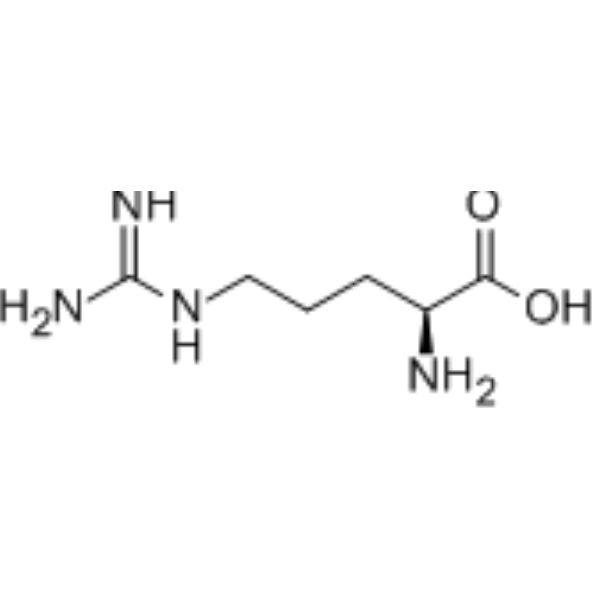
HY-N0455
L-ArginineeNOS Substrate -
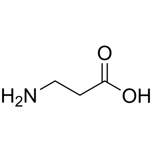
HY-N0230
β-AlanineEndogenous Metabolite -
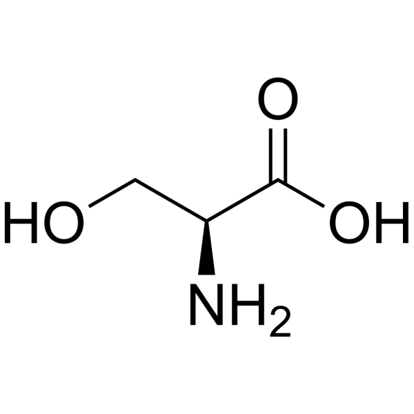
HY-N0650
L-SerineAmino Acid -
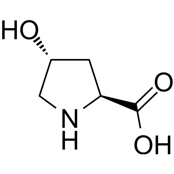
HY-40135
L-HydroxyprolineEndogenous Metabolite -
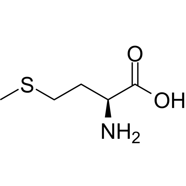
HY-N0326
L-MethionineEndogenous Metabolite -
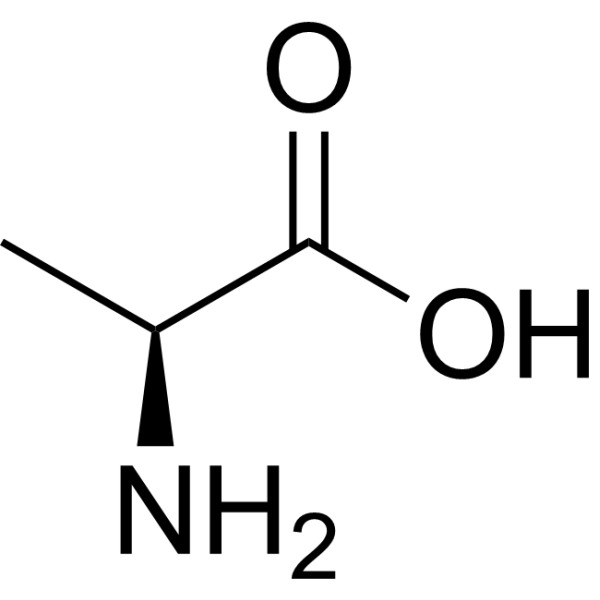
HY-N0229
L-AlanineEndogenous Metabolite -
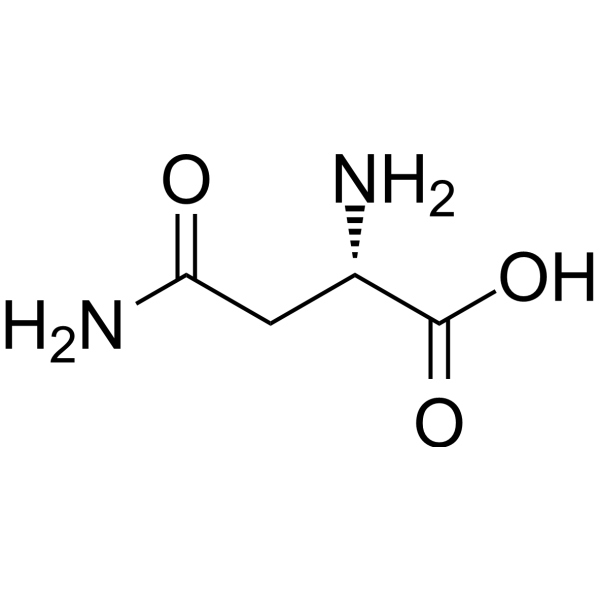
HY-N0667
L-AsparagineEndogenous Metabolite


Biological Activity
Description
In Vitro
L-Citrulline (GMP) (1-2 mM) protects mouse C2C12 myoblasts against 43 °C heat stress-induced mitochondrial dysfunction and cell injury via a nitric oxide-mediated inhibition of DRP1 activation[1].
L-Citrulline GMP maintains normal autophagic flux in primary human airway epithelial cells by mitigating asymmetric dimethylarginine-induced nitrative stress via restored nitric oxide bioavailability[1].
MedChemExpress (MCE) has not independently confirmed the accuracy of these methods. They are for reference only. Further protocols information, click here.
In Vivo
L-Citrulline GMP (1 g/kg; p.o.; once daily; 21 days post-irradiation), in combination with PEG-G-CSF, significantly increases 30-day survival (42% survival rate) in a mouse model of radiation combined injury, accelerates body weight recovery, improves bone marrow clonogenicity, and enhances intestinal crypt recovery[3].
L-Citrulline GMP (1 g/kg; p.o.; once daily; 21 days post-irradiation) does not significantly improve 30-day survival in mice with total-body irradiation alone, but improves bone marrow clonogenicity and partially reduces irradiation-induced splenomegaly[3].
L-Citrulline GMP (1 g/kg; p.o.; once daily; 21 days post-irradiation), in combination with PEG-G-CSF, does not significantly improve 30-day survival in mice with total-body irradiation alone, but improves bone marrow clonogenicity and fully inhibits irradiation-induced splenomegaly[3].
MedChemExpress (MCE) has not independently confirmed the accuracy of these methods. They are for reference only.
-
Animal Model:B6D2F1/J (female, 14-15 weeks old, average weight 24-25 g, radiation combined injury model)[3]
-
Dosage:1 g/kg
-
Administration:p.o.; once daily; 21 days post-irradiation
-
Result:Produced a non-significant trend toward enhanced 30-day survival (40% survival, median survival time 17.5 days, P=0.057 versus vehicle).
Significantly accelerated wound healing, resulting in smaller wound areas on day 14 (P=0.0436) and day 21 (P=0.0002), and higher percentage wound closure on day 14 (P=0.01) and day 21 (P<0.0001) compared to vehicle.
Significantly increased bone marrow colony-forming unit-granulocyte-monocyte (CFU-GM) counts (P=0.032) and total colony counts (CFU-Total, P=0.0491) on day 30 post-irradiation compared to vehicle.
Increased serum citrulline levels to a moderate degree (not statistically significant versus vehicle).\nSignificantly increased 30-day survival to 42% (median survival time 18 days, P=0.007 versus vehicle), and delayed onset of death to after day 13 post-irradiation (versus 6-10 days for other treatments).
Significantly increased body weight on day 21 post-irradiation (P=0.0071 versus vehicle).
Significantly increased bone marrow CFU-GM (P<0.0001), CFU-granulocyte-erythrocyte-monocyte-megakaryocyte (CFU-GEMM, P=0.0476), and CFU-Total (P=0.0029) counts on day 30 post-irradiation compared to vehicle.
Significantly increased serum citrulline levels (P=0.0388 versus vehicle), increased jejunal crypt depth (P=0.0277 versus vehicle), and restored jejunal crypt counts to levels comparable to sham-irradiated mice on day 30 post-irradiation.
-
Animal Model:B6D2F1/J (female, 14-15 weeks old, average weight 24-25 g, acute radiation syndrome model)[3]
-
Dosage:1 g/kg
-
Administration:p.o.; once daily; 21 days post-irradiation
-
Result:Resulted in 35% 30-day survival (median survival time 20 days), which was not significantly different from vehicle control (P=0.45).
Improved bone marrow cellularity and significantly increased bone marrow CFU-GM counts (P=0.0169) on day 30 post-irradiation compared to vehicle.
Partially inhibited irradiation-induced splenomegaly and reduced splenocyte counts in surviving mice.\nResulted in 50% 30-day survival (median survival time 26 days), which was not significantly different from vehicle control (P=0.19).
Significantly increased bone marrow CFU-GM counts (P=0.0207) on day 30 post-irradiation compared to vehicle.
Fully inhibited irradiation-induced splenomegaly and reduced splenocyte counts in surviving mice.


Chemical Information
-
CAS No. 372-75-8
-
Molecular Weight 175.19
-
Formula C6H13N3O3
-
SMILES
N[C@@H](CCCNC(N)=O)C(O)=O
-
Shipping
Room temperature in continental US; may vary elsewhere.
-
Storage
Please store the product under the recommended conditions in the Certificate of Analysis.

Protocols
-
Research Protocol for Infectious Diseases
Infectious-disease experiments test how pathogens interact with host barriers, innate immune receptors, inflammatory signaling, pathogen replication, and tissue injury; pattern-recognition receptors such as TLRs, RIG-I-like receptors, NOD-like receptors, and inflammasomes detect microbial molecules and activate NF-κB, interferon, and cytokine responses. The central hypothesis is that infection severity reflects the balance between pathogen burden and host response: protective inflammation restricts pathogen growth, whereas excessive or mislocalized inflammation contributes to tissue damage and disease phenotype. Unresolved questions include which host pathways are protective versus pathogenic, why some infection models fail to translate to human disease, and which combined readouts best predict clinically relevant infection outcomes.
-
ROS/oxidative-stress fluorescent staining
ROS/oxidative-stress fluorescent staining uses cell-permeant fluorogenic probes that become fluorescent after oxidation inside cells or tissues; commonly used examples include DCFH-DA/DCFDA for broad cellular oxidant detection, DHE for superoxide-related signal detection, MitoSOX for mitochondrial superoxide-related signal detection, and CellROX probes for oxidative-stress-associated fluorescence readouts. The assay detects probe oxidation rather than a single ROS species unless the probe and analysis method have been chemically validated for that species. DCFH-DA enters cells, is deacetylated by intracellular esterases to DCFH, and produces fluorescent DCF after oxidation, so the readout is used as an operational measure of total cellular oxidative stress rather than a species-specific ROS measurement. DHE and MitoSOX can report superoxide-related oxidation, but red fluorescence alone can include non-specific ethidium-like oxidation products; HPLC or optimized spectral approaches are


Purity & Documentation
References
[1]. Qin Y, et al.. Heat stress-mediated oxidative damage in male germ cells: potential protective effects of L-citrulline. Frontiers in endocrinology. 2026;17:1800681. [Content Brief]
[2]. Lange SM, et al.. l-Citrulline Metabolism in Mice Augments CD4 T Cell Proliferation and Cytokine Production , and Accumulation in the Mycobacteria-Infected Lung. Frontiers in immunology. 2017;8:1561. [Content Brief]
[3]. Wang L, et al.. PEG-G-CSF and L-Citrulline Combination Therapy for Mitigating Skin Wound Combined Radiation Injury in a Mouse Model. Radiation research. 2021 Jul 01;196(1):113-127. [Content Brief]

Calculators
Concentration (start) × Volume (start) = Concentration (final) × Volume (final)


L-Citrulline (GMP) Related Classifications
HY-N0391G Related Classifications
 Powered by Bioz