



LacCer (d18:1/16:0)

Based on 1 Customer Validation
LacCer (d18:1/16:0) (C16 Lactosylceramide (d18:1/16:0)) is an endogenous bioactive sphingolipid that can form membrane microdomains with Lyn kinase and the αi subunits of inhibitory G protein-coupled receptors (GPCRs). The levels of LacCer (d18:1/16:0) are elevated in the plasma of insulin-resistant cattle. In a mouse model of Niemann-Pick type C1 disease (a neurodegenerative cholesterol-sphingolipid lysosomal storage disorder), the expression of LacCer (d18:1/16:0) is also upregulated. LacCer (d18:1/16:0) can be used in research on metabolic diseases and neurodegenerative diseases.
For research use only. We do not sell to patients.
- Purity : 99%
- CAS No.: 4201-62-1
- Formula: C46H87NO13
- Molecular Weight:862.18
-
Storage:Powder -20°C, 3 years ; In solvent -80°C, 6 months , -20°C, 1 month


To place orders, for customer services and technical support, please contact: MedChemExpress USA
Tel: 609-228-6898 E-mail: [email protected] [email protected]


-
 Biological Activity
-
Chemical Information
- Protocols
- Purity & Documentation
- References
-
 Help & FAQs


-
HY-141629
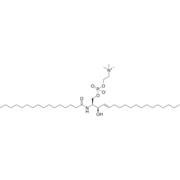
N-Palmitoyl-D-sphingomyelinComponent Of Liposomes -
HY-113423
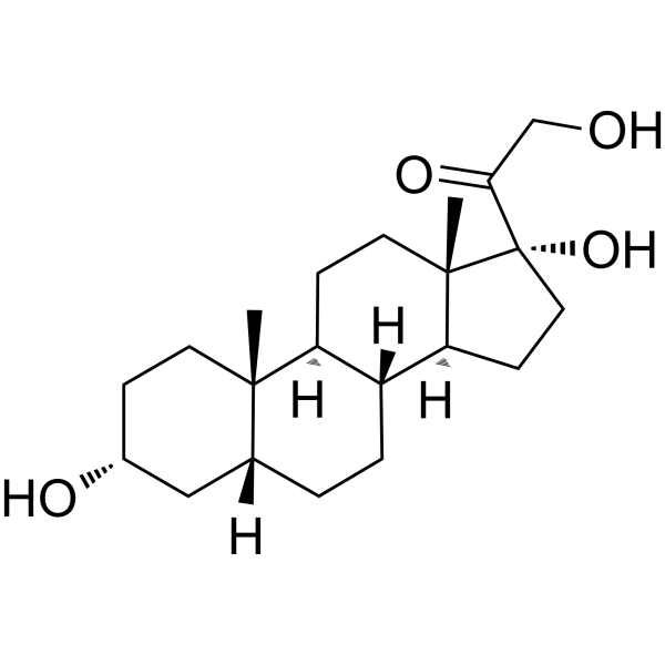
Tetrahydro-11-deoxycortisolEndogenous Metabolite -
HY-116215
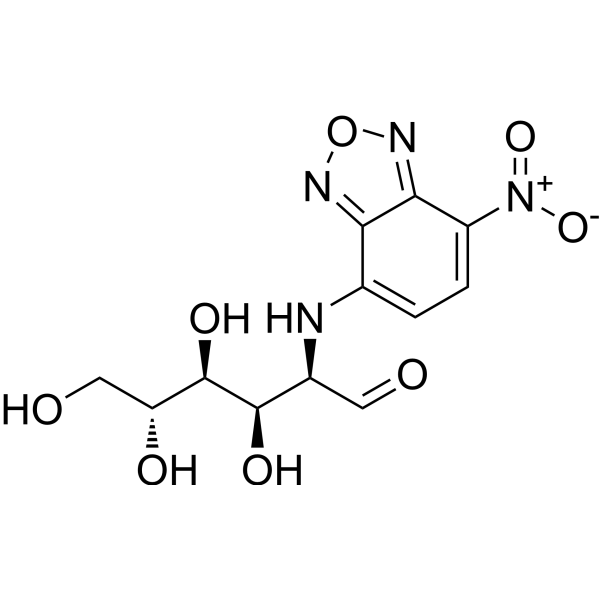
2-NBDGFluorescent Indicator -

HY-165035
Glucosylceramide (gaucher's spleen)Sphingolipid/Gaucher Disease Biomarker -

HY-156206
C24 Dihydro ceramide (d18:0/24:0)Ester Product -

HY-145549
C20 Sphingomyelin (d18:1/20:0) -

HY-DY1019
2-NBDG (solution)Fluorescent Indicator -
HY-120657
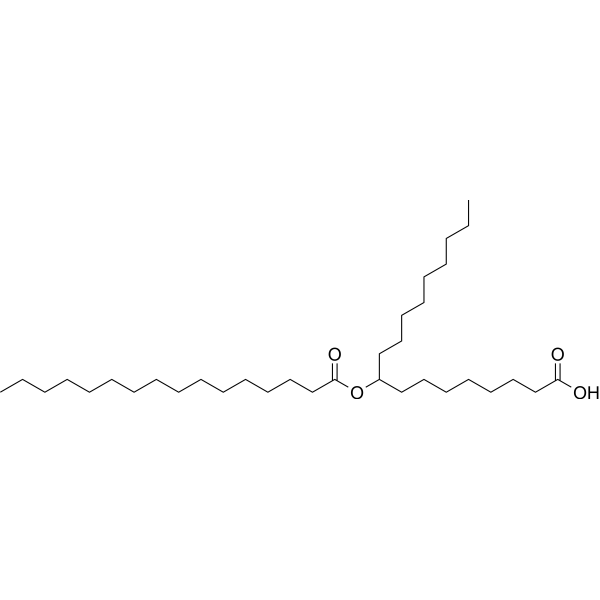
9-PAHSAGPR120 Agonist -

HY-W717504
Glucosylceramide, plantAnti-inflammatory Agent -
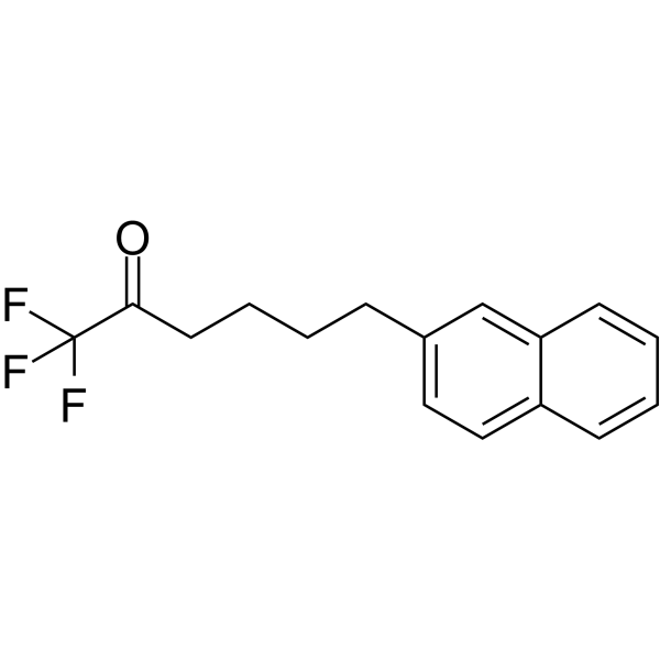
HY-115403
FKGK18GVIA iPLA2 Inhibitor -

HY-122056
1,2-Diheptanoyl-SN-glycero-3-phosphocholineShort-Chain Phospholipid -
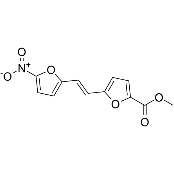
HY-123538
GRK2 Inhibitor 1GRK2 Inhibitor
All Endogenous Metabolite Isoforms
More

Biological Activity
Description


Chemical Information
-
CAS No. 4201-62-1
-
Appearance Solid
-
Molecular Weight 862.18
-
Formula C46H87NO13
-
Color White to off-white
-
SMILES
OC[C@@H](O[C@H]([C@@H]([C@H]1O)O)OC[C@@H]([C@H](O)/C=C/CCCCCCCCCCCCC)NC(CCCCCCCCCCCCCCC)=O)[C@H]1O[C@@H]2O[C@@H]([C@@H]([C@@H]([C@H]2O)O)O)CO
-
Synonyms
C16 Lactosylceramide (d18:1/16:0)
-
Structure Classification
-
Initial Source
Candida albicans
-
Shipping
Room temperature in continental US; may vary elsewhere.
-
Storage
Powder -20°C 3 years In solvent -80°C 6 months -20°C 1 month

Protocols
-
RNA extraction experimental
By lysing cells, releasing RNA, and removing impurities such as proteins and DNA, high-purity RNA products are finally obtained. The commonly used traditional method is the guanidine isothiocyanate/phenol/chloroform method (Trizol), which is suitable for a variety of animal materials including animal tissues, microorganisms, cultured cells, etc., and most plant materials.
-
Research Protocol for Endocrine Diseases
Endocrine diseases often arise from disrupted hormone production, hormone signaling, or target-tissue responsiveness; for diabetes-focused endocrine disease models, insulin signaling regulates glucose uptake, hepatic glucose output, lipid metabolism, and β-cell compensation. Type 2 diabetes develops through interacting defects in insulin resistance, β-cell dysfunction, adipose inflammation, hepatic glucose overproduction, altered incretin signaling, and ectopic lipid metabolism. A major unresolved question is whether endocrine dysfunction is driven primarily by target-tissue insulin resistance, intrinsic β-cell failure, immune/inflammatory stress, or combined multi-organ failure that differs by disease stage.
-
Lysosome and acidic-vesicle live-cell staining
Lysosome and acidic-vesicle live-cell staining detects acidic intracellular compartments by using membrane-permeant acidotropic probes that accumulate in low-pH vesicles, including lysosomes, late endosomes, autolysosomes, and acidic phagosomes. LysoTracker staining is commonly used as an intensity-based readout of acidic lysosomal compartment abundance or enlargement, while acridine orange produces green fluorescence in less concentrated compartments and red fluorescence after concentration-dependent accumulation in acidic vesicular organelles. Loss or reduction of acridine-orange red signal can be used as a readout of lysosomal membrane permeabilization or reduced acidic-vesicle integrity. This protocol is designed for live cultured cells and can be adapted for fluorescence microscopy, high-content imaging, plate-reader readout, or flow cytometry when the selected literature supports the readout. Because these dyes report acidotropic accumulation rather than lysosome identity alone,
-
How to Choose the Right Model Animal
Choosing the right model animal is a validity-driven decision in which the species, strain, sex, age, genetic background, disease-induction method, outcome measures, and welfare burden must match the scientific question rather than laboratory tradition or convenience. A model should be selected by judging face validity, construct validity, and predictive validity: whether it resembles the human phenotype, whether it reproduces relevant mechanisms, and whether results are likely to predict human biology or treatment response. Animal studies often fail to translate because of species differences, weak disease resemblance, poor experimental design, inadequate reporting, publication bias, and underuse of randomization, blinding, and sample-size justification. Unresolved questions include how to rank competing models objectively, how much human-disease complexity must be reproduced for a given objective, and when non-animal systems such as organoids, ex vivo tissue, or computational models


Purity & Documentation
-
Data Sheet (265 KB)
-
SDS (254 KB)
- English - EN (254 KB)
- Français - FR (254 KB)
- Deutsch - DE (254 KB)
- Norwegian - NO (254 KB)
- Español - ES (254 KB)
- Swedish - SV (254 KB)
- Italian - IT (254 KB)
- Korean - KR (254 KB)
- Portuguese - PT (254 KB)
-
Handling Instructions (2659 KB)
References
[1]. Rico JE, et al. Plasma ceramides are elevated in overweight Holstein dairy cows experiencing greater lipolysis and insulin resistance during the transition from late pregnancy to early lactation. J Dairy Sci. 2015 Nov;98(11):7757-70. [Content Brief]
[2]. Praggastis M, et al. A murine Niemann-Pick C1 I1061T knock-in model recapitulates the pathological features of the most prevalent human disease allele. J Neurosci. 2015 May 27;35(21):8091-106. [Content Brief]
[3]. Iwabuchi K, et al. Role of Ceramide from Glycosphingolipids and Its Metabolites in Immunological and Inflammatory Responses in Humans. Mediators Inflamm. 2015;2015:120748. [Content Brief]

Calculators
Concentration (start) × Volume (start) = Concentration (final) × Volume (final)


LacCer (d18:1/16:0) Related Classifications
HY-153830 Related Classifications
 Powered by Bioz