



Lucidadiol

Based on 1 Customer Validation
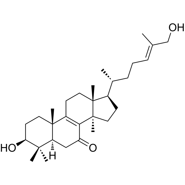
Lucidadiol is a natural compound isolated from Ganoderma lucidum. Lucidadiol exhibits acetylcholinesterase-inhibitory activity, with IC50 values of 31 μM. Lucidadiol shows antiviral activity against influenza virus type A and HSV type 1.
商品は「研究用試薬」です。人や動物の医療用・臨床診断用・食品用の製品ではありません。
研究用途以外に使用した場合、当社は一切の責任を負いかねます。
- 純度 : 98.0%
- CAS 番号: 252351-95-4
- 分子式: C30H48O3
- 分子量:456.70
-
保管条件:
-20°C, sealed storage, away from moisture
* In solvent : -80°C, 6 months; -20°C, 1 month (sealed storage, away from moisture)

To place orders, for customer services and technical support, please contact: MedChemExpress USA
Tel: 609-228-6898 E-mail: [email protected] [email protected]


-
 生物活性
生物活性
-
化学情報
- プロトコル
- 純度とドキュメンテーション
- 参考文献
-
 Help & FAQs




生物活性
製品説明
IC50 & Target
[1]|
AChE 31.03 μM (IC50) |
BChE 156.27 μM (IC50) |
Cellular Effect
|
Cell Line

|
Type | Value | Description | References |
|---|---|---|---|---|
| HeLa | IC50 |
5 μM
Compound: 58
|
Cytotoxicity against human HeLa cells
Cytotoxicity against human HeLa cells
|
[PMID: 23092389] |


化学情報
-
CAS 番号 252351-95-4
-
性状 Solid
-
分子量 456.70
-
分子式 C30H48O3
-
Color White to off-white
-
SMILES
CC(C)([C@](C1)2[H])[C@@H](O)CC[C@]2(C)C3=C([C@@]4(CC[C@@H]([C@]4(CC3)C)[C@H](C)CC/C=C(C)/CO)C)C1=O
-
Structure Classification
-
Initial Source
Ganoderma lucidum
-
輸送条件
Room temperature in continental US; may vary elsewhere.
-
保管条件
-20°C, sealed storage, away from moisture
* In solvent : -80°C, 6 months; -20°C, 1 month (sealed storage, away from moisture)


純度とドキュメンテーション
-
データシート (269 KB)
-
SDS (252 KB)
- English - EN (252 KB)
- Français - FR (252 KB)
- Deutsch - DE (252 KB)
- Norwegian - NO (252 KB)
- Español - ES (252 KB)
- Swedish - SV (252 KB)
- Italian - IT (252 KB)
- Korean - KR (252 KB)
- Portuguese - PT (252 KB)
-
取扱説明書 (2659 KB)
参考文献
[1]. Iksoo Lee, et al. Selective cholinesterase inhibition by lanostane triterpenes from fruiting bodies of Ganoderma lucidum. Bioorg Med Chem Lett. 2011 Nov 1;21(21):6603-7. [Content Brief]
[2]. Lee I,et al. Selective cholinesterase inhibition by lanostane triterpenes from fruiting bodies of Ganoderma lucidum [published correction appears in Bioorg Med Chem Lett. 2013 Nov 15;23(22):6199-200]. Bioorg Med Chem Lett. 2011;21(21):6603-6607. [Content Brief]
[3]. Mothana RA,et al. Antiviral lanostanoid triterpenes from the fungus Ganoderma pfeifferi. Fitoterapia. 2003;74(1-2):177-180. [Content Brief]

Calculators
濃度 (開始) × 体積 (開始) = 濃度 (終了) × 体積 (終了)


Lucidadiol 関連分類
HY-N7346 関連分類
 Powered by Bioz