



Maresin 2

Based on 1 Customer Validation
Maresin 2 is an anti-inflammatory and pro-resolving mediator. Maresin 2 drives intestinal epithelial cell migration by activating the focal cell-matrix adhesion signaling pathway in primary human intestinal epithelial cells, thereby promoting mucosal wound repair. Maresin 2 alleviates nociceptive and anxiety-like behaviors in rats with type 1 diabetes by inhibiting IL-1β in the spinal cord and prefrontal cortex. Maresin 2 attenuates allergic airway inflammation in mice by inhibiting the activation of the NLRP3 inflammasome, Th2-type immune responses, and oxidative stress. Maresin 2 inhibits inflammatory and neuropathic trigeminal neuralgia and reduces neuronal activation in the trigeminal ganglion. Maresin 2 promotes inflammation resolution and mucosal repair after DSS-induced colitis or biopsy-induced colonic mucosal injury.
For research use only. We do not sell to patients.
- Purity : 97.2%
- CAS No.: 1639809-46-3
- Formula: C22H32O4
- Molecular Weight:360.49
-
Storage:
Solution, -20°C, 2 years

To place orders, for customer services and technical support, please contact: MedChemExpress USA
Tel: 609-228-6898 E-mail: [email protected] [email protected]


-
 Biological Activity
-
Chemical Information
- Protocols
- Purity & Documentation
- References
-
 Help & FAQs


-
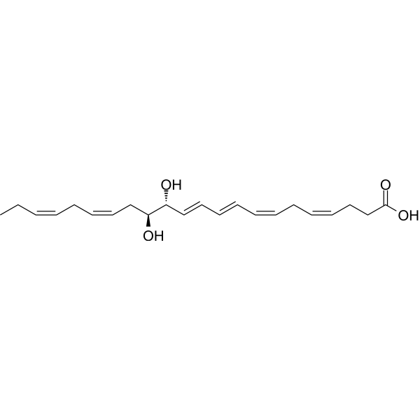
HY-130413
Protectin D1Neuroprotectin D1 -
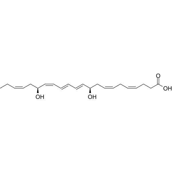
HY-113509
Lipoxin A4Anti-inflammatory Agent -
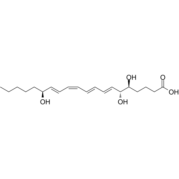
HY-136540
Resolvin D3Antiinflammatory Agent -

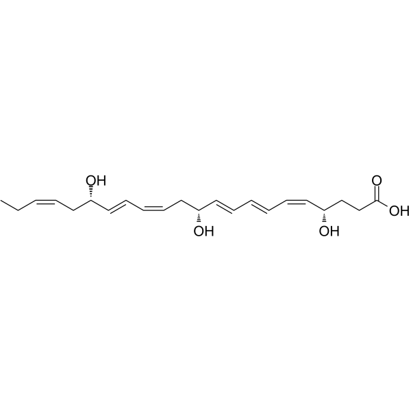
HY-B1521
Aluminum HydroxideAntacid Aegnt -
HY-N0139
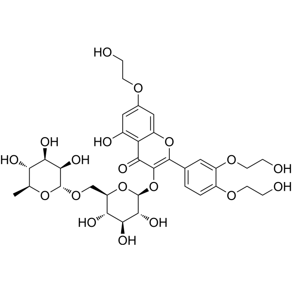
TroxerutinNOD-like Receptor (NLR) Inhibitor -
HY-116429
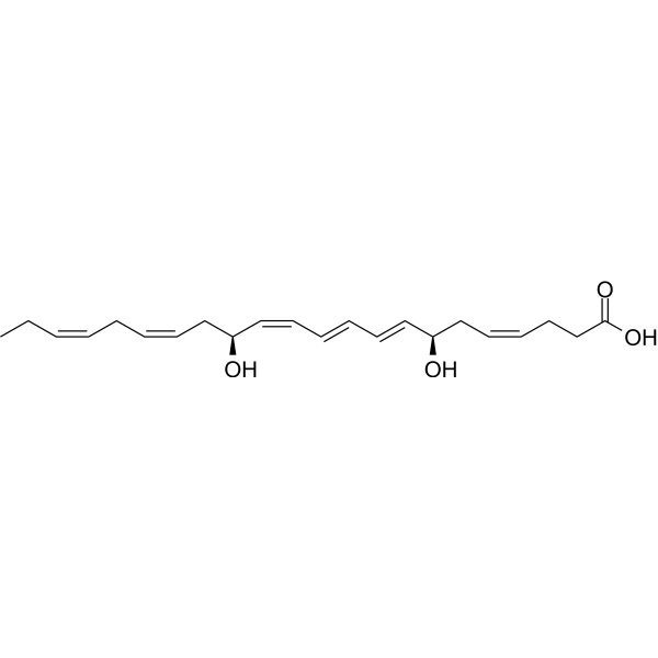
Maresin 1Ca2+ Stimulator -

HY-134123
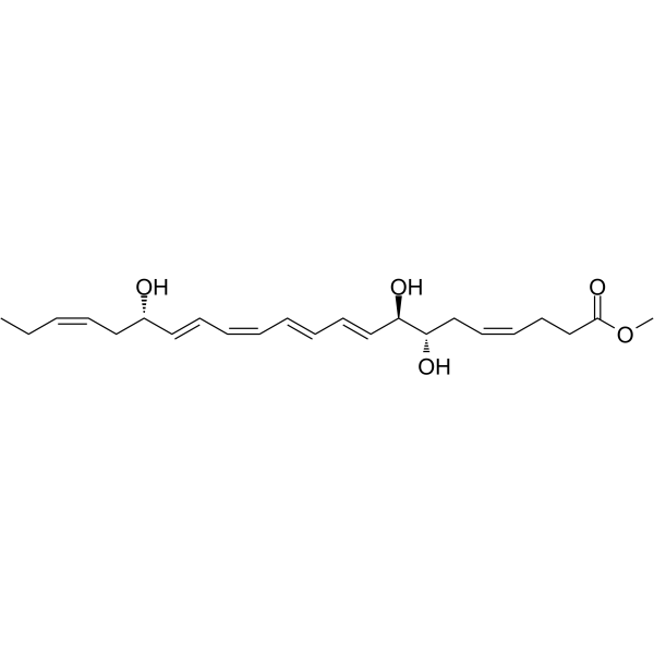
Lipoxin B4 methyl ester -

HY-W116335A
Dried aluminum hydroxide gel,United States Pharmacopeia (USP) Reference StandardAntacid -
HY-113533
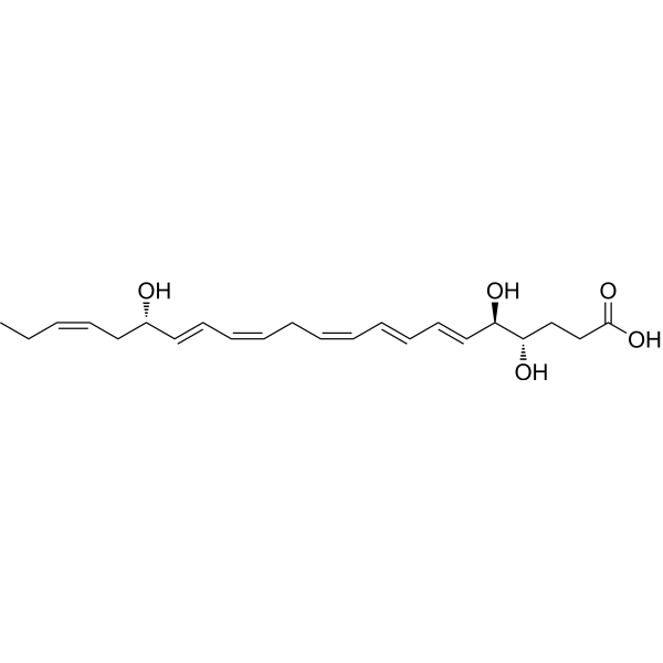
Resolvin D4Proresolving Mediator -
HY-116124
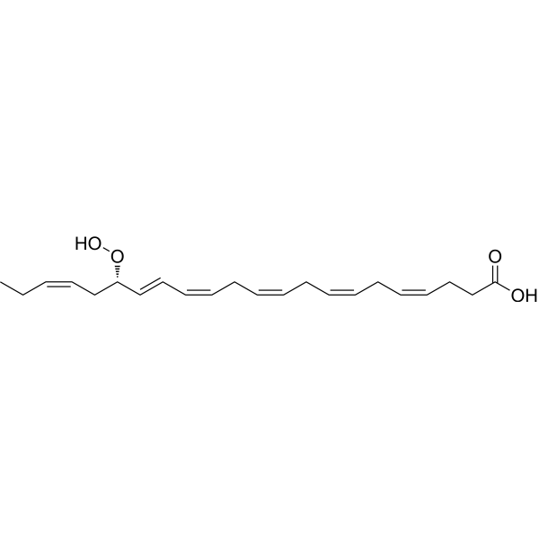
17(S)-HpDHAHydrolyzate -
HY-128031
Resolvin D1 methyl esterTriglyceride Inhibitor -
HY-116429A
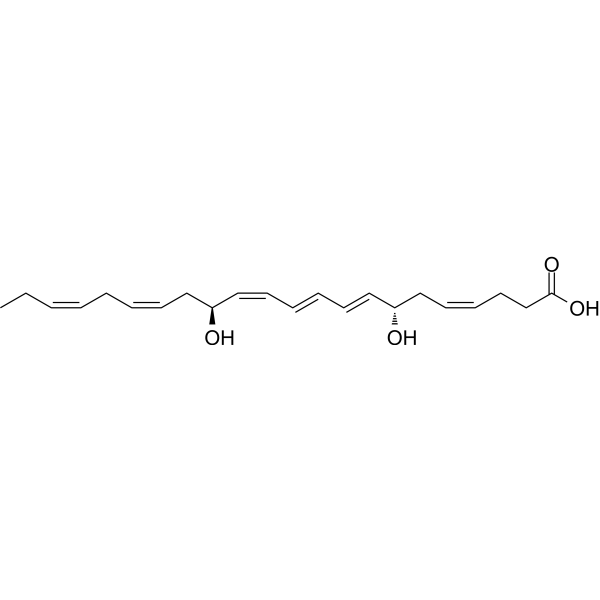
7(S)-Maresin 1Maresin 1 Exomer


Biological Activity
Description
IC50 & Target
[1]|
NLRP3 inflammasome |
IL-1β |
In Vitro
Maresin 2 (200 nM; 16-24 h) enhances scratch wound closure and migration efficiency in HT29/B6 human intestinal epithelial cells when combined with 10 ng/mL TNFα/IFNγ, but has no effect on unstimulated cells[1].
Maresin 2 (200 nM; 16 h) activates the FAK-Src-paxillin and talin-vinculin focal adhesion signaling axes in HT29/B6 human intestinal epithelial cells when combined with 10 ng/mL TNFα/IFNγ, promoting pro-migration signaling at wound leading edges[1].
MedChemExpress (MCE) has not independently confirmed the accuracy of these methods. They are for reference only. Further protocols information, click here.
-
Cell Line:HT29/B6 human intestinal epithelial cells
-
Concentration:200 nM
-
Incubation Time:16 h
-
Result:Increased numbers of vinculin-containing focal adhesions at the leading edge of spreading cells significantly relative to TNFα/IFNγ alone (P < 0.01).
In Vivo
Maresin 2 (2 ng/g body weight; intraperitoneal injection; administered on days 0, 2, 4, 6, and 8 for a total of 5 times) significantly reduces disease activity and colonic mucosal injury in the 5% DSS-induced colitis model of C57BL/6 mice, with a histological colitis score of 3.951[1].
Maresin 2 (1-10 ng per rat; i.p.; administered daily for 2 consecutive days followed by every other day; on days 14-32 after streptozotocin induction) significantly alleviates mechanical hyperalgesia in streptozotocin-induced male Wistar diabetic rats (at doses of 1, 3 and 10 ng/rat), improves anxiety-like behavior at a dose of 3 ng/rat, and normalizes IL-1β levels in the spinal cord and prefrontal cortex[2].
Maresin 2 (1 ng per mouse; intravenous injection; for 4 consecutive days) exerts a protective effect on ovalbumin-induced asthma in female BALB/c mice, and alleviates airway inflammation, Th2 immune response, NLRP3 inflammasome activation and oxidative stress[3].
Maresin 2 (1-10 ng; intrathecal; single injection) significantly attenuates the acute and inflammatory phases of formalin-induced orofacial nociception in rats[5].
Maresin 2 (10 ng; administered twice on postoperative day 1 and day 3) prevents the development of thermal hyperalgesia and mechanical hyperalgesia in a rat model of postoperative orofacial pain[5].
MedChemExpress (MCE) has not independently confirmed the accuracy of these methods. They are for reference only.
-
Animal Model:C57BL/6 (10 to 12 weeks old, male and female, biopsy-induced colonic mucosal injury)[1]
-
Dosage:2 ng/g body weight
-
Administration:i.p.; single injection; 24 h post-wounding
-
Result:Increased colonic mucosal wound healing to 47.67% at 72 h post-wounding compared to 34.58% in vehicle control mice (P < 0.01).
-
Animal Model:C57BL/6 (10 to 12 weeks old, male and female, 5% DSS-induced colitis)[1]
-
Dosage:2 ng/g body weight
-
Administration:i.p.; 5 doses on days 0, 2, 4, 6, and 8
-
Result:Reduced disease activity index (DAI) scores at multiple time points (P < 0.01, P < 0.001, P < 0.0001).
Reduced histological colitis score to 3.951 compared to 5.474 in vehicle control mice (P < 0.05).
Decreased mucosal ulceration/erosion and infiltrating immune cells.
-
Animal Model:Wistar rats (male, 180-240 g, streptozotocin-induced type 1 diabetes mellitus)[2]
-
Dosage:1 ng/rat; 3 ng/rat; 10 ng/rat
-
Administration:i.p.; daily for 2 consecutive days, then alternate days; days 14-32 post-streptozotocin induction
-
Result:Failed to alter mechanical threshold in diabetic rats after acute treatment.
Showed significant improvement in mechanical threshold at 3 ng dose on day 20 post-streptozotocin induction (p = 0.0017).
Showed significant improvement in mechanical threshold at all doses (1, 3, 10 ng) on day 26 post-streptozotocin induction (p < 0.05) compared to vehicle-treated diabetic rats.
Significantly increased time spent in open arms (p = 0.05) and decreased time spent in closed arms (p < 0.05) in elevated plus-maze test at 3 ng dose compared to vehicle-treated diabetic rats.
Did not alter depressive-like behavior in modified forced swimming test or locomotor/exploratory behavior in open-field test at any dose.
Reversed elevated IL-1β levels in spinal cord at 3 ng (p = 0.0170) and 10 ng (p = 0.0445) doses compared to vehicle-treated diabetic rats.
Reversed elevated IL-1β levels in prefrontal cortex at all doses (1 ng, p = 0.0151; 3 ng, p = 0.0266; 10 ng, p = 0.0201) compared to vehicle-treated diabetic rats.
Did not affect IL-1β levels in hippocampus, hyperglycemia, or low weight gain in diabetic rats at any dose.
-
Animal Model:BALB/c (female, 6-8 weeks old, 20-25 g, specific pathogen-free, ovalbumin-induced asthma model)[3]
-
Dosage:1 ng per mouse
-
Administration:i.v.; daily; 4 days
-
Result:Reduced airway inflammation score (P < 0.05) and goblet cell proliferation score (P < 0.05).
Decreased lung tissue expression of MPO (P < 0.01) and Ly-6G (P < 0.05).
Lowered total inflammatory cell count (P < 0.05), neutrophil count (P < 0.05), and eosinophil count (P < 0.05) in BALF.
Reduced BALF levels of Th2 cytokines IL-4 (P < 0.01), IL-5 (P < 0.01), and IL-13 (P < 0.01).
Lowered serum total IgE (P < 0.01) and ovalbumin-specific IgE (P < 0.05).
Decreased lung tissue expression of NLRP3 (P < 0.05), ASC (P < 0.05), and Caspase-1 (P < 0.05).
Reduced BALF levels of IL-1β (P < 0.05) and IL-18 (P < 0.05).
Decreased lung tissue MDA levels (P < 0.05) and increased GSH (P < 0.05) and SOD (P < 0.01) levels.
Reduced lung tissue ICAM-1 expression (P < 0.01).


Chemical Information
-
CAS No. 1639809-46-3
-
Appearance Liquid
-
Molecular Weight 360.49
-
Formula C22H32O4
-
Color Colorless to light yellow
-
SMILES
CC/C=C\C/C=C\C[C@@H]([C@@H](/C=C/C=C/C=C\C/C=C\CCC(O)=O)O)O
-
Shipping
Room temperature in continental US; may vary elsewhere.
-
Storage
Solution, -20°C, 2 years

Protocols
-
Scratch/Wound-Healing Migration Assay
The scratch/wound-healing migration assay measures collective migration of adherent cells into an experimentally created cell-free gap in a confluent monolayer. The readout is generated by imaging the gap immediately after scratching and at later time points, then quantifying reduction in wound area, wound width, or percentage closure as cells move into the denuded region. Gap closure reflects cell migration but may also include cell proliferation, so interpretation should distinguish migration-focused conditions from proliferation-driven closure when possible, such as by using short assay windows, serum-controlled conditions, cell counting, or proliferation controls reported in published protocols.
-
Naïve CD4+ T-cell subset differentiation/polarization
Naïve CD4+ T-cell subset differentiation/polarization is an in vitro assay in which purified naïve CD4+ T cells are activated through TCR and CD28 costimulation and cultured with defined cytokines and neutralizing antibodies to generate Th0, Th1, Th2, Th17, or induced Treg-like populations. Differentiation is detected by subset-associated cytokines and transcription factors: IFN-γ/T-bet for Th1, IL-4/GATA3 for Th2, IL-17A/RORγt for Th17, and Foxp3 for induced Treg cells. The assay readout is usually generated by intracellular cytokine staining after restimulation, transcription-factor staining by flow cytometry, ELISA of secreted cytokines, or gene-expression analysis. The result reflects cytokine-directed lineage commitment or polarization rather than antigen-specific immune protection by itself.
-
Cell migration
Cell migration is a method that plays an important role in wound healing, cell differentiation, embryonic development, etc.
-
ROS/oxidative-stress fluorescent staining
ROS/oxidative-stress fluorescent staining uses cell-permeant fluorogenic probes that become fluorescent after oxidation inside cells or tissues; commonly used examples include DCFH-DA/DCFDA for broad cellular oxidant detection, DHE for superoxide-related signal detection, MitoSOX for mitochondrial superoxide-related signal detection, and CellROX probes for oxidative-stress-associated fluorescence readouts. The assay detects probe oxidation rather than a single ROS species unless the probe and analysis method have been chemically validated for that species. DCFH-DA enters cells, is deacetylated by intracellular esterases to DCFH, and produces fluorescent DCF after oxidation, so the readout is used as an operational measure of total cellular oxidative stress rather than a species-specific ROS measurement. DHE and MitoSOX can report superoxide-related oxidation, but red fluorescence alone can include non-specific ethidium-like oxidation products; HPLC or optimized spectral approaches are
-
Neural Crest/Neuronal Cell Migration Explant Assay
Neural crest (NC) and neuronal cell migration explant assays are in vitro systems in which neural tube-derived tissues are cultured to allow premigratory or newly emigrated neural crest cells to undergo epithelial-to-mesenchymal transition (EMT), migrate away from the explant, and form a measurable radial outgrowth that reflects migratory capacity and environmental responsiveness. These assays typically quantify migration by measuring the expansion of cell outgrowth from neural tube or neural plate border explants over time, often comparing early and later timepoints to derive a migration index such as a radius ratio, which reflects net cell dispersal from the explant core. Neural tube explant cultures preserve key aspects of neural crest behavior, including EMT, migration, and early differentiation, making them suitable for assessing intrinsic migratory ability and extrinsic cue dependence. However, studies emphasize that migratory outgrowth from neural tube explants may include non-n
-
Ovalbumin-Induced Allergic Airway Inflammation
Ovalbumin-induced allergic airway inflammation is a mouse model in which systemic sensitization to ovalbumin, usually with aluminum hydroxide adjuvant, is followed by airway ovalbumin challenge to induce allergic airway inflammation, eosinophil recruitment, mucus production, serum antigen-specific IgE, Th2 cytokine responses, and airway hyperresponsiveness to methacholine. The model is used to study allergen-driven airway inflammation and asthma-like immune responses, but it does not reproduce every feature of human asthma. The main readouts are bronchoalveolar lavage fluid cellularity, lung histopathology, airway hyperresponsiveness, serum OVA-specific IgE, and cytokines such as IL-4, IL-5, and IL-13 in bronchoalveolar lavage fluid or lung samples. Eosinophilia and Th2 cytokines reflect allergic type 2 inflammation, while methacholine responsiveness provides a functional airway-reactivity endpoint.
-
Research Protocol for Inflammation-related Diseases
The NLRP3 inflammasome is a cytosolic innate immune signaling platform that integrates priming signals and danger-signal activation to promote caspase-1 activation, maturation of IL-1β and IL-18, and gasdermin D-mediated pyroptotic cell death. The core experimental logic is to determine whether inflammatory disease phenotypes are driven by increased NLRP3 expression, ASC-containing inflammasome assembly, caspase-1 cleavage, GSDMD cleavage, and extracellular release of IL-1β/IL-18 rather than by nonspecific cell injury alone. The pathway is strongly linked to inflammation-related disease phenotypes because monosodium urate crystals activate NALP3/NLRP3 inflammasome signaling in gout-like crystal inflammation, cholesterol crystals activate NLRP3 inflammasomes in atherogenesis models, and DSS-induced intestinal inflammation has been reported to involve NLRP3 inflammasome activity. However, experimental colitis studies also show context-dependent protective effects of NLRP3 inflammasome co
-
Research Protocol for Metabolic Diseases
AMP-activated protein kinase, AMPK, is a conserved cellular energy sensor that responds to reduced cellular energy status and coordinates metabolism by increasing ATP-generating catabolic pathways while suppressing ATP-consuming anabolic processes. In metabolic disease research, the AMPK pathway is experimentally relevant because it regulates hepatic lipid synthesis, fatty acid oxidation, glucose production, skeletal-muscle glucose disposal, mTORC1-linked biosynthesis, autophagy, mitochondrial homeostasis, and whole-body energy balance. The central pathway logic is that energy stress, metformin, exercise-like stimulation, or direct AMPK activators increase AMPKα Thr172 phosphorylation and downstream substrate phosphorylation, including ACC and RAPTOR. Phosphorylation of ACC suppresses lipogenesis and supports fatty acid oxidation, whereas phosphorylation of RAPTOR suppresses mTORC1 signaling and links cellular energy status to growth and protein synthesis control. The pathway is linked
-
Chemotaxis Gradient Chamber Assay 1
The chemotaxis gradient chamber assay is based on the principle of observing directional cell migration in response to a stable, linear or quasi-linear concentration gradient of a chemoattractant formed between two fluid reservoirs separated by a narrow observation chamber. Cells placed within the chamber respond to the gradient by polarized movement toward higher chemoattractant concentrations, allowing quantification of chemotactic behavior in real time under microscopy. The classic Zigmond chamber design enables simultaneous visualization of gradient formation and individual cell trajectories, making it suitable for studying leukocyte chemotaxis and other motile cell types in vitro.
-
Under-Agarose Cell Migration Assay
The under-agarose cell migration assay is a classical in vitro chemotaxis method designed to evaluate directed cell movement through a semi-solid agarose matrix toward soluble chemoattractant gradients, enabling visualization and quantification of leukocyte or motile cell migration in a confined 2D-like environment. In this system, cells and chemoattractants are placed in separate wells cut into an agarose gel, allowing diffusion-driven gradient formation that guides directional migration, which is typically assessed by measuring migration distance, cell morphology changes, and accumulation toward the chemoattractant source. This assay has been widely used to study neutrophil and leukocyte chemotaxis as a simple alternative to filter-based migration systems and allows direct microscopic observation of migrating cells under near-physiological confinement conditions.
-
DSS-Induced Colitis
Dextran sulfate sodium (DSS)-induced colitis is generated by administering DSS in mouse drinking water, producing epithelial injury, barrier disruption, weight loss, diarrhea, fecal blood, colon shortening, histologic mucosal damage, and inflammatory mediator changes; the model is mainly used to study acute or chronic intestinal inflammation resembling selected features of ulcerative colitis. DSS injury is interpreted through clinical and tissue readouts rather than a single molecular endpoint: daily body weight, stool consistency, and bleeding are combined into a disease activity index, while colon length, histology, cytokines, myeloperoxidase activity, intestinal permeability, and tight-junction markers provide complementary measures of inflammation and barrier damage.
-
Transwell/Boyden Chamber Migration Assay
The Transwell/Boyden chamber migration assay measures cell movement through a porous membrane separating an upper and lower chamber, usually after a chemoattractant gradient is established by placing cells in the upper chamber and chemoattractant-containing medium in the lower chamber. The readout is generated by quantifying cells that traverse the membrane and appear on the lower membrane surface or in the lower chamber, depending on whether the cell type is adherent or non-adherent. This assay reflects chemotactic or haptotactic migration rather than matrix invasion unless an extracellular-matrix barrier is added to the membrane.
-
TNBS-Induced Colitis
TNBS-induced colitis is produced by intrarectal delivery of 2,4,6-trinitrobenzene sulfonic acid in ethanol, where ethanol disrupts the mucosal barrier and TNBS haptenates colonic proteins, generating immune-mediated colonic inflammation with weight loss, diarrhea, ulceration, transmural injury, inflammatory-cell infiltration, and cytokine responses. The model is used as an experimental intestinal inflammation model with Crohn’s disease–like features, especially when Th1-type responses, IL-12–dependent inflammation, chronic relapsing inflammation, or fibrosis-related endpoints are studied.
-
3D Collagen/Hydrogel Matrix Migration Assay
The 3D collagen/hydrogel matrix migration assay is based on embedding cells within or on top of a fibrillar collagen type I-rich three-dimensional matrix to model in vivo-like extracellular matrix (ECM) architecture, enabling analysis of cell migration through a physically and biochemically relevant scaffold. In contrast to 2D migration systems, cells in 3D matrices interact with fibrillar collagen networks, requiring coordinated adhesion remodeling and proteolytic or non-proteolytic deformation mechanisms to move through confined spaces, thereby providing a more physiologically relevant readout of invasive and migratory behavior in tissue-like environments. Cell movement in 3D collagen matrices is typically quantified by tracking single-cell trajectories, invasion depth, or matrix penetration over time, reflecting combined effects of cytoskeletal dynamics, cell-ECM adhesion turnover, and ECM remodeling. These systems are widely used to study tumor cell invasion and stromal cell motili
-
Cell-Exclusion Zone Migration Assay
The Cell-Exclusion Zone (CEZ) migration assay is an in vitro 2D cell migration method in which a defined cell-free area is created using removable physical barriers such as silicone stoppers, allowing cells to be seeded around the barrier and subsequently migrate into the cleared zone after barrier removal. This approach enables quantification of collective cell migration by monitoring repopulation of the initially cell-free region over time using microscopy-based imaging. Compared with scratch-based wound healing assays, barrier-based exclusion methods are designed to avoid mechanical damage to the extracellular matrix and reduce injury-induced effects on boundary cells, thereby improving interpretability of migration behavior in vitro. The assay readout is typically the progressive reduction in the cell-free area or the number of cells invading the exclusion zone, reflecting coordinated cell motility relevant to physiological processes such as wound healing, epithelial repair, and ca
-
Chemotaxis Gradient Chamber Assay 2
Chemotaxis gradient chamber assays measure directional cell migration in response to a soluble chemical gradient by imaging cells as they move across a defined observation region; the readout is generated from time-lapse cell trajectories, displacement toward the gradient, forward migration index, trajectory plots, rose/polar plots, and statistical tests of non-random directionality. The Dunn chamber is a direct-viewing glass chamber in which cells migrate across a bridge between control and chemoattractant wells, allowing observation of cells in a linear concentration gradient; related direct-viewing formats include the Insall chamber, which supports defined unidirectional gradients and high numerical-aperture microscopy, and the μ-Slide Chemotaxis chamber, which supports long-term live-cell imaging and gradient characterization with fluorescent dye.
-
Pyroptosis Solutions
Pyroptosis is a lytic inflammatory cell-death pathway executed by gasdermin pores, most classically through inflammasome-mediated activation of caspase-1, cleavage of gasdermin D, membrane pore formation, LDH release, and secretion of IL-1β and IL-18. The canonical pathway is commonly modeled by priming cells with an inflammatory signal such as LPS to induce pro-IL-1β and inflammasome components, followed by an activation signal such as ATP or nigericin to activate NLRP3, ASC speck formation, caspase-1 cleavage, GSDMD cleavage, cytokine release, and pyroptotic membrane rupture. The non-canonical pathway is triggered when cytosolic LPS activates mouse caspase-11 or human caspase-4/5, leading to GSDMD cleavage and pyroptosis, and this can secondarily activate NLRP3-dependent IL-1β release. Pyroptosis is linked to inflammatory injury, infection, cancer, liver disease, ocular disease, placental inflammation, and other disease phenotypes, but unresolved questions include which gasdermin fam


Purity & Documentation
-
Data Sheet (274 KB)
-
SDS (393 KB)
- English - EN (393 KB)
- Français - FR (393 KB)
- Deutsch - DE (393 KB)
- Norwegian - NO (393 KB)
- Español - ES (393 KB)
- Swedish - SV (393 KB)
- Italian - IT (393 KB)
- Korean - KR (393 KB)
- Portuguese - PT (393 KB)
-
Handling Instructions (2659 KB)
References
[1]. Miranda J, et al. Maresin-2 promotes mucosal repair and has therapeutic potential when encapsulated in thermostable nanoparticles. Proc Natl Acad Sci U S A. 2023;120(4):e2218162120. [Content Brief]
[3]. Yu CX, et al. Maresin-2 alleviates allergic airway inflammation in mice by inhibiting the activation of NLRP3 inflammasome, Th2 type immune response and oxidative stress. Mol Immunol. 2022;146:78-86. [Content Brief]
[4]. Deng B, et al. Maresin biosynthesis and identification of maresin 2, a new anti-inflammatory and pro-resolving mediator from human macrophages. PLoS One. 2014;9(7):e102362. Published 2014 Jul 18. [Content Brief]
[5]. Lopes RV, et al. Maresin-2 inhibits inflammatory and neuropathic trigeminal pain and reduces neuronal activation in the trigeminal ganglion. Curr Res Neurobiol. 2023;4:100093. Published 2023 Jun 8. [Content Brief]

Calculators
Concentration (start) × Volume (start) = Concentration (final) × Volume (final)


Maresin 2 Related Classifications
HY-119684 Related Classifications
 Powered by Bioz
Keywords