



MitoTam bromide, hydrobromide
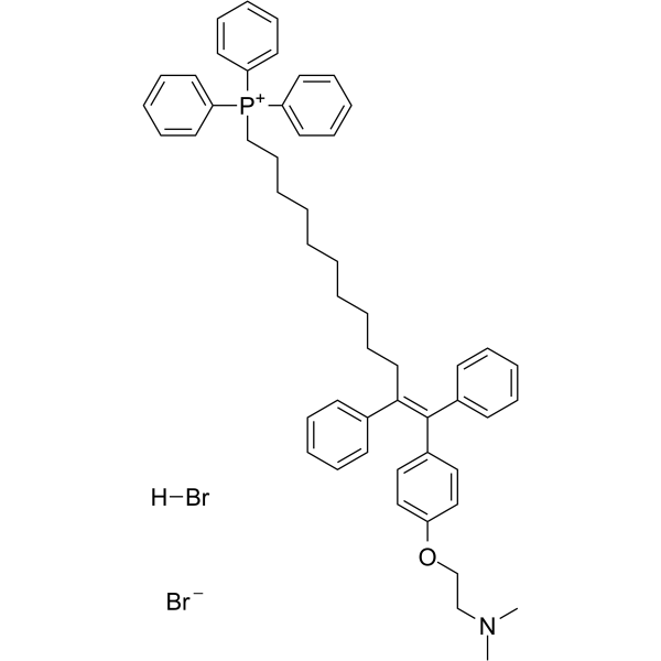
MitoTam bromide, hydrobromide, a Tamoxifen derivative, is an electron transport chain (ETC) inhibitor. MitoTam bromide, hydrobromide reduces mitochondrial membrane potential in senescent cells and affects mitochondrial morphology. MitoTam bromide, hydrobromide is an effective anticancer agent, suppresses respiratory complexes (CI-respiration) and disrupts respiratory supercomplexes (SCs) formation in breast cancer cells.
For research use only. We do not sell to patients.
- CAS No.: 1634624-73-9
- Formula: C52H60Br2NOP
- Molecular Weight:905.82
-
Storage:
4°C, stored under nitrogen, away from moisture
* In solvent : -80°C, 6 months; -20°C, 1 month (stored under nitrogen, away from moisture)

To place orders, for customer services and technical support, please contact: MedChemExpress USA
Tel: 609-228-6898 E-mail: [email protected] [email protected]


-
 Biological Activity
-
Chemical Information
-
Solvent & Solubility
- Protocols
- Purity & Documentation
- References
-
 Help & FAQs




Biological Activity
Description
In Vitro
MitoTam (0.5 μM-56 μM; 24 hours) kills breast cancer cell Lines and nonmalignant cells with an IC50 range from 0.65 μM to 55.9 μM[1].MitoTam (2.5 μM; 2-24 hours) results in stronger activation of the apoptotic pathway in MCF7 Her2 high cells compared with mock MCF7 cells[1].MitoTam (0.05 μM-1 μM; 3 days) causes a concentration-dependent induction of apoptosis in breast cancer cells, while there was no effect for non-malignant breast epithelial cells[2]
MedChemExpress (MCE) has not independently confirmed the accuracy of these methods. They are for reference only. Further protocols information, click here.
-
Cell Line:Breast Cancer Cell Lines: BT474, MCF7, MCF7 Her2high, MCF7 Her2low, MDA-MB-231, MDA-MB-436, MDA-MB-453, SK-BR-3, T47D; NeuTL cells; Nonmalignant Cells: A014578, H9c2 cells
-
Concentration:0.5 μM-56 μM
-
Incubation Time:24 hours
-
Result:Killed breast cancer cells MCF7, MCF7 Her2high, MCF7 Her2low with IC50 values of 1.25 μM, 0.65 μM and 1.45 μM respectively.
-
Cell Line:MCF7 mock cells, MCF7 Her2high cells
-
Concentration:2.5 μM
-
Incubation Time:2 hours, 4 hours, 8 hours, 16 hours, 24 hours
-
Result:Revealed accelerated cleavage of procaspase-9, Parp1/2 and proapoptotic Bax, decreased the antiapoptotic Bcl-2 protein in Her2high cells.
-
Cell Line:MCF-7 cells, 4T1 cells and MCF-10a cells
-
Concentration:0.05 μM-1 μM
-
Incubation Time:3 days
-
Result:Resulted in apoptosis in MCF7 and 4T1 cells.
In Vivo
MedChemExpress (MCE) has not independently confirmed the accuracy of these methods. They are for reference only.
-
Animal Model:18-month-old or 2-month-old FVB/N mice[2]
-
Dosage:2 μg/g
-
Administration:Intraperitoneal injection; 2 μg/g; once a week; 4 weeks
-
Result:Eliminated senescent cells also in vivo.
-
Animal Model:FVB/N c-neu mouse[1]
-
Dosage:0.54 μmol/mouse
-
Administration:Intraperitoneal injection; 0.54 μmol/mouse; twice a week; 2 weeks
-
Result:Suppressed Her2high breast carcinomas.
-
Animal Model:Balb/c nude mice with MCF7 mock or MCF7 Her2high cells[1]
-
Dosage:0.25 μmol/mouse/dose
-
Administration:Intraperitoneal injection; 0.25 μmol/mouse/dose; twice a week; 2 weeks
-
Result:Prevented reaching the ethical endpoint in all situations, slowed down the growth of MCF7 mock tumors and suppressed Her2high carcinomas decreased.


Chemical Information
-
CAS No. 1634624-73-9
-
Appearance Solid
-
Molecular Weight 905.82
-
Formula C52H60Br2NOP
-
Color White to off-white
-
SMILES
CN(CCOC1=CC=C(/C(C2=CC=CC=C2)=C(C3=CC=CC=C3)/CCCCCCCCCC[P+](C4=CC=CC=C4)(C5=CC=CC=C5)C6=CC=CC=C6)C=C1)C.Br[H].[Br-]
-
Shipping
Room temperature in continental US; may vary elsewhere.
-
Storage
4°C, stored under nitrogen, away from moisture
* In solvent : -80°C, 6 months; -20°C, 1 month (stored under nitrogen, away from moisture)


Solvent & Solubility
In Vitro:
DMSO : 240 mg/mL (264.95 mM; Need ultrasonic; Hygroscopic DMSO has a significant impact on the solubility of product, please use newly opened DMSO)
Please refer to the solubility information to select the appropriate solvent. Once prepared, please aliquot and store the solution to prevent product inactivation from repeated freeze-thaw cycles.
Storage method and period of stock solution: -80°C, 6 months; -20°C, 1 month (stored under nitrogen, away from moisture). When stored at -80°C, please use it within 6 months. When stored at -20°C, please use it within 1 month.
Please refer to the solubility information to select the appropriate solvent. Once prepared, please aliquot and store the solution to prevent product inactivation from repeated freeze-thaw cycles.
Storage method and period of stock solution: -80°C, 6 months; -20°C, 1 month (stored under nitrogen, away from moisture). When stored at -80°C, please use it within 6 months. When stored at -20°C, please use it within 1 month.
Concentration (start) × Volume (start) = Concentration (final) × Volume (final)

Protocols
-
Mitochondrial membrane-potential fluorescent assay
Mitochondrial membrane potential fluorescent assays estimate ΔΨm in living cells using lipophilic cationic dyes such as TMRM, TMRE, rhodamine 123, and JC-1, which accumulate in mitochondria according to membrane polarization; loss of signal after FCCP or CCCP treatment is interpreted as mitochondrial depolarization. TMRM/TMRE and rhodamine 123 are commonly used for semi-quantitative live-cell microscopy or flow cytometry, while JC-1 can report a shift from red aggregate fluorescence to green monomer fluorescence during depolarization; interpretation requires controls because dye concentration, quenching mode, cell type, dye efflux, and mitochondrial mass can affect fluorescence independently of ΔΨm.
-
Fluorescent plasma-membrane potential dye assay
Fluorescent plasma-membrane potential dye assays measure changes in cell membrane potential using voltage-sensitive dyes whose fluorescence changes when cells depolarize or hyperpolarize. Anionic bis-oxonol dyes such as DiBAC4(3) enter depolarized cells more readily and show increased fluorescence after intracellular binding, while hyperpolarization reduces dye accumulation and fluorescence. FMP/FLIPR membrane-potential dyes are used for faster, homogeneous microplate assays of ion-channel or receptor-mediated membrane-potential changes.
-
Mitochondrial membrane-potential and mitochondrial mass staining
Mitochondrial membrane potential staining measures the electrochemical polarization across the mitochondrial inner membrane in live cells using lipophilic cationic fluorescent probes; early rhodamine-based work showed that selective mitochondrial dye accumulation is lost when the mitochondrial transmembrane potential is dissipated. JC-1 reports mitochondrial polarization by shifting from green monomer fluorescence to red J-aggregate fluorescence as dye concentration increases within energized mitochondria; therefore, the red/green fluorescence ratio is used as a relative readout of mitochondrial membrane potential. TMRE or TMRM staining provides a single-channel relative readout because these cationic rhodamine esters accumulate in polarized mitochondria, and lower fluorescence indicates reduced mitochondrial polarization when acquisition and dye-loading conditions are controlled. Mitochondrial mass staining is commonly performed with MitoTracker Green FM or related MitoTracker dyes as
-
Breast Cancer Modeling
Breast cancer is a heterogeneous cancer, and it has been distinguished into four subtypes: luminal A, luminal B, HER2-positive and basal-like. Molecular mutations, epigenetic alterations, hormone exposure and immune microenvironment are related to the progression of breast cancer.
-
Senescence-associated β-galactosidase staining
Senescence-associated β-galactosidase staining detects β-galactosidase activity that is histochemically visible at pH 6. 0 in senescent cells, where X-gal cleavage produces an insoluble blue precipitate observable by bright-field microscopy. This activity reflects increased lysosomal β-galactosidase/lysosomal mass rather than a senescence-essential enzyme, because GLB1 depletion or genetic lysosomal β-galactosidase deficiency can abolish SA-β-gal staining while cells still undergo senescence. SA-β-gal was originally reported in senescent but not presenescent fibroblasts and keratinocytes, absent from quiescent fibroblasts and terminally differentiated keratinocytes, and increased with donor age in human skin samples. Because SA-β-gal can also appear in some non-senescent or tissue-specific contexts, interpretation should be paired with experimental controls and, when possible, independent senescence markers.


Purity & Documentation
-
Data Sheet (277 KB)
-
SDS (252 KB)
- English - EN (252 KB)
- Français - FR (252 KB)
- Deutsch - DE (252 KB)
- Norwegian - NO (252 KB)
- Español - ES (252 KB)
- Swedish - SV (252 KB)
- Italian - IT (252 KB)
- Korean - KR (252 KB)
- Portuguese - PT (252 KB)
-
Handling Instructions (2659 KB)
References
[1]. Rohlenova K, et al. Selective Disruption of Respiratory Supercomplexes as a New Strategy to Suppress Her2highBreast Cancer. Antioxid Redox Signal. 2017 Jan 10;26(2):84-103. [Content Brief]
[2]. Hubackova S, et al. Selective elimination of senescent cells by mitochondrial targeting is regulated by ANT2. Cell Death Differ. 2019 Jan;26(2):276-290. [Content Brief]


Complete Stock Solution Preparation Table
Please refer to the solubility information to select the appropriate solvent. Once prepared, please aliquot and store the solution to prevent product inactivation from repeated freeze-thaw cycles.
Storage method and period of stock solution: -80°C, 6 months; -20°C, 1 month (stored under nitrogen, away from moisture). When stored at -80°C, please use it within 6 months. When stored at -20°C, please use it within 1 month.
| Optional Solvent | Concentration Solvent Mass | 1 mg | 5 mg | 10 mg | 25 mg |
|---|---|---|---|---|---|
| DMSO | 1 mM | 1.1040 mL | 5.5199 mL | 11.0397 mL | 27.5993 mL |
| 5 mM | 0.2208 mL | 1.1040 mL | 2.2079 mL | 5.5199 mL | |
| 10 mM | 0.1104 mL | 0.5520 mL | 1.1040 mL | 2.7599 mL | |
| 15 mM | 0.0736 mL | 0.3680 mL | 0.7360 mL | 1.8400 mL | |
| 20 mM | 0.0552 mL | 0.2760 mL | 0.5520 mL | 1.3800 mL | |
| 25 mM | 0.0442 mL | 0.2208 mL | 0.4416 mL | 1.1040 mL | |
| 30 mM | 0.0368 mL | 0.1840 mL | 0.3680 mL | 0.9200 mL | |
| 40 mM | 0.0276 mL | 0.1380 mL | 0.2760 mL | 0.6900 mL | |
| 50 mM | 0.0221 mL | 0.1104 mL | 0.2208 mL | 0.5520 mL | |
| 60 mM | 0.0184 mL | 0.0920 mL | 0.1840 mL | 0.4600 mL | |
| 80 mM | 0.0138 mL | 0.0690 mL | 0.1380 mL | 0.3450 mL | |
| 100 mM | 0.0110 mL | 0.0552 mL | 0.1104 mL | 0.2760 mL |


MitoTam bromide, hydrobromide Related Classifications
HY-126222 Related Classifications
 Powered by Bioz