



MitoTracker Orange CMTMRos
Based on 1 publication(s) in Google Scholar
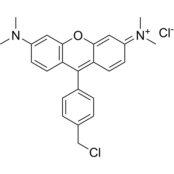
MitoTracker Orange CMTMRos is a fluorescent dye that labels mitochondria within live cells utilizing the mitochondrial membrane potential (Ex/Em: 551/576 nm).
For research use only. We do not sell to patients.
- Purity : 94.19%
- CAS No.: 199116-50-2
- Formula: C24H24Cl2N2O
- Molecular Weight:427.37
-
Storage:
4°C, sealed storage, away from moisture and light
* In solvent : -80°C, 6 months; -20°C, 1 month (sealed storage, away from moisture and light)

To place orders, for customer services and technical support, please contact: MedChemExpress USA
Tel: 609-228-6898 E-mail: [email protected] [email protected]


-
 Biological Activity
-
Chemical Information
-
Solvent & Solubility
- Protocols
- Purity & Documentation
- References
-
 Help & FAQs


-
HY-135056
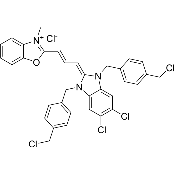
MitoTracker Green FMMitochondrial Dye -
HY-D1783
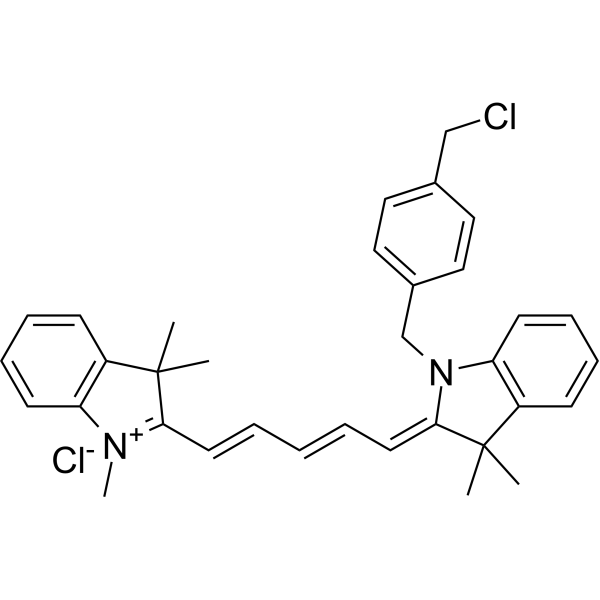
MitoTracker Deep Red FMRed Fluorescent Dye -
HY-D1434
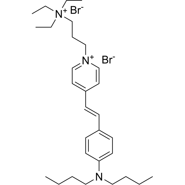
FM1-43Track Endocytosis/Exocytosis -
HY-D1297
ER-Tracker GreenEndoplasmic Reticulum Fluorescent Dye -
HY-D0940
H2DCFDAROS-Sensitive Probe -

HY-D1451
PKH 26Fluorescent -

HY-DY1059
FM1-43 (solution)Track Endocytosis/Exocytosis -

HY-N1446
Oleic acidMonounsaturated Fatty Acid -

HY-13453
BAY 11-7082IκBα/NF-κB Inhibitor -
HY-D1055
MitoSOX RedMitochondrial ROS Indicator -
HY-D1116
MitoMark Red IFluorescent Dye -
HY-D1300
LysoTracker RedCell Death Indicator
Publications Citing Use of MedChemExpress (MCE) MitoTracker Orange CMTMRos
More

Biological Activity
Description
IC50 & Target
Mitochondria[1]
In Vitro
Guide (The following is our recommended experimental protocol. This protocol is for reference only and should be modified according to your specific needs).
Cell labeling[1]:
For live cells, follow steps 1-7. For fixed cells, follow steps 8-13.
1. Prepare a labeling solution at a concentration of 25–500 nM in the desired cell culture medium, and warm it to the cell culture temperature.
2. Remove the culture medium from the cells on the coverslip.
3. Immerse the cells in the labeling medium and incubate at 37°C for 15-45 min.
4. Remove the labeling medium and rinse the cells three times with culture medium.
5. Mount the cells.
6. Examine the cells using a fluorescence microscope. If fluorescence staining is too low, try one of the following:
i. Incubate for an additional 30 min in normal culture medium to allow thiol binding to occur.
ii. Increase the initial concentration of the labeling solution.
7. When fluorescence is sufficient, image the cells.
8. Repeat steps 1-4, but use a labeling solution with a working concentration between 100 and 1000 nM.
9. Wash the cells three times with PBS.
10. Fix the cells in PBS (pH 7.4) containing 3.7% formaldehyde for 10 min.
11. Aspirate and wash the cells three times with PBS, 5 min each.
12. Mount with a coverslip.
13. Image the cells.
MedChemExpress (MCE) has not independently confirmed the accuracy of these methods. They are for reference only. Further protocols information, click here.
Emission (Em)
576
Excitation (Ex)
554


Chemical Information
-
CAS No. 199116-50-2
-
Appearance Solid
-
Molecular Weight 427.37
-
Formula C24H24Cl2N2O
-
Color Brown to black
-
SMILES
CN(C)C(C=C1)=CC2=C1C(C3=CC=C(CCl)C=C3)=C(C=C/4)C(O2)=CC4=[N+](C)/C.[Cl-]
-
Shipping
Room temperature in continental US; may vary elsewhere.
-
Storage
4°C, sealed storage, away from moisture and light
* In solvent : -80°C, 6 months; -20°C, 1 month (sealed storage, away from moisture and light)


Publications (1)
-
Journal Impact Factor
-
Most Recent
-
Mol Plant
A TT1-SCE1 module integrates ubiquitination and SUMOylation to regulate heat tolerance in rice. [Abstract]2024 Dec 2;17(12):1899-1918. PMID: 39552084


Solvent & Solubility
In Vitro:
DMSO : 31.25 mg/mL (73.12 mM; ultrasonic and warming and heat to 60°C; Hygroscopic DMSO has a significant impact on the solubility of product, please use newly opened DMSO)
Please refer to the solubility information to select the appropriate solvent. Once prepared, please aliquot and store the solution to prevent product inactivation from repeated freeze-thaw cycles.
Storage method and period of stock solution: -80°C, 6 months; -20°C, 1 month (sealed storage, away from moisture and light). When stored at -80°C, please use it within 6 months. When stored at -20°C, please use it within 1 month.
Please refer to the solubility information to select the appropriate solvent. Once prepared, please aliquot and store the solution to prevent product inactivation from repeated freeze-thaw cycles.
Storage method and period of stock solution: -80°C, 6 months; -20°C, 1 month (sealed storage, away from moisture and light). When stored at -80°C, please use it within 6 months. When stored at -20°C, please use it within 1 month.
Concentration (start) × Volume (start) = Concentration (final) × Volume (final)

Protocols
-
Mitochondrial membrane-potential fluorescent assay
Mitochondrial membrane potential fluorescent assays estimate ΔΨm in living cells using lipophilic cationic dyes such as TMRM, TMRE, rhodamine 123, and JC-1, which accumulate in mitochondria according to membrane polarization; loss of signal after FCCP or CCCP treatment is interpreted as mitochondrial depolarization. TMRM/TMRE and rhodamine 123 are commonly used for semi-quantitative live-cell microscopy or flow cytometry, while JC-1 can report a shift from red aggregate fluorescence to green monomer fluorescence during depolarization; interpretation requires controls because dye concentration, quenching mode, cell type, dye efflux, and mitochondrial mass can affect fluorescence independently of ΔΨm.
-
Fluorescent plasma-membrane potential dye assay
Fluorescent plasma-membrane potential dye assays measure changes in cell membrane potential using voltage-sensitive dyes whose fluorescence changes when cells depolarize or hyperpolarize. Anionic bis-oxonol dyes such as DiBAC4(3) enter depolarized cells more readily and show increased fluorescence after intracellular binding, while hyperpolarization reduces dye accumulation and fluorescence. FMP/FLIPR membrane-potential dyes are used for faster, homogeneous microplate assays of ion-channel or receptor-mediated membrane-potential changes.
-
Mitochondrial membrane-potential and mitochondrial mass staining
Mitochondrial membrane potential staining measures the electrochemical polarization across the mitochondrial inner membrane in live cells using lipophilic cationic fluorescent probes; early rhodamine-based work showed that selective mitochondrial dye accumulation is lost when the mitochondrial transmembrane potential is dissipated. JC-1 reports mitochondrial polarization by shifting from green monomer fluorescence to red J-aggregate fluorescence as dye concentration increases within energized mitochondria; therefore, the red/green fluorescence ratio is used as a relative readout of mitochondrial membrane potential. TMRE or TMRM staining provides a single-channel relative readout because these cationic rhodamine esters accumulate in polarized mitochondria, and lower fluorescence indicates reduced mitochondrial polarization when acquisition and dye-loading conditions are controlled. Mitochondrial mass staining is commonly performed with MitoTracker Green FM or related MitoTracker dyes as


Purity & Documentation
-
Data Sheet (275 KB)
-
SDS (252 KB)
- English - EN (252 KB)
- Français - FR (252 KB)
- Deutsch - DE (252 KB)
- Norwegian - NO (252 KB)
- Español - ES (252 KB)
- Swedish - SV (252 KB)
- Italian - IT (252 KB)
- Korean - KR (252 KB)
- Portuguese - PT (252 KB)
-
Handling Instructions (2659 KB)
References


Complete Stock Solution Preparation Table
Please refer to the solubility information to select the appropriate solvent. Once prepared, please aliquot and store the solution to prevent product inactivation from repeated freeze-thaw cycles.
Storage method and period of stock solution: -80°C, 6 months; -20°C, 1 month (sealed storage, away from moisture and light). When stored at -80°C, please use it within 6 months. When stored at -20°C, please use it within 1 month.
| Optional Solvent | Concentration Solvent Mass | 1 mg | 5 mg | 10 mg | 25 mg |
|---|---|---|---|---|---|
| DMSO | 1 mM | 2.3399 mL | 11.6995 mL | 23.3989 mL | 58.4973 mL |
| 5 mM | 0.4680 mL | 2.3399 mL | 4.6798 mL | 11.6995 mL | |
| 10 mM | 0.2340 mL | 1.1699 mL | 2.3399 mL | 5.8497 mL | |
| 15 mM | 0.1560 mL | 0.7800 mL | 1.5599 mL | 3.8998 mL | |
| 20 mM | 0.1170 mL | 0.5850 mL | 1.1699 mL | 2.9249 mL | |
| 25 mM | 0.0936 mL | 0.4680 mL | 0.9360 mL | 2.3399 mL | |
| 30 mM | 0.0780 mL | 0.3900 mL | 0.7800 mL | 1.9499 mL | |
| 40 mM | 0.0585 mL | 0.2925 mL | 0.5850 mL | 1.4624 mL | |
| 50 mM | 0.0468 mL | 0.2340 mL | 0.4680 mL | 1.1699 mL | |
| 60 mM | 0.0390 mL | 0.1950 mL | 0.3900 mL | 0.9750 mL |


MitoTracker Orange CMTMRos Related Classifications
HY-D1696 Related Classifications
 Powered by Bioz