



Monobutyl phthalate
Based on 5 publication(s) in Google Scholar
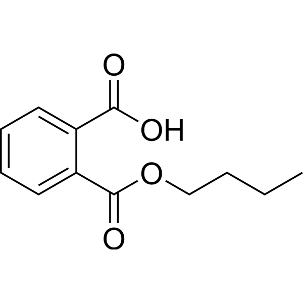
Monobutyl phthalate is an orally active metabolite of Di-n-butyl phthalate (HY-Y0304) and an embryotoxic agent. Monobutyl phthalate regulates mesodermal lineage gene expression, inhibits uterine decidualization, reduces fetal testosterone levels without directly binding to the androgen receptor, and induces teratogenic effects during organogenesis. Monobutyl phthalate induces testicular oxidative stress and sperm abnormalities. Monobutyl phthalate can be used in studies of developmental toxicity and male reproductive toxicity.
For research use only. We do not sell to patients.
- Purity : 99.72%
- CAS No.: 131-70-4
- Formula: C12H14O4
- Molecular Weight:222.24
-
Storage:Powder -20°C, 3 years , 4°C, 2 years ; In solvent -80°C, 6 months , -20°C, 1 month

To place orders, for customer services and technical support, please contact: MedChemExpress USA
Tel: 609-228-6898 E-mail: [email protected] [email protected]


-
 Biological Activity
-
Chemical Information
-
Solvent & Solubility
- Protocols
- Purity & Documentation
- References
-
 Help & FAQs


-
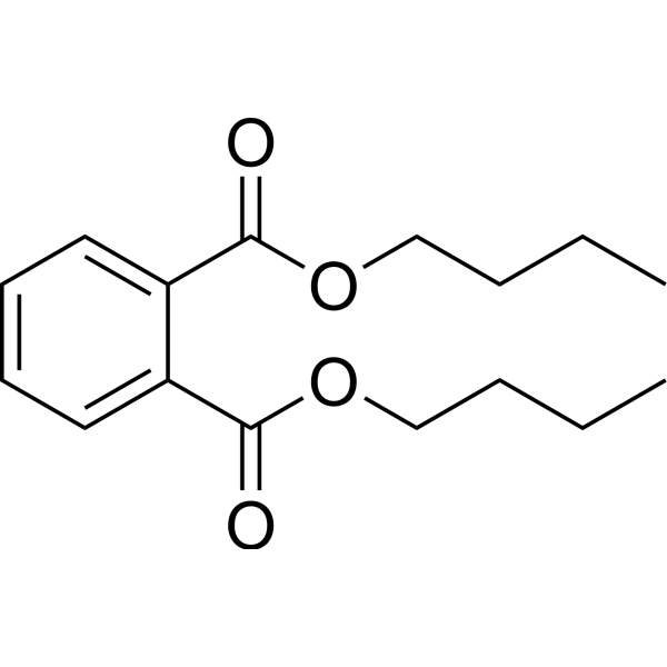
HY-Y0304
Dibutyl phthalatePlasticizer -
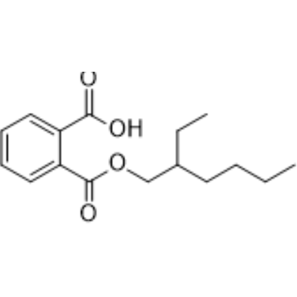
HY-W018392
Mono-(2-ethylhexyl) phthalateEndogenous Metabolite -

HY-N7143R
Monobutyl phthalate (Standard)Antiandrogenic Agent -
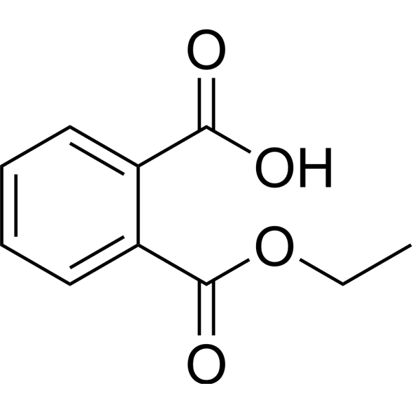
HY-133668
Monoethyl phthalatePDX-1 Activator -
HY-B1945
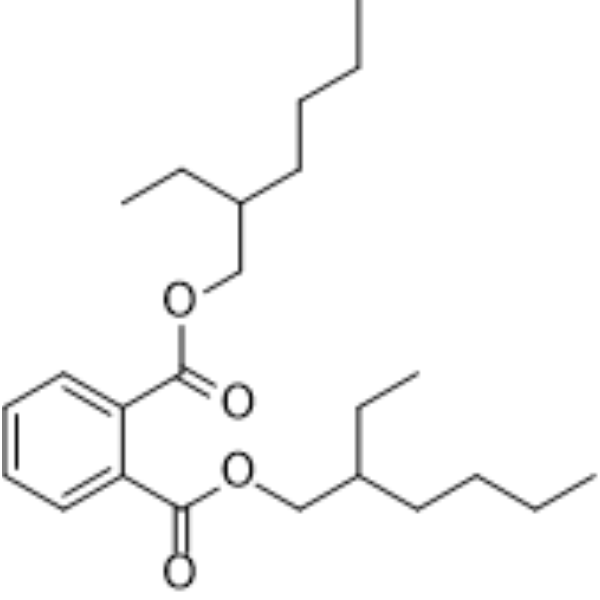
DEHPEndogenous Metabolite -
HY-W011848
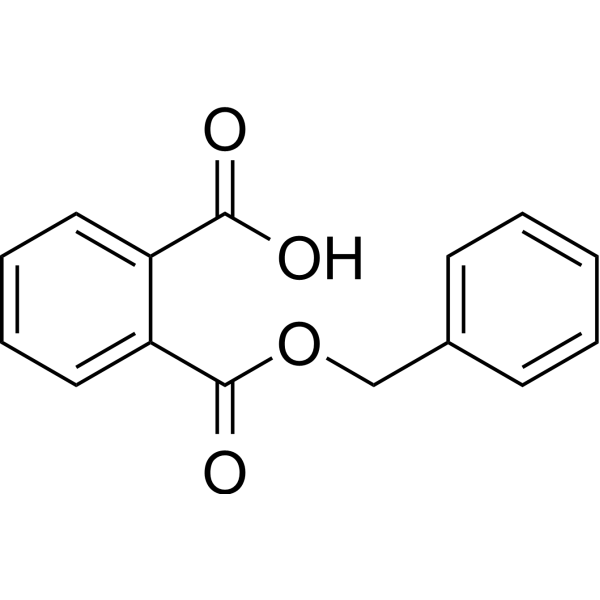
Monobenzyl phthalateDrug Metabolite -
HY-W013570
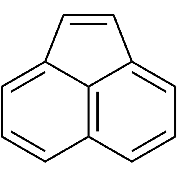
AcenaphthylenePAH -
HY-Y0264
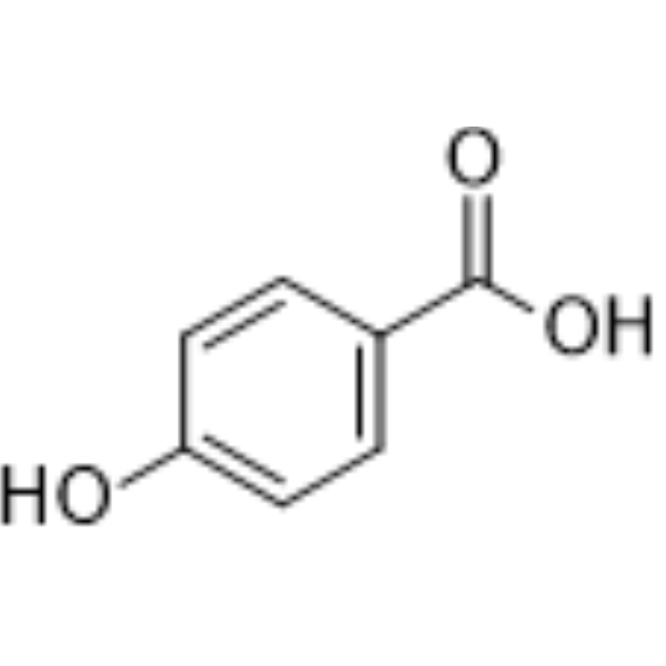
4-Hydroxybenzoic acid -

HY-Y0338
Ethylene glycol,98%Biochemical Assay Reagent -

HY-Y0264R
4-Hydroxybenzoic acid (Standard)
Publications Citing Use of MedChemExpress (MCE) Monobutyl phthalate
More

Biological Activity
Description
Cellular Effect
|
Cell Line

|
Type | Value | Description | References |
|---|---|---|---|---|
| Vero | CC50 |
340.1 μM
Compound: 92
|
Cytotoxicity against African green monkey Vero cells assessed as reduction in cell viability after 4 days by MTT assay
Cytotoxicity against African green monkey Vero cells assessed as reduction in cell viability after 4 days by MTT assay
|
[PMID: 28689975] |
In Vitro
Monobutyl phthalate (0.045-4.5 mM; from suspension culture to day 10 of differentiation) inhibits cardiomyocyte differentiation of mouse D3 ESCs, with an ID50 of 1.35 mM[1].
Monobutyl phthalate (1.35 mM) only causes a slight decrease in the viability of cells derived from mouse D3 ESCs, with a remaining viability of over 90% relative to the control group[1].
Monobutyl phthalate (1.35 mM) upregulates gene sets associated with pluripotency, proliferation, and non-mesodermal differentiation in differentiating mouse D3 ESC embryoid bodies, while specifically downregulating the early cardiomyocyte differentiation gene set 'VAN DARTEL HEARTDIFF 24H', indicating inhibition of mesodermal cardiomyocyte differentiation at the transcriptional level[1].
Monobutyl phthalate (96 h) induces cytotoxicity and inhibits chondrogenic differentiation in limb bud cells of Wistar rat embryos on gestation day 12.5 in a concentration-dependent manner, with an IC50 of 1.38 mM for cytotoxicity and an IC50 of 0.64 mM for differentiation inhibition[3].
MedChemExpress (MCE) has not independently confirmed the accuracy of these methods. They are for reference only. Further protocols information, click here.
-
Cell Line:mouse D3 embryonic stem cell-derived embryoid body cells
-
Concentration:1.35 mM
-
Incubation Time:concurrent with the differentiation assay timeframe starting from day 3 EB stage
-
Result:Measured cell viability via resazurin reduction assay to be 90.7% relative to control, indicating minimal cytotoxicity.
In Vivo
Monobutyl phthalate (250-1000 mg/kg; p.o.; daily; days 0-8 of pseudopregnancy) at 1000 mg/kg suppresses uterine decidualization in pseudopregnant rats[2].
Monobutyl phthalate (500-750 mg/kg; p.o.; daily for 3 consecutive days; on either days 7-9, 10-12, or 13-15 of pregnancy) exerts phase-specific teratogenic effects in rats, with susceptibility windows on days 7-9 and 13-15 of pregnancy, inducing distinct patterns of external and skeletal malformations[2].
Monobutyl phthalate (25-200 mg/kg; p.o.; once daily; 14 days) induces dose-dependent testicular toxicity in male Balb/c mice, with 200 mg/kg causing severe seminiferous tubule degeneration, 27.3% elevated ROS, 28.3% elevated MDA, and significant down-regulation of Sox9 and Dazl mRNA[4].
MedChemExpress (MCE) has not independently confirmed the accuracy of these methods. They are for reference only.
-
Animal Model:Wistar rats (female; pregnant)[2]
-
Dosage:250, 500, 750, 1000 mg/kg
-
Administration:p.o.; daily; days 0-8 of pregnancy
-
Result:Significantly lowered maternal body weight gain and food consumption on days 0-9 of pregnancy at 500 mg/kg and higher.
Significantly increased incidences of preimplantation and postimplantation embryonic loss at 1000 mg/kg.
Significantly lower body weight of live fetuses at 500 mg/kg and higher.
-
Animal Model:Wistar rats (female; pseudopregnant)[2]
-
Dosage:250, 500, 750, 1000 mg/kg
-
Administration:p.o.; daily; days 0-8 of pseudopregnancy
-
Result:Significantly decreased uterine weight on day 9 of pseudopregnancy at 1000 mg/kg, indicating suppression of uterine decidualization.
-
Animal Model:Wistar rats (female; pregnant)[2]
-
Dosage:500, 625, 750 mg/kg
-
Administration:p.o.; daily for 3 consecutive days; on either days 7-9, 10-12, or 13-15 of pregnancy
-
Result:Significantly increased incidence of postimplantation embryonic loss with administration on days 7-9 and days 10-12 at 625 mg/kg and higher, and on days 13-15 at 500 mg/kg and higher.
Significantly increased incidence of fetuses with external malformations after administration on days 7-9 and on days 13-15 at 625 mg/kg and higher.
Significantly increased incidence of fetuses with skeletal malformations with administration on days 7-9 at 500 mg/kg and higher and on days 13-15 at 625 mg/kg and higher.
Cervical vertebrae deformity was frequent after days 7-9 exposure; cleft palate and sternebrae fusion were exclusively found after days 13-15 exposure.
No teratogenicity was detected following days 10-12 administration.
-
Animal Model:Balb/c (male; 6-7 weeks old; 20-25 g; specific pathogen-free)[4]
-
Dosage:25 mg/kg; 50 mg/kg; 100 mg/kg; 200 mg/kg
-
Administration:p.o.; once daily; 14 days
-
Result:Reduced body weight at 50, 100, and 200 mg/kg, with 200 mg/kg producing a final body weight of 21.02 g and a weight change of -9.82%.
Did not significantly alter testis coefficient.
Decreased sperm count in a dose-dependent manner (correlation coefficient r = 0.9917), with significant reductions at 100 and 200 mg/kg.
Increased sperm malformation rate with dose, reaching 12.8% at 100 mg/kg and 13.6% at 200 mg/kg.
Caused no obvious testicular changes at 25 mg/kg, mild testicular changes at 50 mg/kg and 100 mg/kg, and severe histopathological changes including seminiferous tubule degeneration, loss of spermatogenic cells and Sertoli cells, and oligozoospermia in seminiferous tubule lumina at 200 mg/kg.
Significantly increased testicular reactive oxygen species levels by 27.3% and testicular malondialdehyde content by 28.3% at 200 mg/kg.
Decreased glutathione levels in all exposure groups but not significantly.
Significantly decreased Sox9 and Dazl mRNA levels in the 200 mg/kg group.
Showed a slight decreasing trend in Dmrt1 mRNA levels but no significant alteration at any dose.


Chemical Information
-
CAS No. 131-70-4
-
Appearance Solid
-
Molecular Weight 222.24
-
Formula C12H14O4
-
Color White to off-white
-
SMILES
O=C(O)C1=CC=CC=C1C(OCCCC)=O
-
Structure Classification
-
Initial Source
-
Shipping
Room temperature in continental US; may vary elsewhere.
-
Storage
Powder -20°C 3 years 4°C 2 years In solvent -80°C 6 months -20°C 1 month


Publications (5)
-
Journal Impact Factor
-
Most Recent
-
Environ Pollut
Glycine ameliorates MBP-induced meiotic abnormalities and apoptosis by regulating mitochondrial-endoplasmic reticulum interactions in porcine oocytes. [Abstract]2022 Sep 15:309:119756. PMID: 35839969 -
Reprod Toxicol
Effects of Glycine on epigenetic modification and early embryonic development in porcine oocytes exposed to monobutyl phthalate. [Abstract]2024 Oct:129:108684. PMID: 39127149 -
Food Chem Toxicol
Integrated network toxicology and transcriptomics analysis reveals the pathogenic mechanisms in DBP/MBP-induced MASLD/MASH. [Abstract]2026 Jun:212:116039. PMID: 41771476 -
Reprod Sci
DBP Exposure Affects Oocyte Fertilization Via Extracellular Vesicles-Derived miR-116-5p in Ovarian Granulosa Cells Through Downregulating FOXO3a Expression. [Abstract]2024 Dec;31(12):3858-3869. PMID: 38858331 -
J Toxicol Sci
The mechanism of monobutyl phthalate -induced ferroptosis via TNF/IL6/STAT3 signal pathway in TM-3 cells. [Abstract]2023;48(5):299-310. PMID: 37121744


Solvent & Solubility
In Vitro:
DMSO : 100 mg/mL (449.96 mM; Need ultrasonic; Hygroscopic DMSO has a significant impact on the solubility of product, please use newly opened DMSO)
Please refer to the solubility information to select the appropriate solvent. Once prepared, please aliquot and store the solution to prevent product inactivation from repeated freeze-thaw cycles.
Storage method and period of stock solution: -80°C, 6 months; -20°C, 1 month. When stored at -80°C, please use it within 6 months. When stored at -20°C, please use it within 1 month.
Please refer to the solubility information to select the appropriate solvent. Once prepared, please aliquot and store the solution to prevent product inactivation from repeated freeze-thaw cycles.
Storage method and period of stock solution: -80°C, 6 months; -20°C, 1 month. When stored at -80°C, please use it within 6 months. When stored at -20°C, please use it within 1 month.
Concentration (start) × Volume (start) = Concentration (final) × Volume (final)
In Vivo:
Select the appropriate dissolution method based on your experimental animal and administration route.
- For the following dissolution methods, please ensure to first prepare a clear stock solution using an In Vitro approach and then sequentially add co-solvents:
- To ensure reliable experimental results, the clarified stock solution can be appropriately stored based on storage conditions. As for the working solution for In Vivo experiments, it is recommended to prepare freshly and use it on the same day.
- The percentages shown for the solvents indicate their volumetric ratio in the final prepared solution. If precipitation or phase separation occurs during preparation, heat and/or sonication can be used to aid dissolution.
Add each solvent one by one: 10% DMSO 40% PEG300 5% Tween-80 45% Saline
Solubility: ≥ 2.5 mg/mL (11.25 mM); Clear solution
This protocol yields a clear solution of ≥ 2.5 mg/mL (saturation unknown).
Taking 1 mL working solution as an example, add 100 μL DMSO stock solution (25.0 mg/mL) to 400 μL PEG300, and mix evenly; then add 50 μL Tween-80 and mix evenly; then add 450 μL Saline to adjust the volume to 1 mL.
Preparation of Saline: Dissolve 0.9 g sodium chloride in ddH₂O and dilute to 100 mL to obtain a clear Saline solution.
Add each solvent one by one: 10% DMSO 90% (20% SBE-β-CD in Saline)
Solubility: ≥ 2.5 mg/mL (11.25 mM); Clear solution
This protocol yields a clear solution of ≥ 2.5 mg/mL (saturation unknown).
Taking 1 mL working solution as an example, add 100 μL DMSO stock solution (25.0 mg/mL) to 900 μL 20% SBE-β-CD in Saline, and mix evenly.
Preparation of 20% SBE-β-CD in Saline (4°C, storage for one week): 2 g SBE-β-CD powder is dissolved in 10 mL Saline, completely dissolve until clear.
In Vivo Dissolution Calculator
Please enter the basic information of animal experiments:
-
-
-
-
Recommended: Prepare an additional quantity of animals to account for potential losses during experiments.
Please enter your animal formula composition:
-
%DMSO +
Recommended: Keep the proportion of DMSO in working solution below 2% if your animal is weak.
-
%+
-
+%Tween-80 + +
-
%Saline +
The co-solvents required include: DMSO, . All of co-solvents are available by MedChemExpress (MCE). , Tween 80. All of co-solvents are available by MedChemExpress (MCE).
Working solution concentration: 0.22 mg/mL
Method for preparing stock solution: mg drug dissolved in μL DMSO. Stock solution concentration: mg/mL.
1. Take μL DMSO stock solution;
2. Add μL .
μL , mix evenly;
3. Then add μL Tween 80, mix evenly;
4. Then add μL
Please ensure that the stock solution in the first step is dissolved to a clear state, and add co-solvents in sequence. You can use ultrasonic heating (ultrasonic cleaner, recommended frequency 20-40 kHz), vortexing, etc. to assist dissolution.

Protocols
-
RT-PCR
Reverse transcription technology uses RNA as a template to synthesize DNA. RT-PCR is simple, specific and sensitive, and can be used to detect gene expression levels and expression differences in cells; detect RNA virus content; clone cDNA sequences of specific genes.
-
Reproductive and Developmental Toxicity Study
Reproductive and developmental toxicity studies detect adverse effects of prenatal or peri/postnatal exposure on maternal condition, pregnancy maintenance, embryo-fetal survival, fetal growth, structural development, and offspring reproductive or developmental endpoints; classic rat protocols generate readouts by comparing treated groups with vehicle, pair-fed, or untreated controls for implantation, resorption, fetal weight, crown-rump length, external morphology, visceral morphology, skeletal ossification, anogenital distance, nipple/areola retention, and postnatal cohort outcomes.
-
RNA extraction experimental
By lysing cells, releasing RNA, and removing impurities such as proteins and DNA, high-purity RNA products are finally obtained. The commonly used traditional method is the guanidine isothiocyanate/phenol/chloroform method (Trizol), which is suitable for a variety of animal materials including animal tissues, microorganisms, cultured cells, etc., and most plant materials.
-
ROS/oxidative-stress fluorescent staining
ROS/oxidative-stress fluorescent staining uses cell-permeant fluorogenic probes that become fluorescent after oxidation inside cells or tissues; commonly used examples include DCFH-DA/DCFDA for broad cellular oxidant detection, DHE for superoxide-related signal detection, MitoSOX for mitochondrial superoxide-related signal detection, and CellROX probes for oxidative-stress-associated fluorescence readouts. The assay detects probe oxidation rather than a single ROS species unless the probe and analysis method have been chemically validated for that species. DCFH-DA enters cells, is deacetylated by intracellular esterases to DCFH, and produces fluorescent DCF after oxidation, so the readout is used as an operational measure of total cellular oxidative stress rather than a species-specific ROS measurement. DHE and MitoSOX can report superoxide-related oxidation, but red fluorescence alone can include non-specific ethidium-like oxidation products; HPLC or optimized spectral approaches are
-
Research Protocol for Endocrine Diseases
Endocrine diseases often arise from disrupted hormone production, hormone signaling, or target-tissue responsiveness; for diabetes-focused endocrine disease models, insulin signaling regulates glucose uptake, hepatic glucose output, lipid metabolism, and β-cell compensation. Type 2 diabetes develops through interacting defects in insulin resistance, β-cell dysfunction, adipose inflammation, hepatic glucose overproduction, altered incretin signaling, and ectopic lipid metabolism. A major unresolved question is whether endocrine dysfunction is driven primarily by target-tissue insulin resistance, intrinsic β-cell failure, immune/inflammatory stress, or combined multi-organ failure that differs by disease stage.
-
Real Time qPCR (Q-PCR)
Real-time quantitative PCR (qPCR) quantifies an amplifiable nucleic-acid target by monitoring fluorescence during PCR cycling rather than measuring product only after amplification. The increase in fluorescence tracks accumulation of PCR product, and the quantification cycle (Cq; historically also Ct/CP) is related to the initial amount of target: samples containing more starting target generally reach the defined fluorescence threshold in fewer cycles.


Purity & Documentation
-
Data Sheet (292 KB)
-
SDS (393 KB)
- English - EN (393 KB)
- Français - FR (393 KB)
- Deutsch - DE (393 KB)
- Norwegian - NO (393 KB)
- Español - ES (393 KB)
- Swedish - SV (393 KB)
- Italian - IT (393 KB)
- Korean - KR (393 KB)
- Portuguese - PT (393 KB)
-
Handling Instructions (2659 KB)
References
[1]. van Dartel DA, et al. Early gene expression changes during embryonic stem cell differentiation into cardiomyocytes and their modulation by monobutyl phthalate. Reproductive toxicology (Elmsford, N.Y.). 2009 Apr;27(2):93-102. [Content Brief]
[2]. Ema M, et al. Antiandrogenic effects of dibutyl phthalate and its metabolite, monobutyl phthalate, in rats. Congenital anomalies. 2002 Dec;42(4):297-308. [Content Brief]


Complete Stock Solution Preparation Table
Please refer to the solubility information to select the appropriate solvent. Once prepared, please aliquot and store the solution to prevent product inactivation from repeated freeze-thaw cycles.
Storage method and period of stock solution: -80°C, 6 months; -20°C, 1 month. When stored at -80°C, please use it within 6 months. When stored at -20°C, please use it within 1 month.
| Optional Solvent | Concentration Solvent Mass | 1 mg | 5 mg | 10 mg | 25 mg |
|---|---|---|---|---|---|
| DMSO | 1 mM | 4.4996 mL | 22.4982 mL | 44.9964 mL | 112.4910 mL |
| 5 mM | 0.8999 mL | 4.4996 mL | 8.9993 mL | 22.4982 mL | |
| 10 mM | 0.4500 mL | 2.2498 mL | 4.4996 mL | 11.2491 mL | |
| 15 mM | 0.3000 mL | 1.4999 mL | 2.9998 mL | 7.4994 mL | |
| 20 mM | 0.2250 mL | 1.1249 mL | 2.2498 mL | 5.6245 mL | |
| 25 mM | 0.1800 mL | 0.8999 mL | 1.7999 mL | 4.4996 mL | |
| 30 mM | 0.1500 mL | 0.7499 mL | 1.4999 mL | 3.7497 mL | |
| 40 mM | 0.1125 mL | 0.5625 mL | 1.1249 mL | 2.8123 mL | |
| 50 mM | 0.0900 mL | 0.4500 mL | 0.8999 mL | 2.2498 mL | |
| 60 mM | 0.0750 mL | 0.3750 mL | 0.7499 mL | 1.8748 mL | |
| 80 mM | 0.0562 mL | 0.2812 mL | 0.5625 mL | 1.4061 mL | |
| 100 mM | 0.0450 mL | 0.2250 mL | 0.4500 mL | 1.1249 mL |


Monobutyl phthalate Related Classifications
HY-N7143 Related Classifications
 Powered by Bioz