



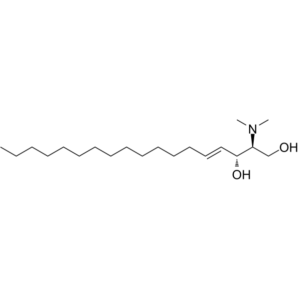
N,N-Dimethylsphingosine
Based on 1 publication(s) in Google Scholar
N,N-Dimethylsphingosine is a sphingosine kinase inhibitor. N,N-Dimethylsphingosine binds competitively to sphingosine kinase and blocks the conversion of sphingosine to sphingosine-1-phosphate. N,N-Dimethylsphingosine inhibits sphingosine-1-phosphate release and aggregation of platelets, and also exerts pro-apoptotic effects by resetting ceramide/sphingosine-1-phosphate homeostasis. N,N-Dimethylsphingosine can be used in cancer-related research.
For research use only. We do not sell to patients.
- Purity : 99.93%
- CAS No.: 119567-63-4
- Formula: C20H41NO2
- Molecular Weight:327.55
-
Storage:
Solution, -20°C, 2 years

To place orders, for customer services and technical support, please contact: MedChemExpress USA
Tel: 609-228-6898 E-mail: [email protected] [email protected]


-
 Biological Activity
-
Chemical Information
- Protocols
- Purity & Documentation
- References
-
 Help & FAQs


-
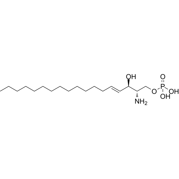
HY-108496
Sphingosine-1-phosphateS1PR Agonist -
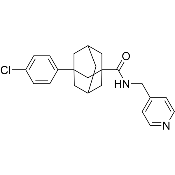
HY-16015
Opaganib -
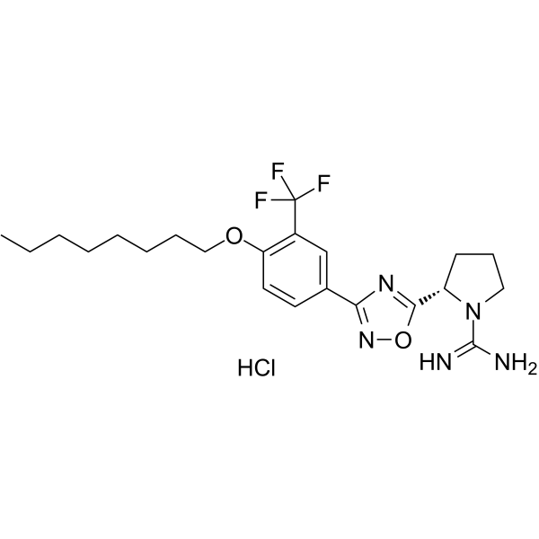
HY-120268A
SLM6031434 hydrochlorideSphK2 Inhibitor -
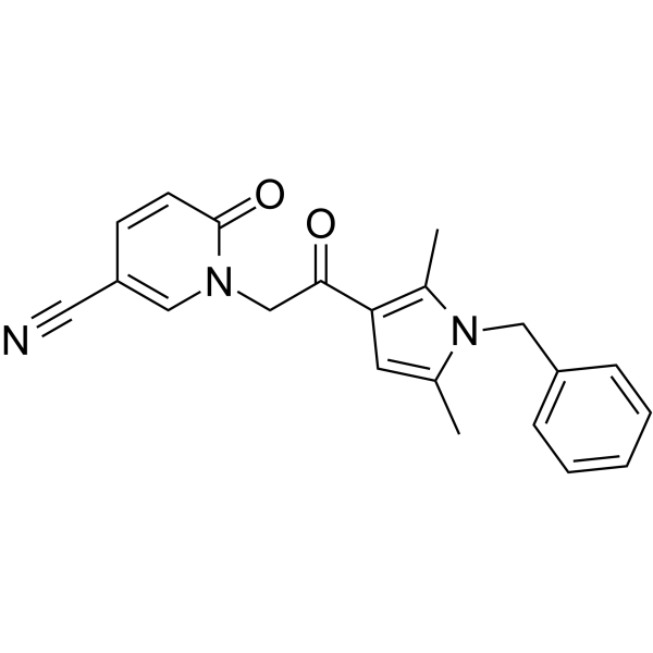
HY-100953
CYM-5520S1PR2 Agonist -
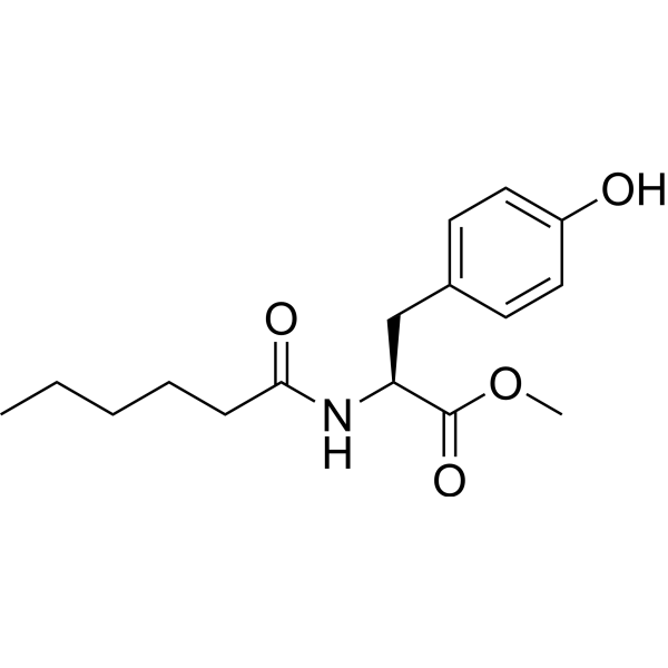
HY-101572
MHPSPHK1 Activator -
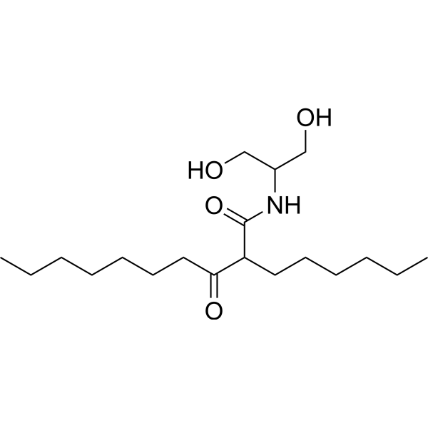
HY-124042
K6PC-5SPHK1 Activator -
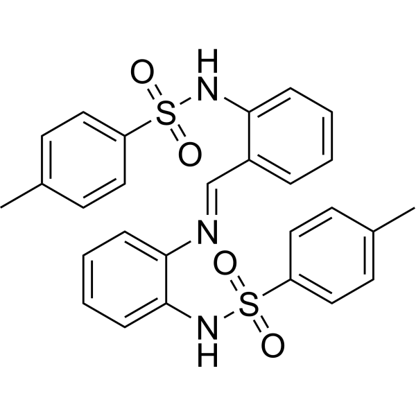
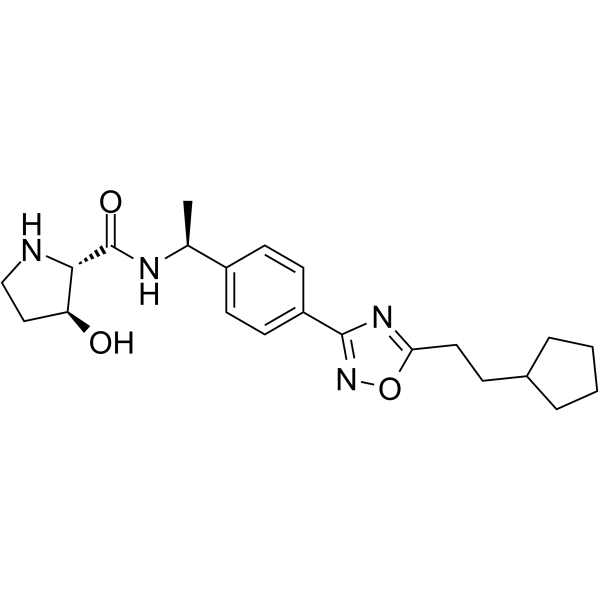
HY-19794
MP-A08SphK Inhibitor -
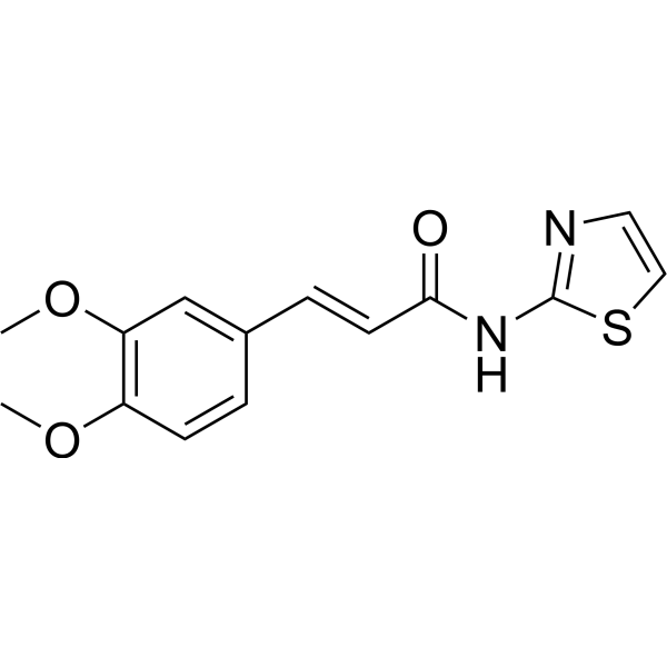
HY-101805
SK1-IN-1SPHK1 Inhibitor -
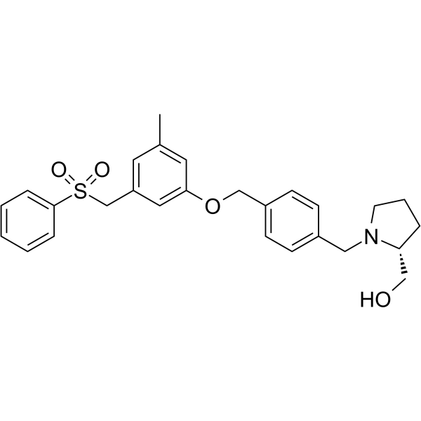
HY-148707
SphK1&2-IN-1SphK Inhibitor -
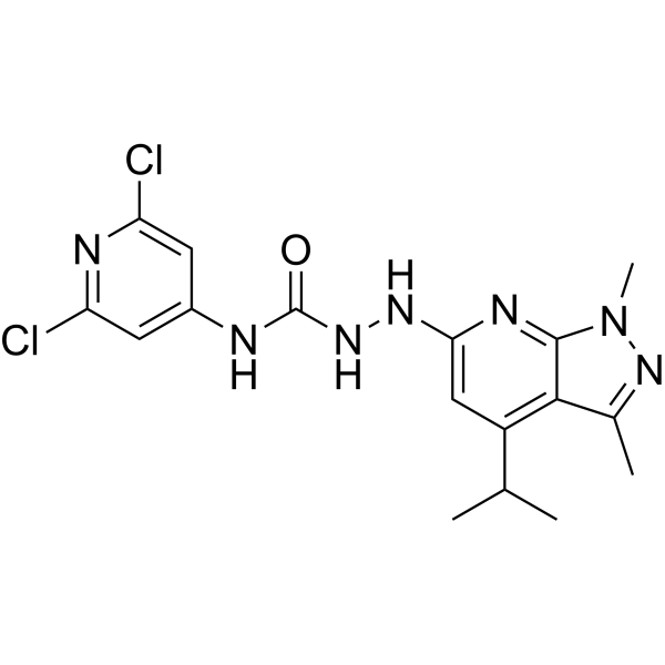
HY-15425
PF-543SPHK1 Inhibitor -
HY-100675
JTE-013S1P2 Antagonist -
HY-W008947
SEW2871S1P1 Agonist
Publications Citing Use of MedChemExpress (MCE) N,N-Dimethylsphingosine
More

Biological Activity
Description
Cellular Effect
|
Cell Line

|
Type | Value | Description | References |
|---|---|---|---|---|
| CHO | IC50 |
5.7 μM
Compound: DMS
|
Inhibition of EGFP-fused human SPHK1 expressed in CHO cells
Inhibition of EGFP-fused human SPHK1 expressed in CHO cells
|
[PMID: 19469544] |
| FL5.12 | IC50 |
0.8 μM
Compound: 8
|
Cytotoxicity against mouse FL5.12A cells after 48 hrs by DAPI staining-based flow cytometric analysis
Cytotoxicity against mouse FL5.12A cells after 48 hrs by DAPI staining-based flow cytometric analysis
|
[PMID: 27475534] |
| RAW264.7 | IC50 |
2.3 μM
Compound: N,N-Dimethylsphingosine
|
Protection against Bacillus anthracis lethal toxin-mediated cytotoxicity in mouse RAW264.7 cells assessed as change in viability after 24 hrs by WST1 dye reduction assay
Protection against Bacillus anthracis lethal toxin-mediated cytotoxicity in mouse RAW264.7 cells assessed as change in viability after 24 hrs by WST1 dye reduction assay
|
[PMID: 17485504] |
In Vitro
N,N-Dimethylsphingosine (0.2-50 µM; 5 min) inhibits sphingosine kinase activity in human platelets, with an IC50 of approximately 10 µM[1].
N,N-Dimethylsphingosine (0.2-50 µM; 1 h) inhibits sphingosine kinase activity in cell-free cytosolic fractions of human platelets, with an IC50 of approximately 5 µM[1].
N,N-Dimethylsphingosine (25 µM; 1 min pretreatment) inhibits TPA (HY-18739)-induced sphingosine-1-phosphate release in human platelets[1].
N,N-Dimethylsphingosine (5-50 µM; 30 min) is a potent competitive inhibitor of sphingosine kinase, with Ki values of 3.1 µM, 2.3 µM, and 6.8 µM in cytoplasmic extracts of U937, Swiss 3T3, and PC12 cells, respectively[2].
N,N-Dimethylsphingosine (10-20 μM; 4-6 h) induces apoptosis in human leukemia cell lines CMK-7, HL60 and U937[3].
N,N-Dimethylsphingosine (5-20 μM; 6-12 h) induces apoptosis in human cancer cell lines A431, MKN74, HT29, HRT18 and COLO205[3].
MedChemExpress (MCE) has not independently confirmed the accuracy of these methods. They are for reference only. Further protocols information, click here.
-
Cell Line:CMK-7, HL60, U937 human leukemic cell lines
-
Concentration:10 μM, 20 μM
-
Incubation Time:4 h, 6 h
-
Result:Induced apoptosis in 65% of CMK-7 cells, 85% of HL60 cells, and 50% of U937 cells after treatment with 20 μM for 6 h.
Revealed condensed/fragmented nuclei, apoptotic bodies, and membrane blebbing in cells treated with 20 μM for 4 h.
Demonstrated clear internucleosomal ladder formation in all three cell lines treated with 10 μM or 20 μM for 4 h or 6 h, with nearly complete DNA fragmentation observed by 6 h.


Chemical Information
-
CAS No. 119567-63-4
-
Appearance Liquid
-
Molecular Weight 327.55
-
Formula C20H41NO2
-
Color Colorless to light yellow
-
SMILES
CCCCCCCCCCCCC/C=C/[C@@H](O)[C@@H](N(C)C)CO
-
Shipping
Room temperature in continental US; may vary elsewhere.
-
Storage
Solution, -20°C, 2 years


Publications (1)
-
Journal Impact Factor
-
Most Recent
-
J Adv Res
Targeting YTHDF2 inhibits tumorigenesis of diffuse large B-cell lymphoma through ACER2-mediated ceramide catabolism. [Abstract]2024 Sep:63:17-33. PMID: 37865189

Protocols
-
Apoptosis
Apoptosis, also called programmed cell death, is generally characterized by distinct morphological characteristics.
-
Apoptosis Solutions
Apoptosis is a regulated, generally non-lytic cell-death pathway that removes unwanted, damaged, infected, or abnormal cells through coordinated morphological changes, caspase activation, DNA fragmentation, and membrane remodeling. The intrinsic apoptosis pathway is controlled mainly by mitochondrial outer membrane permeabilization, BCL-2 family proteins, cytochrome c release, apoptosome formation, caspase-9 activation, and downstream executioner caspase-3/7 activation. The extrinsic apoptosis pathway is initiated by death receptors such as Fas, TNFR, and TRAIL receptors, which recruit adaptor proteins and activate caspase-8 before engaging executioner caspases or mitochondrial amplification through BID cleavage. Apoptosis is linked to many phenotypes, including cancer cell killing, tissue homeostasis, immune regulation, neurodegeneration, infection response, and treatment-induced cytotoxicity; unresolved questions include how apoptosis interacts with necroptosis, pyroptosis, ferroptos


Purity & Documentation
-
Data Sheet (270 KB)
-
SDS (570 KB)
- English - EN (570 KB)
- Français - FR (570 KB)
- Deutsch - DE (570 KB)
- Norwegian - NO (570 KB)
- Español - ES (570 KB)
- Swedish - SV (570 KB)
- Italian - IT (570 KB)
- Korean - KR (570 KB)
- Portuguese - PT (570 KB)
-
Handling Instructions (2659 KB)
References

Calculators
Concentration (start) × Volume (start) = Concentration (final) × Volume (final)


N,N-Dimethylsphingosine Related Classifications
HY-108491 Related Classifications
 Powered by Bioz