



Nagrestipen
Nagrestipen, a human macrophage inflammatory protein-1 alpha (MIP-1α) variant, also known as ECI 301. Nagrestipen has antitumor activity and can be used in therapeutic trials to study cancer, tumors, metastases, radiation oncology, and tumor metastasis.
Nos produits utilisent uniquement pour la recherche. Nous ne vendons pas aux patients.
- CAS No.: 166089-33-4
- Formule: C338H516N88O108S4
- Masse moléculaire:7668.48
-
Stockage:
Please store the product under the recommended conditions in the Certificate of Analysis.

To place orders, for customer services and technical support, please contact: MedChemExpress USA
Tel: 609-228-6898 E-mail: [email protected] [email protected]


-
 Activité biologique
-
Chemical Information
- Protocole
- Pureté et documentation
- Références
-
 Help & FAQs




Activité biologique
Description
In Vivo
Nagrestipen (ECI 301) (i.v; 0.08, 0.4, 2 μg; day 1,8,15) can inhibit tumor growth in a dose-dependent manner at both irradiated and unirradiated sites of female C57BL/6 with LLC cells[1].
MedChemExpress (MCE) has not independently confirmed the accuracy of these methods. They are for reference only.
-
Animal Model:Male BALB/c mice with Colon26 cells and MethA cells[1]
-
Dosage:2 μg
-
Administration:Intravenous injection; daily for 5 days
-
Result:Significantly inhibited tumor size without significant toxicity.
Stopped tumor growth in the colon not only at the irradiated site but also at the unirradiated.


Chemical Information
-
CAS No. 166089-33-4
-
Masse moléculaire 7668.48
-
Formule C338H516N88O108S4
-
Sequence Shortening
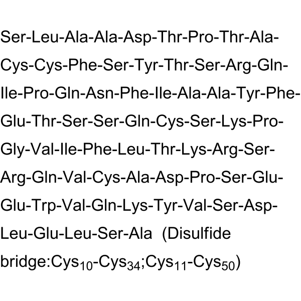
SLAADTPTACCFSYTSRQIPQNFIAAYFETSSQCSKPGVIFLTKRSRQVCADPSEEWVQKYVSDLELSA (Disulfide bridge:Cys10-Cys34;Cys11-Cys50)
-
Livraison
Room temperature in continental US; may vary elsewhere.
-
Stockage
Please store the product under the recommended conditions in the Certificate of Analysis.

Protocole
-
Phagocytosis Functional Assay
A phagocytosis functional assay measures the ability of phagocytic cells, such as neutrophils, macrophages, monocytes, or microglia/macrophages, to bind and internalize particulate targets including bacteria, yeast particles, beads, or myelin particles. Fluorescent flow-cytometry assays detect target uptake as fluorescence associated with gated phagocytes, while pH-sensitive dyes such as pHrodo increase signal in acidic phagosomal compartments and therefore preferentially report internalized particles rather than particles remaining outside the cell. Microscopy or high-content imaging can be used to confirm intracellular localization and, in some protocols, to follow uptake kinetics.
-
Subcutaneous Cell-Line-Derived Xenograft
Subcutaneous cell-line-derived xenograft (CDX) models are established by implanting cultured human cancer cell lines into immunodeficient mice, where the injected cells form localized tumors that can be monitored in vivo as a measure of tumorigenic potential, growth kinetics, and treatment response. These models are widely used in oncology research because they allow reproducible tumor formation and enable comparative assessment of tumor growth between different cell lines or genetic manipulations in a controlled in vivo microenvironment. Subcutaneous implantation of cancer cells in immunodeficient mice is a standard approach for evaluating tumor growth behavior and therapeutic response across multiple cancer types, including prostate, esophageal, pancreatic, and colon cancer models.
-
Orthotopic Cell-Line Xenograft
Orthotopic cell-line xenograft models involve implantation of human cancer cell lines into the anatomically corresponding organ of immunodeficient mice to reproduce tumor growth within a native microenvironment, enabling more clinically relevant tumor behavior compared with subcutaneous models. These models are widely used because orthotopic placement better recapitulates tumor progression, including invasion and metastatic spread, which are often underrepresented in heterotopic implantation systems. Compared with conventional xenografts, orthotopic implantation is described as more technically complex but provides improved simulation of tumor-microenvironment interactions and metastatic behavior, making it particularly valuable for translational oncology research. Surgical orthotopic implantation approaches have been emphasized as enabling faithful reproduction of clinical cancer features, including metastasis and disease progression patterns that align with the tumor’s organ of origi


Pureté et documentation
Références

Calculators
Concentration (start) × Volume (start) = Concentration (final) × Volume (final)


Nagrestipen Related Classifications
HY-P3627 Related Classifications
 Powered by Bioz