



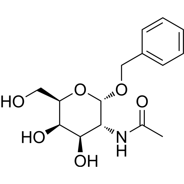
Benzyl-α-GalNAc
Based on 13 publication(s) in Google Scholar
Benzyl-α-GalNAc is a potent O-glycosylation inhibitor. Benzyl-α-GalNAc effectively inhibits the proliferation and activation of LX-2 cells and suppresses the expression of collagen I/III, which has good potential for investigation in liver fibrosis. Benzyl-α-GalNAc also significantly enhances the anti-tumour activity of 5-Fluorouracil (HY-90006) (e.g. pancreatic cancer) by inhibiting O-glycosylation.
For research use only. We do not sell to patients.
- Purity : 99.93%
- CAS No.: 3554-93-6
- Formula: C15H21NO6
- Molecular Weight:311.33
-
Storage:Powder -20°C, 3 years , 4°C, 2 years ; In solvent -80°C, 2 years , -20°C, 1 year

To place orders, for customer services and technical support, please contact: MedChemExpress USA
Tel: 609-228-6898 E-mail: [email protected] [email protected]


-
 Biological Activity
-
Chemical Information
-
Solvent & Solubility
- Protocols
- Purity & Documentation
- References
-
 Help & FAQs


-
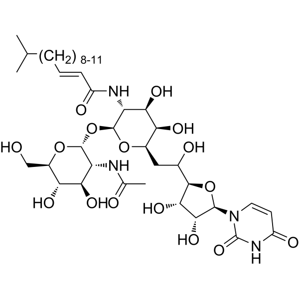
HY-A0098
TunicamycinER Stress Inducer -
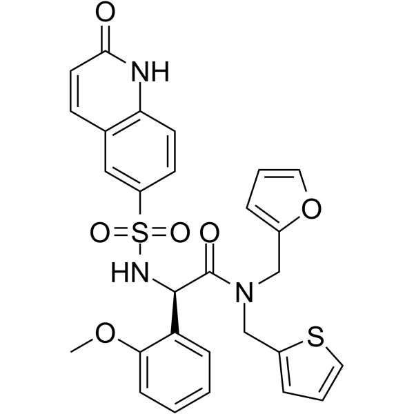
HY-119738
OSMI-1O-GlcNAc Transferase Inhibitor -

HY-13259
MG-132Proteasome Inhibitor -

HY-10071
Y-27632ROCK Inhibitor -
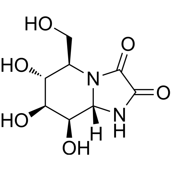
HY-19332
Kifunensineα-Mannosidases Inhibitor -

HY-10583
Y-27632 dihydrochlorideROCK Inhibitor -
HY-110288
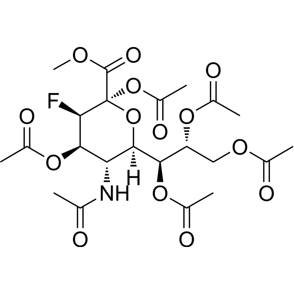
3FAx-Neu5AcSialyltransferase Inhibitor -

HY-15141
StaurosporinePKC/PKA Inhibitor -
HY-N0162
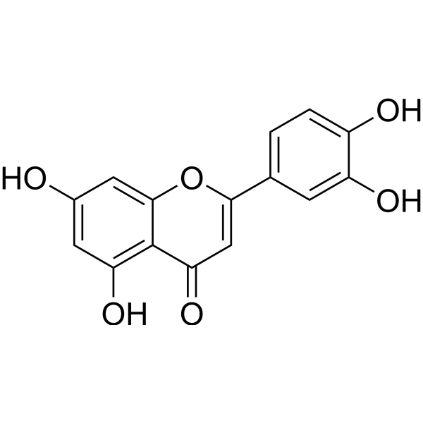
LuteolinNrf2 Inhibitor -

HY-18739
Phorbol 12-myristate 13-acetatePKC Activator -

HY-15371
ForskolinAdenylate Cyclase Activator -

HY-B0015
PaclitaxelAntitumor Agent
Publications Citing Use of MedChemExpress (MCE) Benzyl-α-GalNAc
More- Cancer Discov. 2026 May 13. [Abstract]
- Acta Pharm Sin B. 2025 Oct 24.
- Adv Sci (Weinh). 2026 Apr;13(19):e16106. [Abstract]
- Adv Sci (Weinh). 2025 Apr;12(15):e2415941. [Abstract]
- Cell Death Differ. 2026 May 25. [Abstract]
- Cell Commun Signal. 2026 Jun 4. [Abstract]
- Cell Mol Gastroenterol Hepatol. 2026 Jan 7;20(5):101721. [Abstract]
- Cell Rep. 2025 Jun 25;44(7):115919. [Abstract]
- Br J Pharmacol. 2026 Aug;183(16):4989-5017. [Abstract]
- Commun Biol. 2025 Apr 17;8(1):626. [Abstract]
- Glycobiology. 2026 May 22;36(7):cwag041. [Abstract]
- Virology. 2024 Apr:592:110008. [Abstract]
- bioRxiv. 2026 Jun 18.
 Customer Validation & Images

-
WB
-
IF
-
In Vivo Efficacy Study
-
Histological Imaging/Staining
-
RT-PCR


Biological Activity
Description
IC50 & Target
In Vitro
Benzyl-α-GalNAc (5 mM; 72 h) inhibits O-glycosylation of mucin in SUIT-2 cells[1].
Benzyl-α-GalNAc (2, 4 mM; 48 h) inhibits the proliferation and activation of LX-2 cells[2].
Benzyl-α-GalNAc (2, 4 mM; 48 h) decreases collagen expression in LX-2 cells[2].
MedChemExpress (MCE) has not independently confirmed the accuracy of these methods. They are for reference only. Further protocols information, click here.
-
Cell Line:SUIT-2 cells
-
Concentration:5 mM
-
Incubation Time:72 h
-
Result:Inhibited mucin O-glycosylation.
-
Cell Line:LX-2 cells
-
Concentration:2, 4 mM
-
Incubation Time:48 h
-
Result:Remarkably decreased the expression of α-SMA in a dose-dependent manner (α-SMA is the marker for the activation of HSCs, which means that quiescent cells transform into myofibroblasts).
-
Cell Line:LX-2 cells (PDGF-BB-induced)
-
Concentration:2, 4 mM
-
Incubation Time:48 h
-
Result:Partly reversed PDGF-BB-induced proliferation in cells.
-
Cell Line:LX-2 cells
-
Concentration:2, 4 mM
-
Incubation Time:48 h
-
Result:Downregulated the mRNA levels of collagens I and III in a dose-dependent manner.
In Vivo
MedChemExpress (MCE) has not independently confirmed the accuracy of these methods. They are for reference only.
-
Animal Model:Female severe combined immunodeficient (SCID) mice (6 to 8-week-old; capan-1 tumour model)[3].
-
Dosage:1 mg/mice (in combination with 5-FU)
-
Administration:Tumoural injection; single daily; days 4, 6, 8 and 10 after tumour size reaches 50-70 mm3
-
Result:Significantly lowered neoplastic cells compared to 5-FU alone.


Chemical Information
-
CAS No. 3554-93-6
-
Appearance Solid
-
Molecular Weight 311.33
-
Formula C15H21NO6
-
Color White to off-white
-
SMILES
O[C@@H]([C@H]([C@@H](CO)O1)O)[C@@H](NC(C)=O)[C@H]1OCC2=CC=CC=C2
-
Shipping
Room temperature in continental US; may vary elsewhere.
-
Storage
Powder -20°C 3 years 4°C 2 years In solvent -80°C 2 years -20°C 1 year


Publications (13)
-
Journal Impact Factor
-
Most Recent
-
Cancer Discov
Mismatch Repair-Proficient Colorectal Cancer can evade Immune Surveillance Through an Intrinsic Suppressive Program. [Abstract]2026 May 13. PMID: 42126206 -
-
Adv Sci (Weinh)
GALNT10 Affects O-Glycosylation of IGFBP7 to Promote Tumor Vascular Remodeling and Metastasis of Ovarian Cancer. [Abstract]2026 Apr;13(19):e16106. PMID: 41637621 -
Adv Sci (Weinh)
LIMA1 O-GlcNAcylation Promotes Hepatic Lipid Deposition through Inducing β-catenin-Regulated FASn Expression in Metabolic Dysfunction-Associated Steatotic Liver Disease. [Abstract]2025 Apr;12(15):e2415941. PMID: 39921472
Benzyl-α-GalNAc purchased from MedChemExpress. Usage Cited in: Adv Sci (Weinh). 2025 Apr;12(15):e2415941. [Abstract]
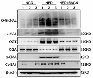
Benzyl-α‐GalNAc (BAGN; 25 mg/kg; i.v.). Western blot analysis of O‐GlcNAc, LIMA1, OGT, OGA, α‐SMA, and Col3A1 in livers from NCD‐fed mice, HFD‐fed mice, and HFD‐fed mice injected with BAGN.
Benzyl-α-GalNAc purchased from MedChemExpress. Usage Cited in: Adv Sci (Weinh). 2025 Apr;12(15):e2415941. [Abstract]
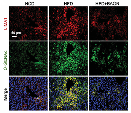
Benzyl-α‐GalNAc (BAGN; 25 mg/kg; i.v.). Immunofluorescence showing LIMA1 (red) and O‐GlcNAc (green) colocalization in livers from NCD‐fed mice, HFD‐fed mice, and HFD‐fed mice injected with BAGN.
Benzyl-α-GalNAc purchased from MedChemExpress. Usage Cited in: Adv Sci (Weinh). 2025 Apr;12(15):e2415941. [Abstract]
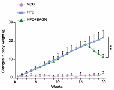
Benzyl-α-GalNAc (BAGN; 25 mg/kg; i.v.). Changes in body weight of NCD‐fed mice, HFD‐fed mice, and HFD‐fed mice injected with BAGN.
Benzyl-α-GalNAc purchased from MedChemExpress. Usage Cited in: Adv Sci (Weinh). 2025 Apr;12(15):e2415941. [Abstract]
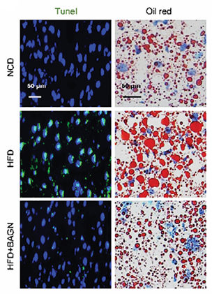
Benzyl-α-GalNAc (BAGN; 25 mg/kg; i.v.). Tunel and Oil red O staining in livers from NCD‐fed mice, HFD‐fed mice, and HFD‐fed mice injected with BAGN.
Benzyl-α-GalNAc purchased from MedChemExpress. Usage Cited in: Adv Sci (Weinh). 2025 Apr;12(15):e2415941. [Abstract]
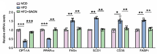
Benzyl-α-GalNAc (BAGN; 25 mg/kg; i.v.). Lipometabolic mRNA expression in the liver of NCD‐fed mice, HFD‐fed mice, and HFD‐fed mice injected with BAGN.
-
Cell Death Differ
Targeting ADAR1 lactylation activates innate immune and overcomes chemoresistance in ovarian cancer. [Abstract]2026 May 25. PMID: 42185630 -
Cell Commun Signal
Heterodimerization with signaling-inert TLR8b converts TLR8a from proviral to antiviral sensor in Ctenopharyngodon idella. [Abstract]2026 Jun 4. PMID: 42243884 -
Cell Mol Gastroenterol Hepatol
OGT-enriched Hepatocyte-derived Extracellular Vesicles Promote Capillarization of Liver Sinusoidal Endothelial Cells in Metabolic Dysfunction-associated Steatotic Liver Disease. [Abstract]2026 Jan 7;20(5):101721. PMID: 41513003 -
Cell Rep
Inhibiting UPF1 methylation enhances tumor immunotherapy sensitivity by reducing nonsense-mediated mRNA decay. [Abstract]2025 Jun 25;44(7):115919. PMID: 40570371 -
Br J Pharmacol
Enzyme-responsive 612-AYR nanoparticles for targeted inhibition of B4GALT3-mediated ITGB1 glycosylation in hepatocellular carcinoma. [Abstract]2026 Aug;183(16):4989-5017. PMID: 42157415 -
Commun Biol
The role of ladderlectin in spermatogenesis and ovarian sperm storage in the black rockfish (Sebastes schlegelii). [Abstract]2025 Apr 17;8(1):626. PMID: 40247087 -
Glycobiology
Sialylation profile and Siglec-E expression across tissues in the B16OVA melanoma mouse model. [Abstract]2026 May 22;36(7):cwag041. PMID: 42223497 -
Virology
Proprotein convertase cleavage of Ictalurid herpesvirus 1 spike-like protein ORF46 is modulated by N-glycosylation. [Abstract]2024 Apr:592:110008. PMID: 38335866 -


Solvent & Solubility
In Vitro:
DMSO : ≥ 100 mg/mL (321.20 mM; Hygroscopic DMSO has a significant impact on the solubility of product, please use newly opened DMSO)
H2O : 7 mg/mL (22.48 mM; ultrasonic and warming and heat to 60°C)
* "≥" means soluble, but saturation unknown.
Please refer to the solubility information to select the appropriate solvent. Once prepared, please aliquot and store the solution to prevent product inactivation from repeated freeze-thaw cycles.
Storage method and period of stock solution: -80°C, 2 years; -20°C, 1 year. When stored at -80°C, please use it within 2 years. When stored at -20°C, please use it within 1 year.
* Note: If you choose water as the stock solution, please dilute it to the working solution, then filter and sterilize it with a 0.22 μm filter before use.
Please refer to the solubility information to select the appropriate solvent. Once prepared, please aliquot and store the solution to prevent product inactivation from repeated freeze-thaw cycles.
Storage method and period of stock solution: -80°C, 2 years; -20°C, 1 year. When stored at -80°C, please use it within 2 years. When stored at -20°C, please use it within 1 year.
* Note: If you choose water as the stock solution, please dilute it to the working solution, then filter and sterilize it with a 0.22 μm filter before use.
Concentration (start) × Volume (start) = Concentration (final) × Volume (final)

Protocols
-
Fibrosis/Collagen Morphometry
Fibrosis and collagen morphometry is based on the quantitative visualization of fibrillar collagen deposition in tissue sections using histochemical stains such as Sirius Red (Picrosirius Red) or Masson's trichrome, followed by image-based or polarization-enhanced analysis to estimate collagen proportional area as a surrogate of extracellular matrix accumulation during fibrotic remodeling. Sirius Red combined with polarized light microscopy enhances detection of collagen fibers due to birefringence properties, enabling more specific visualization of collagen type I and III fibrils compared to conventional bright-field histology, while whole-section or region-restricted digital morphometry reduces field-selection bias in fibrosis assessment. Alternative quantitative approaches include second harmonic generation (SHG) and two-photon excited fluorescence microscopy, which enable label-free detection of fibrillar collagen and have been validated against histological staining and biochemica
-
RNA extraction experimental
By lysing cells, releasing RNA, and removing impurities such as proteins and DNA, high-purity RNA products are finally obtained. The commonly used traditional method is the guanidine isothiocyanate/phenol/chloroform method (Trizol), which is suitable for a variety of animal materials including animal tissues, microorganisms, cultured cells, etc., and most plant materials.
-
Collagen: Sirius Red Staining
Sirius Red or picrosirius red staining is a histochemical method for visualizing collagen-rich extracellular matrix in tissue sections, and collagen fibers are detected as red-stained structures under bright-field microscopy with enhanced birefringence under polarized light. Picrosirius red is useful for assessing total collagen organization, distribution, and fibrosis burden, but polarized color should not be interpreted as a definitive collagen type I versus type III readout because color is affected by fiber orientation, thickness, and packing.
-
Connective Tissue: Masson's Trichrome/Collagen Trichrome Staining
Masson’s Trichrome (collagen/trichrome staining) is a histological technique that differentially stains tissue compartments using sequential acidic dyes to distinguish collagen from muscle and cytoplasmic components based on dye affinity and tissue permeability differences, enabling visualization of fibrosis and connective tissue architecture in histological sections. The classical formulation typically uses Weigert's iron hematoxylin for nuclear staining, Biebrich scarlet-acid fuchsin for cytoplasm and muscle, and aniline blue (or light green variants) for collagen, producing a characteristic blue/green collagen signal contrasted against red cytoplasm and dark nuclei. The staining principle relies on selective displacement of smaller dye molecules by larger anionic dyes in collagen-rich regions under controlled acidified conditions, which enhances collagen-specific dye retention. This property makes the method widely used for fibrosis assessment in organs such as heart, liver, lung, a
-
Cell Viability Determination by MTT Colorimetric Assay
The following protocol uses the MTT colorimetric assay as a classic literature-established method for assessing cell viability/metabolic activity in cultured mammalian cells. MTT[3-(4,5-dimethylthiazol-2-yl)-2,5-diphenyltetrazolium bromide] is reduced by metabolically active cells to a colored formazan product; the amount of formazan is quantified spectrophotometrically and provides an indirect measure of metabolically active viable cells. Importantly, MTT reduction reflects cellular oxidoreductase/metabolic activity rather than an absolute direct count of living cells, so changes in cellular metabolism can alter the signal independently of cell number.
-
Liver Histomorphometry
Liver histomorphometry is a quantitative histological approach used to measure structural alterations in hepatic tissue, including parenchymal loss, steatosis, fibrosis, and vascular remodeling, by combining stained tissue section analysis with stereological or computerized image-based measurements. Classical morphometric frameworks quantify volume fractions of liver compartments and fibrotic regions using systematic sampling and image analysis, enabling objective comparison of pathological changes across experimental groups. These approaches are widely applied in liver cirrhosis and fibrosis studies to reduce subjectivity in histological scoring and improve reproducibility of tissue evaluation. Recent methodological advances integrate automated image analysis and radiomics-based extraction of histological features from standard liver stains (e. g. , H&E and fibrotic stains), enabling quantitative correlation between morphometric features and fibrosis stages in non-alcoholic fatty live


Purity & Documentation
-
Data Sheet (282 KB)
-
SDS (252 KB)
- English - EN (252 KB)
- Français - FR (252 KB)
- Deutsch - DE (252 KB)
- Norwegian - NO (252 KB)
- Español - ES (252 KB)
- Swedish - SV (252 KB)
- Italian - IT (252 KB)
- Korean - KR (252 KB)
- Portuguese - PT (252 KB)
-
Handling Instructions (2659 KB)
References
[1]. Kalra AV, et al. Mucin impedes cytotoxic effect of 5-FU against growth of human pancreatic cancer cells: overcoming cellular barriers for therapeutic gain. Br J Cancer. 2007 Oct 8;97(7):910-8. Epub 2007 Oct 2. [Content Brief]
[2]. Kalra AV, et al. Mucin overexpression limits the effectiveness of 5-FU by reducing intracellular drug uptake and antineoplastic drug effects in pancreatic tumours. Eur J Cancer. 2009 Jan;45(1):164-73. [Content Brief]
[3]. Fan X, et al. Protein O glycosylation regulates activation of hepatic stellate cells. Inflammation. 2013 Dec;36(6):1248-52. https://pubmed.ncbi.nlm.nih.gov/23743764/ [Content Brief]


Complete Stock Solution Preparation Table
Please refer to the solubility information to select the appropriate solvent. Once prepared, please aliquot and store the solution to prevent product inactivation from repeated freeze-thaw cycles.
Storage method and period of stock solution: -80°C, 2 years; -20°C, 1 year. When stored at -80°C, please use it within 2 years. When stored at -20°C, please use it within 1 year.
| Optional Solvent | Concentration Solvent Mass | 1 mg | 5 mg | 10 mg | 25 mg |
|---|---|---|---|---|---|
| H2O / DMSO | 1 mM | 3.2120 mL | 16.0601 mL | 32.1203 mL | 80.3006 mL |
| 5 mM | 0.6424 mL | 3.2120 mL | 6.4241 mL | 16.0601 mL | |
| 10 mM | 0.3212 mL | 1.6060 mL | 3.2120 mL | 8.0301 mL | |
| 15 mM | 0.2141 mL | 1.0707 mL | 2.1414 mL | 5.3534 mL | |
| 20 mM | 0.1606 mL | 0.8030 mL | 1.6060 mL | 4.0150 mL | |
| DMSO | 25 mM | 0.1285 mL | 0.6424 mL | 1.2848 mL | 3.2120 mL |
| 30 mM | 0.1071 mL | 0.5353 mL | 1.0707 mL | 2.6767 mL | |
| 40 mM | 0.0803 mL | 0.4015 mL | 0.8030 mL | 2.0075 mL | |
| 50 mM | 0.0642 mL | 0.3212 mL | 0.6424 mL | 1.6060 mL | |
| 60 mM | 0.0535 mL | 0.2677 mL | 0.5353 mL | 1.3383 mL | |
| 80 mM | 0.0402 mL | 0.2008 mL | 0.4015 mL | 1.0038 mL | |
| 100 mM | 0.0321 mL | 0.1606 mL | 0.3212 mL | 0.8030 mL |
* Note: If you choose water as the stock solution, please dilute it to the working solution, then filter and sterilize it with a 0.22 μm filter before use.


Benzyl-α-GalNAc Related Classifications
HY-129389 Related Classifications
 Powered by Bioz