



OAB-14

Based on 1 Customer Validation
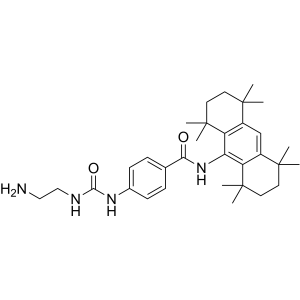
OAB-14, is a Bexarotene (HY-14171) derivative, improves Alzheimer's disease-related pathologies and cognitive impairments by increasing β-amyloid clearance in APP/PS1 mice. OAB-14 effectively ameliorates the dysfunction of the endosomal-autophagic-lysosomal pathway in APP/PS1 transgenic mice.
Nos produits utilisent uniquement pour la recherche. Nous ne vendons pas aux patients.
- Pureté : 99.21%
- CAS No.: 2140911-49-3
- Formule: C32H46N4O2
- Masse moléculaire:518.73
-
Stockage:Powder -20°C, 3 years , 4°C, 2 years ; In solvent -80°C, 6 months , -20°C, 1 month

To place orders, for customer services and technical support, please contact: MedChemExpress USA
Tel: 609-228-6898 E-mail: [email protected] [email protected]


-
 Activité biologique
-
Chemical Information
-
Solvant et solubilité
- Protocole
- Pureté et documentation
- Références
-
 Help & FAQs


-
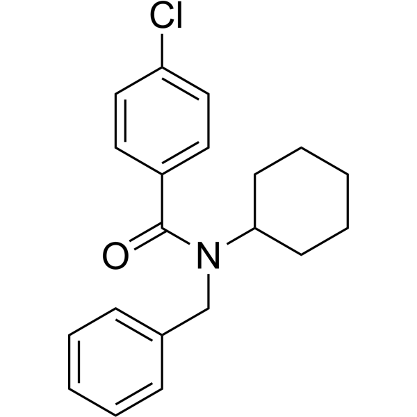
HY-19370
FPS-ZM1Amyloid-β Inhibitor -
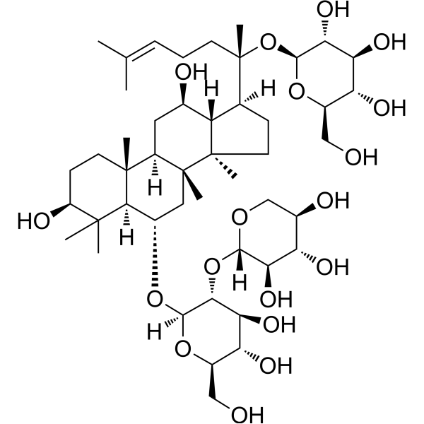
HY-N0615
Notoginsenoside R1Apoptosis Inhibitor -
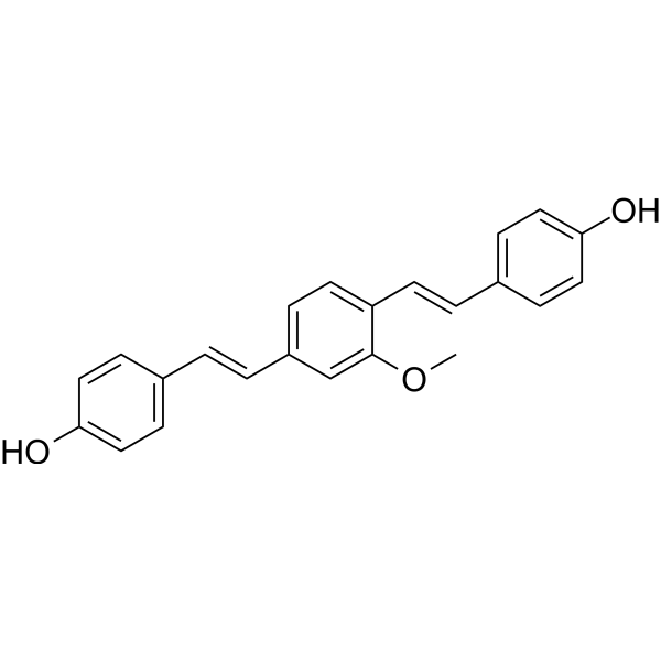
HY-103240
Methoxy-X04Fluorescent Dye -
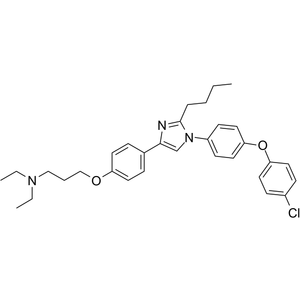
HY-50682
AzeliragonRAGE Inhibitor -
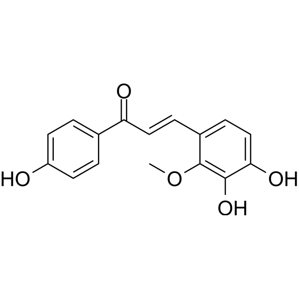
HY-N0373
Licochalcone BApoptosis Inducer -
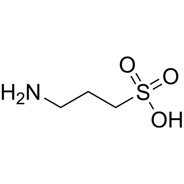
HY-14602
TramiprosateNeuroprotection Agent -
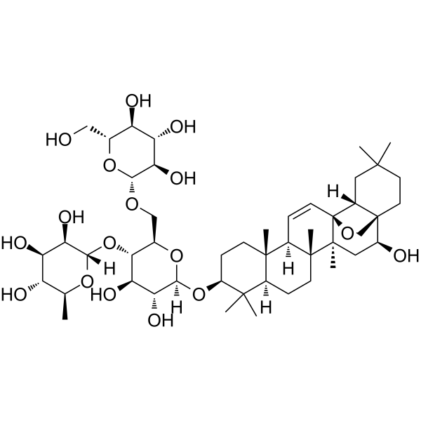
HY-N0249
Saikosaponin CMMP-2 Inducer -
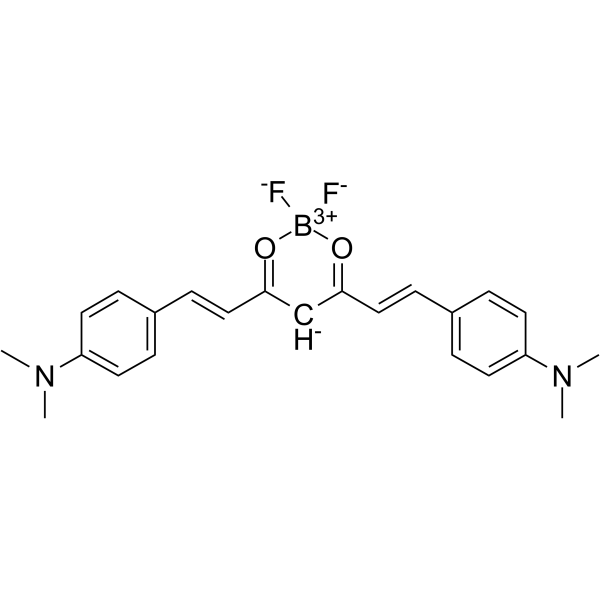
HY-103242
CRANAD-2Fluorescent Probe -
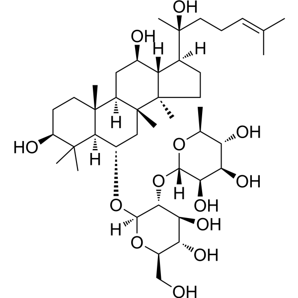
HY-N0602
Ginsenoside Rg2NF-κB Inhibitor -
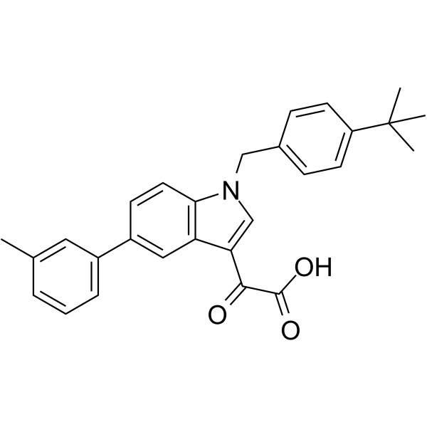
HY-14759
AleplasininPAI-1 Inhibitor -
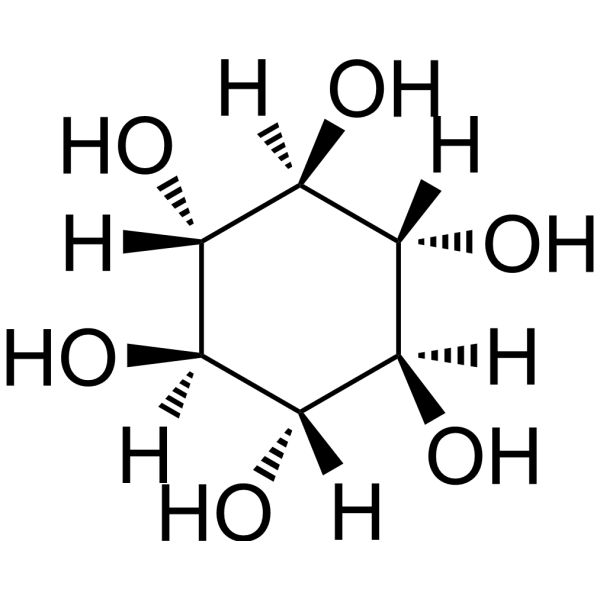
HY-W010041
Scyllo-Inositolα-synuclein/Amyloid-β Aggregation Inhibitor -
HY-18292
ARN2966APP Translation Modulator


Activité biologique
Description
In Vivo
OAB-14 facilitates receptor-mediated endocytosis and restores autophagy flux via the AMPK/mTOR pathway. OAB-14 enhances the lysosomal activity, and reduced Aβ accumulation in lysosomes is observed in OAB-14-treated AD mice[2].
MedChemExpress (MCE) has not independently confirmed the accuracy of these methods. They are for reference only.


Chemical Information
-
CAS No. 2140911-49-3
-
Appearance Solid
-
Masse moléculaire 518.73
-
Formule C32H46N4O2
-
Color White to off-white
-
SMILES
O=C(NCCN)NC1=CC=C(C(NC2=C(C(C)(CCC3(C)C)C)C3=CC4=C2C(C)(C)CCC4(C)C)=O)C=C1
-
Livraison
Room temperature in continental US; may vary elsewhere.
-
Stockage
Powder -20°C 3 years 4°C 2 years In solvent -80°C 6 months -20°C 1 month


Solvant et solubilité
In Vitro:
DMSO : 20 mg/mL (38.56 mM; ultrasonic and warming and adjust pH to 3 with HCl and heat to 60°C; Hygroscopic DMSO has a significant impact on the solubility of product, please use newly opened DMSO)
Please refer to the solubility information to select the appropriate solvent. Once prepared, please aliquot and store the solution to prevent product inactivation from repeated freeze-thaw cycles.
Storage method and period of stock solution: -80°C, 6 months; -20°C, 1 month. When stored at -80°C, please use it within 6 months. When stored at -20°C, please use it within 1 month.
Please refer to the solubility information to select the appropriate solvent. Once prepared, please aliquot and store the solution to prevent product inactivation from repeated freeze-thaw cycles.
Storage method and period of stock solution: -80°C, 6 months; -20°C, 1 month. When stored at -80°C, please use it within 6 months. When stored at -20°C, please use it within 1 month.
Concentration (start) × Volume (start) = Concentration (final) × Volume (final)


Pureté et documentation
-
Fiche technique (271 KB)
-
SDS (251 KB)
- English - EN (251 KB)
- Français - FR (251 KB)
- Deutsch - DE (251 KB)
- Norwegian - NO (251 KB)
- Español - ES (251 KB)
- Swedish - SV (251 KB)
- Italian - IT (251 KB)
- Korean - KR (251 KB)
- Portuguese - PT (251 KB)
-
Instruction de manipulation (2659 KB)
Références
[1]. Guo X, et al. OAB-14 Effectively Ameliorates the Dysfunction of the Endosomal-Autophagic-Lysosomal Pathway in APP/PS1 Transgenic Mice. ACS Chem Neurosci. 2021;12(21):3985-3993. [Content Brief]
[2]. Yuan C, et al. OAB-14, a bexarotene derivative, improves Alzheimer's disease-related pathologies and cognitive impairments by increasing β-amyloid clearance in APP/PS1 mice. Biochim Biophys Acta Mol Basis Dis. 2019;1865(1):161-180. [Content Brief]


Complete Stock Solution Preparation Table
Please refer to the solubility information to select the appropriate solvent. Once prepared, please aliquot and store the solution to prevent product inactivation from repeated freeze-thaw cycles.
Storage method and period of stock solution: -80°C, 6 months; -20°C, 1 month. When stored at -80°C, please use it within 6 months. When stored at -20°C, please use it within 1 month.
| Optional Solvent | Concentration Solvent Mass | 1 mg | 5 mg | 10 mg | 25 mg |
|---|---|---|---|---|---|
| DMSO | 1 mM | 1.9278 mL | 9.6389 mL | 19.2779 mL | 48.1946 mL |
| 5 mM | 0.3856 mL | 1.9278 mL | 3.8556 mL | 9.6389 mL | |
| 10 mM | 0.1928 mL | 0.9639 mL | 1.9278 mL | 4.8195 mL | |
| 15 mM | 0.1285 mL | 0.6426 mL | 1.2852 mL | 3.2130 mL | |
| 20 mM | 0.0964 mL | 0.4819 mL | 0.9639 mL | 2.4097 mL | |
| 25 mM | 0.0771 mL | 0.3856 mL | 0.7711 mL | 1.9278 mL | |
| 30 mM | 0.0643 mL | 0.3213 mL | 0.6426 mL | 1.6065 mL |


OAB-14 Related Classifications
HY-139973 Related Classifications
 Powered by Bioz