



Octacosane

Based on 1 Customer Validation
Octacosane is an endogenous metabolite with antibacterial activity. Octacosane shows high cytotoxicity against murine melanoma B16F10-Nex2 cells besides inducing protection against a grafted subcutaneous melanoma. Octacosane has the larvicidal activity against mosquito Culex quinquefasciatus with the LC50 concentration of 7.2 mg/l.
For research use only. We do not sell to patients.
- Purity : 98.0%
- CAS No.: 630-02-4
- Formula: C28H58
- Molecular Weight:394.76
-
Storage:
4°C, sealed storage, away from moisture
* In solvent : -80°C, 6 months; -20°C, 1 month (sealed storage, away from moisture)

To place orders, for customer services and technical support, please contact: MedChemExpress USA
Tel: 609-228-6898 E-mail: [email protected] [email protected]


-
 Biological Activity
-
Chemical Information
-
Solvent & Solubility
- Protocols
- Purity & Documentation
- References
-
 Help & FAQs


-
HY-N9929
DocosanePlant Resistance Inducer -
HY-N7933
TetracosaneNatural Product -

HY-W131352
HexacosaneEster Product -

HY-W142855
TriacontaneEster Product -
HY-N9600
DotriacontaneAlkane -

HY-W131353
HexatriacontaneEster Product -
HY-N6600
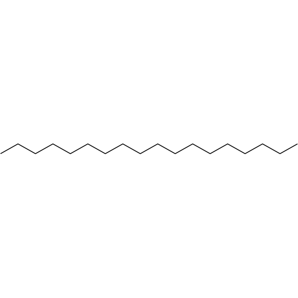
OctadecaneTemperature-Responsive Material -

HY-N5127
NonacosaneBacterial Inhibitor -
HY-W094846
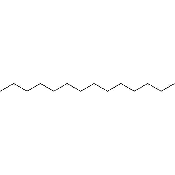
TetradecaneLow-temperature Phase Change Material -
HY-W089845
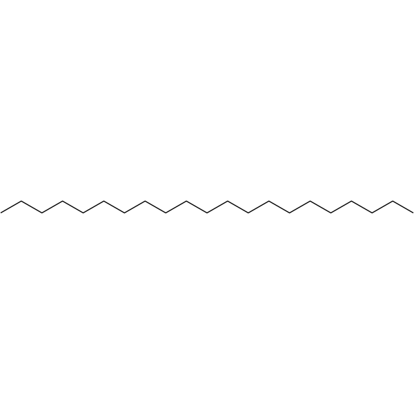
HeneicosaneSex Pheromone -

HY-W269368
HeptacosaneEster Product -
HY-N9971
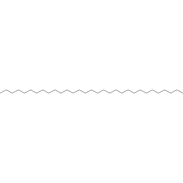
HentriacontaneAnti-inflammatory agent
All Endogenous Metabolite Isoforms
More

Biological Activity
Description
|
Human Endogenous Metabolite |
In Vitro
Octacosane (12.5-100 μg/ml; 18 h) displays strong cytotoxic activity on B16F10-Nex2 cells, with an IC50 value of 41.08 μg/ml[1].
MedChemExpress (MCE) has not independently confirmed the accuracy of these methods. They are for reference only. Further protocols information, click here.
-
Cell Line:B16F10-Nex2 cells
-
Concentration:12.5, 25, 50, 100 μg/ml
-
Incubation Time:18 h
-
Result:Displayed strong cytotoxic activity on B16F10-Nex2 cells, with an IC50 value of 41.08 μg/ml
In Vivo
MedChemExpress (MCE) has not independently confirmed the accuracy of these methods. They are for reference only.
-
Animal Model:C57Bl/6 mice with B16F10-Nex2 Cellsl[1]
-
Dosage:500 μg
-
Administration:Injected at peripheral sites in relation to the original cell grafting; daily; 35 days
-
Result:Resulted in a significant delay of tumor progression with a significant antitumor effect.
The survival rate of treated groups was significantly increased.


Chemical Information
-
CAS No. 630-02-4
-
Appearance Solid
-
Molecular Weight 394.76
-
Formula C28H58
-
Color White to off-white
-
SMILES
CCCCCCCCCCCCCCCCCCCCCCCCCCCC
-
Synonyms
n-Octacosane; NSC 5549
-
Structure Classification
-
Initial Source
-
Shipping
Room temperature in continental US; may vary elsewhere.
-
Storage
4°C, sealed storage, away from moisture
* In solvent : -80°C, 6 months; -20°C, 1 month (sealed storage, away from moisture)


Solvent & Solubility
In Vitro:
Ethanol : < 1 mg/mL (insoluble)

Protocols
-
How to Select the Route of Administration for Mammals
Route-of-administration selection in mammals is a pharmacokinetic, pharmacodynamic, formulation, animal-welfare, and translational decision, not a default technical choice. The selected route should match the study goal: intravenous dosing is most useful when complete systemic exposure and rapid onset are required, oral dosing is most translational for orally intended medicines but is affected by absorption and first-pass metabolism, subcutaneous or intramuscular dosing can provide slower systemic exposure, and intraperitoneal dosing can be useful in rodent proof-of-concept studies but may have limited clinical translation. Published route-comparison studies show that the same compound can produce different exposure, onset, bioavailability, tissue distribution, and tolerability depending on route; therefore, route choice should be supported by pilot pharmacokinetic or pharmacodynamic evidence when the literature is insufficient. Unresolved questions include how to standardize route sel
-
Research Protocol for Infectious Diseases
Infectious-disease experiments test how pathogens interact with host barriers, innate immune receptors, inflammatory signaling, pathogen replication, and tissue injury; pattern-recognition receptors such as TLRs, RIG-I-like receptors, NOD-like receptors, and inflammasomes detect microbial molecules and activate NF-κB, interferon, and cytokine responses. The central hypothesis is that infection severity reflects the balance between pathogen burden and host response: protective inflammation restricts pathogen growth, whereas excessive or mislocalized inflammation contributes to tissue damage and disease phenotype. Unresolved questions include which host pathways are protective versus pathogenic, why some infection models fail to translate to human disease, and which combined readouts best predict clinically relevant infection outcomes.
-
Cell Cytotoxicity Assay
Cytotoxicity assays are usually based on the assessment of cell membrane damage, which can also be indirectly detected by measuring cell viability. Detection methods include MTT assay, CKK-8 assay, LDH assay and ATP assay, etc.
-
Mammalian live/dead viability and cytotoxicity staining
Live/dead viability and cytotoxicity staining assays are based on the simultaneous detection of intracellular esterase activity in metabolically active (viable) cells and membrane integrity loss in non-viable cells. In commonly used dual-staining approaches, membrane-permeant fluorogenic substrates are converted by intracellular esterases into fluorescent products in live cells, while impermeant DNA-binding dyes selectively enter cells with compromised plasma membranes and label nucleic acids in dead or dying cells, enabling discrimination between viable and non-viable populations by fluorescence microscopy or flow cytometry.
-
Bacterial live/dead nucleic-acid viability staining
The LIVE/DEAD bacterial viability staining method is based on differential permeability of nucleic-acid-binding fluorescent dyes, most commonly SYTO 9 and propidium iodide (PI), which enables discrimination of bacterial populations with intact versus compromised cytoplasmic membranes. SYTO 9 penetrates both intact and damaged bacterial membranes and binds nucleic acids to produce green fluorescence, whereas propidium iodide penetrates only cells with compromised membranes and fluoresces red while also reducing SYTO 9 signal through competitive binding and fluorescence interactions. The resulting fluorescence pattern is interpreted as a proxy for membrane integrity, which is widely used as an indicator of bacterial viability in microscopy, flow cytometry, and spectroscopic platforms. However, mechanistic studies show that SYTO 9 and PI interactions involve displacement and fluorescence resonance energy transfer effects, which can influence signal interpretation depending on dye ratios a
-
Cell Viability Determination by MTT Colorimetric Assay
The following protocol uses the MTT colorimetric assay as a classic literature-established method for assessing cell viability/metabolic activity in cultured mammalian cells. MTT[3-(4,5-dimethylthiazol-2-yl)-2,5-diphenyltetrazolium bromide] is reduced by metabolically active cells to a colored formazan product; the amount of formazan is quantified spectrophotometrically and provides an indirect measure of metabolically active viable cells. Importantly, MTT reduction reflects cellular oxidoreductase/metabolic activity rather than an absolute direct count of living cells, so changes in cellular metabolism can alter the signal independently of cell number.


Purity & Documentation
-
Data Sheet (265 KB)
-
SDS (562 KB)
- English - EN (562 KB)
- Français - FR (562 KB)
- Deutsch - DE (562 KB)
- Norwegian - NO (562 KB)
- Español - ES (562 KB)
- Swedish - SV (562 KB)
- Italian - IT (562 KB)
- Korean - KR (562 KB)
- Portuguese - PT (562 KB)
-
Handling Instructions (2659 KB)
References
[1]. Carlos R Figueiredo, et al. Pyrostegia venusta heptane extract containing saturated aliphatic hydrocarbons induces apoptosis on B16F10-Nex2 melanoma cells and displays antitumor activity in vivo. Pharmacogn Mag. 2014 Apr;10(Suppl 2):S363-76. [Content Brief]
[2]. S Rajkumar, et al. Mosquitocidal activities of octacosane from Moschosma polystachyum Linn (lamiaceae). J Ethnopharmacol. 2004 Jan;90(1):87-9. [Content Brief]
[3]. Sameh S M Soliman, et al. Effective targeting of breast cancer cells (MCF7) via novel biogenic synthesis of gold nanoparticles using cancer-derived metabolites. PLoS One. 2020 Oct 6;15(10):e0240156. [Content Brief]

Calculators
Concentration (start) × Volume (start) = Concentration (final) × Volume (final)


Octacosane Related Classifications
HY-W272217 Related Classifications
 Powered by Bioz