



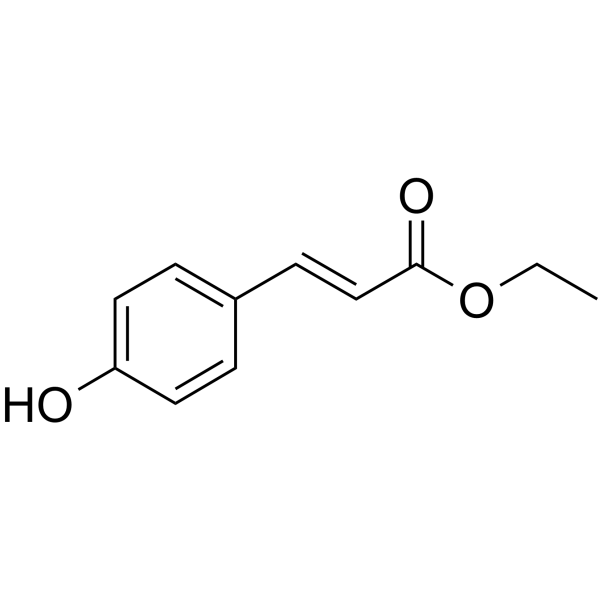
p-Coumaric Acid Ethyl Ester

Based on 1 Customer Validation
p-Coumaric Acid Ethyl Ester (Ethyl (E)-p-hydroxycinnamate; Ethyl trans-4-hydroxycinnamate) is a tyrosinase (Tyrosinase) inhibitor (IC50 = 4.89 μg/mL; Ki = 1.83 μg/mL). p-Coumaric Acid Ethyl Ester exhibits antitumor, antifungal, anti-inflammatory, and antioxidant activities. p-Coumaric Acid Ethyl Ester inhibits melanoma proliferation by inducing cell cycle arrest, and inhibits melanin synthesis by non-competitively altering tyrosinase conformation to hinder substrate binding. p-Coumaric Acid Ethyl Ester can be used in research on melanoma, whitening cosmetics, and fruit preservation.
Nos produits utilisent uniquement pour la recherche. Nous ne vendons pas aux patients.
- Pureté : 99.19%
- CAS No.: 7362-39-2
- Formule: C11H12O3
- Masse moléculaire:192.21
-
Stockage:Powder -20°C, 3 years , 4°C, 2 years ; In solvent -80°C, 6 months , -20°C, 1 month

To place orders, for customer services and technical support, please contact: MedChemExpress USA
Tel: 609-228-6898 E-mail: [email protected] [email protected]


-
 Activité biologique
-
Chemical Information
-
Solvant et solubilité
- Protocole
- Pureté et documentation
- Références
-
 Help & FAQs




Activité biologique
Description
Cellular Effect
|
Cell Line

|
Type | Value | Description | References |
|---|---|---|---|---|
| Platelet | IC50 |
130.4 μM
Compound: 4-OH-ethyl cinnamate
|
Anti-platelet activity in rabbit platelet-rich plasma assessed as inhibition of ADP-induced platelet aggregation incubated for 5 mins at 37 degC by aggregometry
Anti-platelet activity in rabbit platelet-rich plasma assessed as inhibition of ADP-induced platelet aggregation incubated for 5 mins at 37 degC by aggregometry
|
10.1039/C4MD00022F |
In Vitro
p-Coumaric Acid Ethyl Ester (Ethyl (E)-p-hydroxycinnamate; Ethyl trans-4-hydroxycinnamate) (0.06-1 mM; 4-24 h) exerts concentration- and time-dependent cytotoxic effects on mouse B16-F10 and human SK-MEL-25 melanoma cells[7].
p-Coumaric Acid Ethyl Ester (0.1 mM; 24-72 h) inhibits B16-F10 cell proliferation by inducing cell cycle arrest at the G0/G1 phase and preventing colony formation[7].
p-Coumaric Acid Ethyl Ester (0.1 mM; 24-72 h) inhibits SK-MEL-25 cell proliferation by inducing cell cycle arrest at S phase and preventing colony formation[7].
p-Coumaric Acid Ethyl Ester is a potent non-competitive tyrosinase inhibitor with an IC50 of 4.89 μg/mL, a Ki of 1.83 μg/mL, and a Km of 0.52 mM[1].
MedChemExpress (MCE) has not independently confirmed the accuracy of these methods. They are for reference only.
-
Cell Line:B16-F10
-
Concentration:0.06, 0.1, 0.2, 0.5, 1 mM (cytotoxicity); 0.5 mM (flow cytometry)
-
Incubation Time:4, 24 h (cytotoxicity); 2 h (flow cytometry)
-
Result:Induced significant cytotoxicity at concentrations >0.1 mM.
Induced significant LDH release at 0.2-1 mM after 4 h.
Induced significant LDH release at 0.5-1.0 mM after 24 h.
Decreased the frequency of viable cells starting from 0.06 mM.
Induced a significant increase in the frequency of dead cells after 2 h.
-
Cell Line:B16-F10
-
Concentration:0.1 mM
-
Incubation Time:24, 72 h
-
Result:Diminished the CFSE decay over time.
Induced cell cycle arrest at the G0/G1 phase.
Failed to form significant cell colonies.
-
Cell Line:SK-MEL-25
-
Concentration:0.06, 0.1, 0.2, 0.5, 1 mM (cytotoxicity); 0.5 mM (flow cytometry)
-
Incubation Time:4, 24 h (cytotoxicity); 2 h (flow cytometry)
-
Result:Induced LDH release at 0.5 and 1.0 mM at both 4 h and 24 h.
Significantly impacted cell viability at 4 h.
Induced a significant increase in the frequency of dead cells after 2 h.
-
Cell Line:SK-MEL-25
-
Concentration:0.1 mM
-
Incubation Time:24, 48, 72 h
-
Result:Efficiently inhibited SK-MEL-25 proliferation over 72 h.
Induced cell cycle arrest at the S phase.
Prevented colony formation.


Chemical Information
-
CAS No. 7362-39-2
-
Appearance Solid
-
Masse moléculaire 192.21
-
Formule C11H12O3
-
Color Light yellow to light brown
-
SMILES
CCOC(/C=C/C1=CC=C(O)C=C1)=O
-
Synonyms
Ethyl (E)-p-hydroxycinnamate; Ethyl trans-4-hydroxycinnamate
-
Structure Classification
-
Initial Source
-
Livraison
Room temperature in continental US; may vary elsewhere.
-
Stockage
Powder -20°C 3 years 4°C 2 years In solvent -80°C 6 months -20°C 1 month


Solvant et solubilité
In Vitro:
DMSO : 100 mg/mL (520.26 mM; Need ultrasonic; Hygroscopic DMSO has a significant impact on the solubility of product, please use newly opened DMSO)
Please refer to the solubility information to select the appropriate solvent. Once prepared, please aliquot and store the solution to prevent product inactivation from repeated freeze-thaw cycles.
Storage method and period of stock solution: -80°C, 6 months; -20°C, 1 month. When stored at -80°C, please use it within 6 months. When stored at -20°C, please use it within 1 month.
Please refer to the solubility information to select the appropriate solvent. Once prepared, please aliquot and store the solution to prevent product inactivation from repeated freeze-thaw cycles.
Storage method and period of stock solution: -80°C, 6 months; -20°C, 1 month. When stored at -80°C, please use it within 6 months. When stored at -20°C, please use it within 1 month.
Concentration (start) × Volume (start) = Concentration (final) × Volume (final)


Pureté et documentation
-
Fiche technique (275 KB)
-
SDS (393 KB)
- English - EN (393 KB)
- Français - FR (393 KB)
- Deutsch - DE (393 KB)
- Norwegian - NO (393 KB)
- Español - ES (393 KB)
- Swedish - SV (393 KB)
- Italian - IT (393 KB)
- Korean - KR (393 KB)
- Portuguese - PT (393 KB)
-
Instruction de manipulation (2659 KB)
Références
[1]. Li L, et al. Tyrosinase inhibition by -coumaric acid ethyl ester identified from camellia pollen. Food science & nutrition. 2021 Jan;9(1):389-400. [Content Brief]
[6]. Fernandes DC, et al. Myeloperoxidase inhibitory and radical scavenging activities of flavones from Pterogyne nitens. Chemical & pharmaceutical bulletin. 2008 May;56(5):723-6. [Content Brief]


Complete Stock Solution Preparation Table
Please refer to the solubility information to select the appropriate solvent. Once prepared, please aliquot and store the solution to prevent product inactivation from repeated freeze-thaw cycles.
Storage method and period of stock solution: -80°C, 6 months; -20°C, 1 month. When stored at -80°C, please use it within 6 months. When stored at -20°C, please use it within 1 month.
| Optional Solvent | Concentration Solvent Mass | 1 mg | 5 mg | 10 mg | 25 mg |
|---|---|---|---|---|---|
| DMSO | 1 mM | 5.2026 mL | 26.0132 mL | 52.0264 mL | 130.0661 mL |
| 5 mM | 1.0405 mL | 5.2026 mL | 10.4053 mL | 26.0132 mL | |
| 10 mM | 0.5203 mL | 2.6013 mL | 5.2026 mL | 13.0066 mL | |
| 15 mM | 0.3468 mL | 1.7342 mL | 3.4684 mL | 8.6711 mL | |
| 20 mM | 0.2601 mL | 1.3007 mL | 2.6013 mL | 6.5033 mL | |
| 25 mM | 0.2081 mL | 1.0405 mL | 2.0811 mL | 5.2026 mL | |
| 30 mM | 0.1734 mL | 0.8671 mL | 1.7342 mL | 4.3355 mL | |
| 40 mM | 0.1301 mL | 0.6503 mL | 1.3007 mL | 3.2517 mL | |
| 50 mM | 0.1041 mL | 0.5203 mL | 1.0405 mL | 2.6013 mL | |
| 60 mM | 0.0867 mL | 0.4336 mL | 0.8671 mL | 2.1678 mL | |
| 80 mM | 0.0650 mL | 0.3252 mL | 0.6503 mL | 1.6258 mL | |
| 100 mM | 0.0520 mL | 0.2601 mL | 0.5203 mL | 1.3007 mL |


p-Coumaric Acid Ethyl Ester Related Classifications
HY-N3103 Related Classifications
 Powered by Bioz