



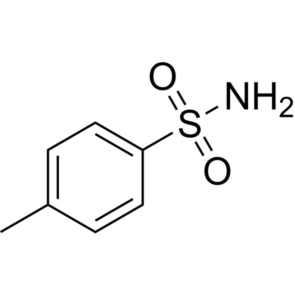
p-Toluenesulfonamide

Based on 1 Customer Validation
p-Toluenesulfonamide is a small-molecule anticancer agent and plasticizer. p-Toluenesulfonamide exerts antitumor activity by inducing lysosomal membrane permeabilization, cathepsin B release and lysosome-mediated cell death. p-Toluenesulfonamide modulates cholesterol distribution in lipid rafts of tumor cell membranes and the Akt/mTOR/p70S6K pathway. p-Toluenesulfonamide shows activity against various cancers including hepatocellular carcinoma, non-small cell lung cancer and tongue squamous cell carcinoma; intrapleural injection effectively reduces malignant pleural effusion without causing pleural adhesion. p-Toluenesulfonamide is also the main degradation product of the disinfectant Chloramine-T (HY-B0959) in water. p-Toluenesulfonamide facilitates the localization of fluorescent probes to the endoplasmic reticulum. p-Toluenesulfonamide can be used in cancer-related research.
For research use only. We do not sell to patients.
- Purity : 99.98%
- CAS No.: 70-55-3
- Formula: C7H9NO2S
- Molecular Weight:171.22
-
Storage:
Store at room temperature 3 years.
In solvent -80°C, 2 years , -20°C, 1 year

To place orders, for customer services and technical support, please contact: MedChemExpress USA
Tel: 609-228-6898 E-mail: [email protected] [email protected]


-
 Biological Activity
-
Chemical Information
-
Solvent & Solubility
- Protocols
- Purity & Documentation
- References
-
 Help & FAQs





Biological Activity
Description
In Vitro
p-Toluenesulfonamide (3333 μg/plate; 2 days) shows no mutagenicity in *Salmonella typhimurium* strains TA98, TA100 or TA102 with or without exogenous metabolic activation[2].
p-Toluenesulfonamide (1.3-2.0 mg/mL) does not induce chromosome aberrations in cultured Chinese hamster lung cells with or without exogenous metabolic activation, but exhibits cytotoxicity at high concentrations[2].
p-Toluenesulfonamide (25-250 µg/L; 7 days) can be reliably quantified in wastewater from a sewage treatment plant in Athens, Greece, using the developed SPE-LC-MS/MS pseudo SRM analytical method, with an LOD of 11 ng/L and an LOQ of 35 ng/L[4].
MedChemExpress (MCE) has not independently confirmed the accuracy of these methods. They are for reference only. Further protocols information, click here.
In Vivo
p-Toluenesulfonamide (750-30,000 ppm; oral administration; ad libitum feeding; 2 weeks) causes significant dose-dependent reductions in body weight and body weight gain in F344/N rats, along with an increase in relative kidney weight without associated histopathological lesions[2].
p-Toluenesulfonamide (750-30,000 ppm; oral administration; ad libitum access; 15 days) causes a significant decrease in body weight in both male and female B6C3F1/N mice in the high-dose group; meanwhile, starting from the 1,500 ppm dose, the absolute and relative kidney weights of female mice increase in a dose-dependent manner, without associated histopathological lesions[2].
p-Toluenesulfonamide (625-10,000 ppm; oral administration; ad libitum access; 14 weeks) induces dose-dependent body weight changes in F344/NTac rats. Male rats exhibit increased relative kidney weights starting from the 2,500 ppm dose, without associated histopathological lesions[2].
p-Toluenesulfonamide (625-10,000 ppm; oral administration; ad libitum access; 14 weeks) via feed to B6C3F1/N mice for 14 weeks increases the relative lung weight in high-dose male B6C3F1/N mice, and elevates the absolute kidney weight, relative kidney weight and relative liver weight in female mice[2].
MedChemExpress (MCE) has not independently confirmed the accuracy of these methods. They are for reference only.
-
Animal Model:C57BL/6 (male, 6-12 weeks old, 20-25 g, intrapleural injection of LLC-Luc cells)[1]
-
Dosage:82.5 mg/kg; 123.75 mg/kg; 165 mg/kg
-
Administration:intrapleural; 3 doses (days 4, 7, 10 post-cell injection)
-
Result:Significantly reduced malignant pleural effusion volume (P < 0.01 compared with PBS control) at 123.75 mg/kg and 165 mg/kg doses.
Significantly reduced pleural tumor weight (P < 0.05 compared with PBS control) at 123.75 mg/kg and 165 mg/kg doses.
Showed no pleural adhesions to the chest wall across all treatment groups.
-
Animal Model:F344/N (male and female, 5 weeks old)[2]
-
Dosage:750 ppm (equivalent to 95 mg/kg/day male, 80 mg/kg/day female); 1,500 ppm (equivalent to 185 mg/kg/day male, 170 mg/kg/day female); 3,000 ppm (equivalent to 370 mg/kg/day male, 335 mg/kg/day female); 10,000 ppm (equivalent to 1,170 mg/kg/day male, 1,050 mg/kg/day female); 30,000 ppm (equivalent to 3,135 mg/kg/day male, 2,645 mg/kg/day female)
-
Administration:p.o.; ad libitum; two weeks
-
Result:Allowed all animals to survive to study end.
Reduced final mean body weights to 91% of control in 10,000 ppm males, 71% of control in 30,000 ppm males, and 83% of control in 30,000 ppm females.
Decreased mean body weight gains significantly in 10,000 and 30,000 ppm males, and 10,000 and 30,000 ppm females.
Reduced feed consumption in 10,000 and 30,000 ppm males and 30,000 ppm females.
Decreased absolute kidney weights by ~19% in 30,000 ppm males.
Increased relative kidney weights in 3,000 ppm or greater males and 10,000 and 30,000 ppm females.
Showed no clinical observations or histopathologic findings attributed to exposure.
-
Animal Model:B6C3F1/N (male and female, 5 to 6 weeks old)[2]
-
Dosage:750 ppm (equivalent to 150 mg/kg/day male, 125 mg/kg/day female); 1,500 ppm (equivalent to 300 mg/kg/day male, 280 mg/kg/day female); 3,000 ppm (equivalent to 700 mg/kg/day male, 635 mg/kg/day female); 10,000 ppm (equivalent to 2,035 mg/kg/day male, 2,410 mg/kg/day female); 30,000 ppm (equivalent to 7,690 mg/kg/day male, 6,000 mg/kg/day female)
-
Administration:p.o.; ad libitum; 15 days
-
Result:Allowed all animals to survive to study end.
Reduced final mean body weights to 86% of control in 30,000 ppm males and 85% of control in 30,000 ppm females.
Decreased mean body weight gains in 30,000 ppm male mice and all exposed female mice, with 30,000 ppm groups losing weight during the study.
Maintained feed consumption generally similar to controls across all exposed groups.
Increased absolute and relative kidney weights significantly in 10,000 and 30,000 ppm female mice.
Increased relative kidney weights significantly in 1,500 and 3,000 ppm female mice.
Showed no clinical observations or histopathologic findings attributed to exposure.
-
Animal Model:F344/NTac (male and female, 5 to 6 weeks old)[2]
-
Dosage:625 ppm (equivalent to 50 mg/kg/day male, 30 mg/kg/day female); 1,250 ppm (equivalent to 100 mg/kg/day male, 110 mg/kg/day female); 2,500 ppm (equivalent to 200 mg/kg/day male, 210 mg/kg/day female); 5,000 ppm (equivalent to 380 mg/kg/day male, 400 mg/kg/day female); 10,000 ppm (equivalent to 725 mg/kg/day male, 780 mg/kg/day female)
-
Administration:p.o.; ad libitum; 14 weeks
-
Result:Allowed all animals to survive to study end.
Reduced final mean body weights to 93% of control in 10,000 ppm males and 92% of control in 10,000 ppm females.
Decreased mean body weight gains significantly in 2,500 ppm or greater males and 5,000 and 10,000 ppm females.
Increased final mean body weight and weight gain significantly in 1,250 ppm females.
Reduced feed consumption in 5,000 ppm males and 10,000 ppm males and females early in the study, with recovery to near control values later.
Reduced absolute and relative thymus weights significantly in 10,000 ppm males.
Increased relative kidney weights significantly in 2,500 ppm or greater males.
Showed no clinical observations, histopathologic findings, or clinically significant changes in hematology, clinical chemistry, or reproductive parameters attributed to exposure.
-
Animal Model:B6C3F1/N (male and female, 7 to 8 weeks old)[2]
-
Dosage:625 ppm (equivalent to 120 mg/kg/day male, 90 mg/kg/day female); 1,250 ppm (equivalent to 230 mg/kg/day male, 210 mg/kg/day female); 2,500 ppm (equivalent to 420 mg/kg/day male, 380 mg/kg/day female); 5,000 ppm (equivalent to 770 mg/kg/day male, 780 mg/kg/day female); 10,000 ppm (equivalent to 1,760 mg/kg/day male, 1,890 mg/kg/day female)
-
Administration:p.o.; ad libitum; 14 weeks
-
Result:Allowed all male mice to survive to study end; caused one 10,000 ppm female mouse to die during week 6.
Maintained final mean body weights similar to controls across all exposed groups.
Increased feed consumption in 625 and 1,250 ppm male mice early in the study, with return to near control values later.
Increased relative lung weights significantly in 10,000 ppm males.
Increased absolute and relative kidney weights and relative liver weights significantly in 10,000 ppm females.
Showed no clinical observations, histopathologic findings, or clinically significant changes in hematology or reproductive parameters attributed to exposure in surviving animals.


Chemical Information
-
CAS No. 70-55-3
-
Appearance Solid
-
Molecular Weight 171.22
-
Formula C7H9NO2S
-
Color White to off-white
-
SMILES
O=S(C1=CC=C(C)C=C1)(N)=O
-
Shipping
Room temperature in continental US; may vary elsewhere.
-
Storage
Store at room temperature 3 years
In solvent -80°C 2 years -20°C 1 year


Solvent & Solubility
In Vitro:
DMSO : 175 mg/mL (1022.08 mM; Need ultrasonic; Hygroscopic DMSO has a significant impact on the solubility of product, please use newly opened DMSO)
Please refer to the solubility information to select the appropriate solvent. Once prepared, please aliquot and store the solution to prevent product inactivation from repeated freeze-thaw cycles.
Storage method and period of stock solution: -80°C, 2 years; -20°C, 1 year. When stored at -80°C, please use it within 2 years. When stored at -20°C, please use it within 1 year.
Please refer to the solubility information to select the appropriate solvent. Once prepared, please aliquot and store the solution to prevent product inactivation from repeated freeze-thaw cycles.
Storage method and period of stock solution: -80°C, 2 years; -20°C, 1 year. When stored at -80°C, please use it within 2 years. When stored at -20°C, please use it within 1 year.
Concentration (start) × Volume (start) = Concentration (final) × Volume (final)

Protocols
-
How to Select the Route of Administration for Mammals
Route-of-administration selection in mammals is a pharmacokinetic, pharmacodynamic, formulation, animal-welfare, and translational decision, not a default technical choice. The selected route should match the study goal: intravenous dosing is most useful when complete systemic exposure and rapid onset are required, oral dosing is most translational for orally intended medicines but is affected by absorption and first-pass metabolism, subcutaneous or intramuscular dosing can provide slower systemic exposure, and intraperitoneal dosing can be useful in rodent proof-of-concept studies but may have limited clinical translation. Published route-comparison studies show that the same compound can produce different exposure, onset, bioavailability, tissue distribution, and tolerability depending on route; therefore, route choice should be supported by pilot pharmacokinetic or pharmacodynamic evidence when the literature is insufficient. Unresolved questions include how to standardize route sel
-
Cell Cytotoxicity Assay
Cytotoxicity assays are usually based on the assessment of cell membrane damage, which can also be indirectly detected by measuring cell viability. Detection methods include MTT assay, CKK-8 assay, LDH assay and ATP assay, etc.
-
Mammalian live/dead viability and cytotoxicity staining
Live/dead viability and cytotoxicity staining assays are based on the simultaneous detection of intracellular esterase activity in metabolically active (viable) cells and membrane integrity loss in non-viable cells. In commonly used dual-staining approaches, membrane-permeant fluorogenic substrates are converted by intracellular esterases into fluorescent products in live cells, while impermeant DNA-binding dyes selectively enter cells with compromised plasma membranes and label nucleic acids in dead or dying cells, enabling discrimination between viable and non-viable populations by fluorescence microscopy or flow cytometry.
-
Apoptosis Solutions
Apoptosis is a regulated, generally non-lytic cell-death pathway that removes unwanted, damaged, infected, or abnormal cells through coordinated morphological changes, caspase activation, DNA fragmentation, and membrane remodeling. The intrinsic apoptosis pathway is controlled mainly by mitochondrial outer membrane permeabilization, BCL-2 family proteins, cytochrome c release, apoptosome formation, caspase-9 activation, and downstream executioner caspase-3/7 activation. The extrinsic apoptosis pathway is initiated by death receptors such as Fas, TNFR, and TRAIL receptors, which recruit adaptor proteins and activate caspase-8 before engaging executioner caspases or mitochondrial amplification through BID cleavage. Apoptosis is linked to many phenotypes, including cancer cell killing, tissue homeostasis, immune regulation, neurodegeneration, infection response, and treatment-induced cytotoxicity; unresolved questions include how apoptosis interacts with necroptosis, pyroptosis, ferroptos
-
Lysosome and acidic-vesicle live-cell staining
Lysosome and acidic-vesicle live-cell staining detects acidic intracellular compartments by using membrane-permeant acidotropic probes that accumulate in low-pH vesicles, including lysosomes, late endosomes, autolysosomes, and acidic phagosomes. LysoTracker staining is commonly used as an intensity-based readout of acidic lysosomal compartment abundance or enlargement, while acridine orange produces green fluorescence in less concentrated compartments and red fluorescence after concentration-dependent accumulation in acidic vesicular organelles. Loss or reduction of acridine-orange red signal can be used as a readout of lysosomal membrane permeabilization or reduced acidic-vesicle integrity. This protocol is designed for live cultured cells and can be adapted for fluorescence microscopy, high-content imaging, plate-reader readout, or flow cytometry when the selected literature supports the readout. Because these dyes report acidotropic accumulation rather than lysosome identity alone,
-
Liver Cancer Modeling
Liver cancer can be classified into primary liver cancer and secondary liver cancer. Secondary liver cancer is the metastatic liver cancer. Primary liver cancer includes hepatocellular carcinoma (HCC), intrahepatic cholangiocarcinoma (ICC) and fibrolamellar HCC, of which HCC is the most common form, accounting for approximately 90% of primary liver cancers[1]. HCC mouse models include chemical agent-induced models, transplanted tumor models, and genetic engineered models.
-
Subcutaneous Cell-Line-Derived Xenograft
Subcutaneous cell-line-derived xenograft (CDX) models are established by implanting cultured human cancer cell lines into immunodeficient mice, where the injected cells form localized tumors that can be monitored in vivo as a measure of tumorigenic potential, growth kinetics, and treatment response. These models are widely used in oncology research because they allow reproducible tumor formation and enable comparative assessment of tumor growth between different cell lines or genetic manipulations in a controlled in vivo microenvironment. Subcutaneous implantation of cancer cells in immunodeficient mice is a standard approach for evaluating tumor growth behavior and therapeutic response across multiple cancer types, including prostate, esophageal, pancreatic, and colon cancer models.


Purity & Documentation
-
Data Sheet (295 KB)
-
SDS (393 KB)
- English - EN (393 KB)
- Français - FR (393 KB)
- Deutsch - DE (393 KB)
- Norwegian - NO (393 KB)
- Español - ES (393 KB)
- Swedish - SV (393 KB)
- Italian - IT (393 KB)
- Korean - KR (393 KB)
- Portuguese - PT (393 KB)
-
Handling Instructions (2659 KB)
References
[3]. Tian YM, et al. A p-toluenesulfonamide-modified benzo[h]chromene hydrazone: Fluorescent turn-on detection of hypochlorite and its application to imaging the endoplasmic reticula of living cells and zebrafishes. Spectrochim Acta A Mol Biomol Spectrosc. 2023 Aug 5;296:122669. [Content Brief]
[4]. Ajibola A, et al. Benzosulfonamides in wastewater: method development, occurrence and removal efficiencies. Chemosphere. 2015 Jan;119 Suppl:S21-7. [Content Brief]


Complete Stock Solution Preparation Table
Please refer to the solubility information to select the appropriate solvent. Once prepared, please aliquot and store the solution to prevent product inactivation from repeated freeze-thaw cycles.
Storage method and period of stock solution: -80°C, 2 years; -20°C, 1 year. When stored at -80°C, please use it within 2 years. When stored at -20°C, please use it within 1 year.
| Optional Solvent | Concentration Solvent Mass | 1 mg | 5 mg | 10 mg | 25 mg |
|---|---|---|---|---|---|
| DMSO | 1 mM | 5.8404 mL | 29.2022 mL | 58.4044 mL | 146.0110 mL |
| 5 mM | 1.1681 mL | 5.8404 mL | 11.6809 mL | 29.2022 mL | |
| 10 mM | 0.5840 mL | 2.9202 mL | 5.8404 mL | 14.6011 mL | |
| 15 mM | 0.3894 mL | 1.9468 mL | 3.8936 mL | 9.7341 mL | |
| 20 mM | 0.2920 mL | 1.4601 mL | 2.9202 mL | 7.3005 mL | |
| 25 mM | 0.2336 mL | 1.1681 mL | 2.3362 mL | 5.8404 mL | |
| 30 mM | 0.1947 mL | 0.9734 mL | 1.9468 mL | 4.8670 mL | |
| 40 mM | 0.1460 mL | 0.7301 mL | 1.4601 mL | 3.6503 mL | |
| 50 mM | 0.1168 mL | 0.5840 mL | 1.1681 mL | 2.9202 mL | |
| 60 mM | 0.0973 mL | 0.4867 mL | 0.9734 mL | 2.4335 mL | |
| 80 mM | 0.0730 mL | 0.3650 mL | 0.7301 mL | 1.8251 mL | |
| 100 mM | 0.0584 mL | 0.2920 mL | 0.5840 mL | 1.4601 mL |


p-Toluenesulfonamide Related Classifications
HY-79602 Related Classifications
 Powered by Bioz