



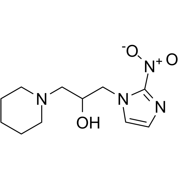
Pimonidazole
Based on 9 publication(s) in Google Scholar
Pimonidazole is a novel hypoxia marker for complementary study of tumor hypoxia and cell proliferation in tumor. Pimonidazole accumulates in hypoxic cells via covalent binding with macromolecules or by forming reductive metabolites after reduction of its nitro group, it can be used for qualitative and quantitative assessment of tumor hypoxia .
Nur für Forschungszwecke. Wir verkaufen nicht an Patienten.
- Reinheit : 99.32%
- CAS. Nr.: 70132-50-2
- Formel: C11H18N4O3
- Molecular Weight:254.29
-
Speicherung:Powder -20°C, 3 years ; In solvent -80°C, 6 months , -20°C, 1 month

To place orders, for customer services and technical support, please contact: MedChemExpress USA
Tel: 609-228-6898 E-mail: [email protected] [email protected]


-
 Biologische Aktivität
-
Chemical Information
-
Lösungsmittel & Löslichkeit
- Protokoll
- Reinheit & Dokumentation
- Verweise
-
 Help & FAQs



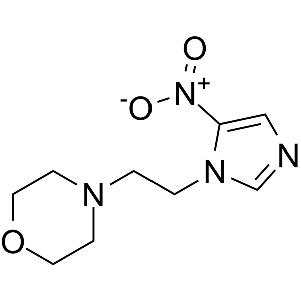
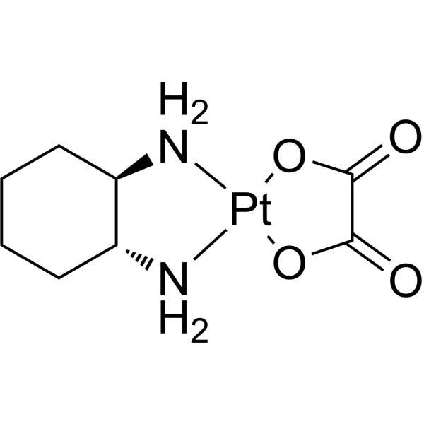
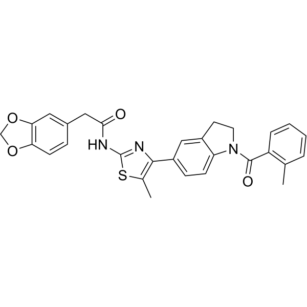


Publications Citing Use of MedChemExpress (MCE) Pimonidazole
More- Theranostics. 2022 Apr 4;12(7):3196-3216. [Abstract]
- Asian J Pharm Sci. 2023 May;18(3):100798. [Abstract]
- Mater Today Bio. 2025 Sep 30:35:102370. [Abstract]
- J Colloid Interface Sci. 2026 Mar 15:706:139602. [Abstract]
- J Colloid Interface Sci. 2025 Aug 28;702(Pt 1):138870. [Abstract]
- J Colloid Interface Sci. 2024 May:661:908-922. [Abstract]
- Sci Rep. 2023 Apr 21;13(1):6528. [Abstract]
- Exp Neurol. 2025 Aug 27:115447. [Abstract]
- Exp Eye Res. 2025 Oct:259:110557. [Abstract]
 Customer Validation & Images

-
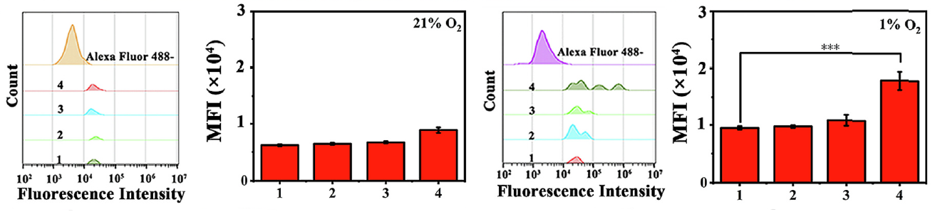
Flow Cytometry
-
Histological Imaging/Staining
-
IF

-
Histological Imaging/Staining
-
Histological Imaging/Staining


Biologische Aktivität
Beschreibung
Cellular Effect
|
Cell Line

|
Type | Value | Description | References |
|---|---|---|---|---|
| CHO-AA8 | IC50 |
3.03 mM
Compound: 2
|
Aerobic growth inhibition in Chinese hamster cell line AA8
Aerobic growth inhibition in Chinese hamster cell line AA8
|
[PMID: 7783125] |
In Vitro
Pimonidazole, the exogenous hypoxia marker, is a 2-nitroimidazole compound, which forms covalent bonds with cellular macromolecules at oxygen levels below 1.3%.
Detection: Hypoxic cells were recognized by immunohistochemical detection of pimonidazole using a mouse monoclonal antibody. Cell proliferation was detected with a commercially available monoclonal antibody for proliferating cell nuclear antigen (PCNA). Assessment of hypoxia and cell proliferation was made qualitatively with light microscopy and quantitatively using point counting and image analysis software methods.
MedChemExpress (MCE) has not independently confirmed the accuracy of these methods. They are for reference only.
- BrdU Incorporation Assay
- Somatic Cell Culture
- CFSE Dye Dilution Proliferation Assay
- Dye-dilution cell tracking and proliferation staining
- CCK-8/WST-8 Cell Proliferation Assay
- Protocol for Cell Cycle
- Cell Counting-Based Growth Curve Assay
- Colony Formation (Clonogenic) Assay
- Cell Viability Determination by MTT Colorimetric Assay
- EdU Incorporation Assay (Click Chemistry-Based DNA Synthesis Measurement)
- Ki-67 Immunostaining Proliferation Assay
- PCNA Immunodetection Proliferation Assay
- Protocol for Cell Counting and Cell Density Analysis
- MTT Cell Proliferation Assay


Chemical Information
-
CAS. Nr. 70132-50-2
-
Appearance Solid
-
Molecular Weight 254.29
-
Formel C11H18N4O3
-
Color Light yellow to yellow
-
SMILES
OC(CN1C=CN=C1[N+]([O-])=O)CN2CCCCC2
-
Versand
Room temperature in continental US; may vary elsewhere.
-
Speicherung
Powder -20°C 3 years In solvent -80°C 6 months -20°C 1 month


Publications (9)
-
Journal Impact Factor
-
Most Recent
-
Theranostics
Neuronal STAT3/HIF-1α/PTRF axis-mediated bioenergetic disturbance exacerbates cerebral ischemia-reperfusion injury via PLA2G4A. [Abstract]2022 Apr 4;12(7):3196-3216. PMID: 35547748
Pimonidazole purchased from MedChemExpress. Usage Cited in: Theranostics. 2022 Apr 4;12(7):3196-3216. [Abstract]
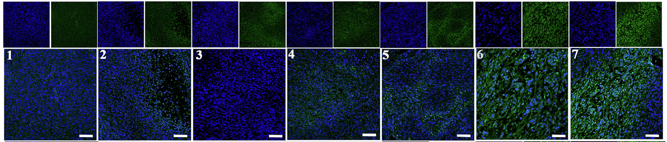
IF was conducted on brain sections from the mice at 24 h post-cerebral I/R injury using anti-PTRF and hypoxyprobe-1 (Pimonidazole).
-
Asian J Pharm Sci
Tumor microenvironment-responsive artesunate loaded Z-scheme heterostructures for synergistic photo-chemodynamic therapy of hypoxic tumor. [Abstract]2023 May;18(3):100798. PMID: 37252037 -
Mater Today Bio
"Top-down and Bottom-up" cholesterol-depleting biomimetic nanoparticle for enhancing sonodynamic therapy against hepatocellular carcinoma. [Abstract]2025 Sep 30:35:102370. PMID: 41104047
Pimonidazole purchased from MedChemExpress. Usage Cited in: Mater Today Bio. 2025 Sep 30:35:102370. [Abstract]
Pimonidazole staining of tumor sections after different treatments.
-
J Colloid Interface Sci
Self-amplifying hypoxia cascade in covalent organic framework/metal organic framework nanoreactors for synergistic cancer therapy. [Abstract]2026 Mar 15:706:139602. PMID: 41353990
Pimonidazole purchased from MedChemExpress. Usage Cited in: J Colloid Interface Sci. 2026 Mar 15:706:139602. [Abstract]
The hypoxia level in 4T1 cells was quantitatively assessed using Pimonidazole hydrochloride (PIMO) (100 μM; 60 min) as a probe.
Pimonidazole purchased from MedChemExpress. Usage Cited in: J Colloid Interface Sci. 2026 Mar 15:706:139602. [Abstract]
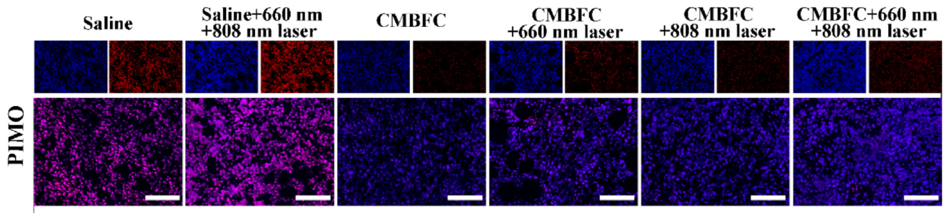
Representative fluorescence images of tumor slices from 4 T1 tumor-bearing mice after various treatments were stained for hypoxia with Pimonidazole hydrochloride (PIMO) (60 mg/kg; i.p.; 90 min).
-
J Colloid Interface Sci
A multifunctional oxidative stress amplifier for synergistic disruption of redox homeostasis and enhanced cancer therapy. [Abstract]2025 Aug 28;702(Pt 1):138870. PMID: 40902490
Pimonidazole purchased from MedChemExpress. Usage Cited in: J Colloid Interface Sci. 2025 Aug 28;702(Pt 1):138870. [Abstract]
Representative fluorescence images of tumor slices from mice bearing 4 T1 tumors stained with Pimonidazole hydrochloride (PIMO) (60 mg/kg; i.p.; 90 min).
-
J Colloid Interface Sci
Covalent organic frameworks-derived carbon nanospheres based nanoplatform for tumor specific synergistic therapy via oxidative stress amplification and calcium overload. [Abstract]2024 May:661:908-922. PMID: 38330663
Pimonidazole purchased from MedChemExpress. Usage Cited in: J Colloid Interface Sci. 2024 May:661:908-922. [Abstract]
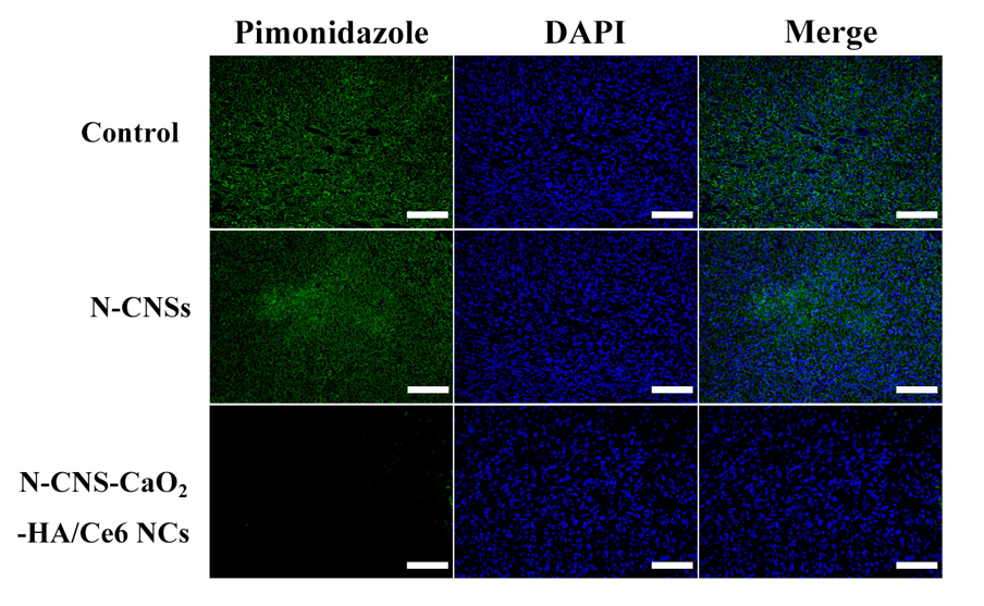
Fluorescence images of tumor slices from mice bearing 4T1 tumors stained with the hypoxia probe Pimonidazole hydrochloride (PIMO) (60 mg/kg; i.p.; 90 min) after different treatments were shown.
-
Sci Rep
Contribution of HIF-1α/BNIP3-mediated autophagy to lipid accumulation during irinotecan-induced liver injury. [Abstract]2023 Apr 21;13(1):6528. PMID: 37085612
Pimonidazole purchased from MedChemExpress. Usage Cited in: Sci Rep. 2023 Apr 21;13(1):6528. [Abstract]
Immunofluorescence was performed on liver sections using pimonidazole (green) to detect hypoxic regions, nuclei were stained with DAPI (blue).
-
Exp Neurol
By inhibiting pyroptosis to reduce neuroinflammation, PEG-bHb may prevent the development of secondary injury after traumatic brain injury. [Abstract]2025 Aug 27:115447. PMID: 40882799 -
Exp Eye Res
2025 Oct:259:110557. PMID: 40738388


Lösungsmittel & Löslichkeit
In Vitro:
DMSO : 50 mg/mL (196.63 mM; ultrasonic and warming and heat to 60°C; Hygroscopic DMSO has a significant impact on the solubility of product, please use newly opened DMSO)
H2O : 1 mg/mL (3.93 mM; Need ultrasonic)
Please refer to the solubility information to select the appropriate solvent. Once prepared, please aliquot and store the solution to prevent product inactivation from repeated freeze-thaw cycles.
Storage method and period of stock solution: -80°C, 6 months; -20°C, 1 month. When stored at -80°C, please use it within 6 months. When stored at -20°C, please use it within 1 month.
* Note: If you choose water as the stock solution, please dilute it to the working solution, then filter and sterilize it with a 0.22 μm filter before use.
Please refer to the solubility information to select the appropriate solvent. Once prepared, please aliquot and store the solution to prevent product inactivation from repeated freeze-thaw cycles.
Storage method and period of stock solution: -80°C, 6 months; -20°C, 1 month. When stored at -80°C, please use it within 6 months. When stored at -20°C, please use it within 1 month.
* Note: If you choose water as the stock solution, please dilute it to the working solution, then filter and sterilize it with a 0.22 μm filter before use.
Konzentration (Stammlösung) × Volumen (Stammlösung) = Konzentration (Ziellösung) × Volumen (Ziellösung)


Reinheit & Dokumentation
-
Data Sheet (274 KB)
-
SDS (392 KB)
- English - EN (392 KB)
- Français - FR (392 KB)
- Deutsch - DE (392 KB)
- Norwegian - NO (392 KB)
- Español - ES (392 KB)
- Swedish - SV (392 KB)
- Italian - IT (392 KB)
- Korean - KR (392 KB)
- Portuguese - PT (392 KB)
-
Handling Instructions (2659 KB)
Verweise
[1]. Varia MA, et al. Pimonidazole: a novel hypoxia marker for complementary study of tumor hypoxia and cell proliferation in cervical carcinoma. Gynecol Oncol. 1998 Nov;71(2):270-7. [Content Brief]
[2]. Masaki Y, et al. Imaging Mass Spectrometry Revealed the Accumulation Characteristics of the 2-Nitroimidazole-Based Agent "Pimonidazole" in Hypoxia. PLoS One. 2016 Aug 31;11(8):e0161639. [Content Brief]


Complete Stock Solution Preparation Table
Please refer to the solubility information to select the appropriate solvent. Once prepared, please aliquot and store the solution to prevent product inactivation from repeated freeze-thaw cycles.
Storage method and period of stock solution: -80°C, 6 months; -20°C, 1 month. When stored at -80°C, please use it within 6 months. When stored at -20°C, please use it within 1 month.
| Optional Solvent | Concentration Solvent Mass | 1 mg | 5 mg | 10 mg | 25 mg |
|---|---|---|---|---|---|
| H2O / DMSO | 1 mM | 3.9325 mL | 19.6626 mL | 39.3252 mL | 98.3130 mL |
| DMSO | 5 mM | 0.7865 mL | 3.9325 mL | 7.8650 mL | 19.6626 mL |
| 10 mM | 0.3933 mL | 1.9663 mL | 3.9325 mL | 9.8313 mL | |
| 15 mM | 0.2622 mL | 1.3108 mL | 2.6217 mL | 6.5542 mL | |
| 20 mM | 0.1966 mL | 0.9831 mL | 1.9663 mL | 4.9156 mL | |
| 25 mM | 0.1573 mL | 0.7865 mL | 1.5730 mL | 3.9325 mL | |
| 30 mM | 0.1311 mL | 0.6554 mL | 1.3108 mL | 3.2771 mL | |
| 40 mM | 0.0983 mL | 0.4916 mL | 0.9831 mL | 2.4578 mL | |
| 50 mM | 0.0787 mL | 0.3933 mL | 0.7865 mL | 1.9663 mL | |
| 60 mM | 0.0655 mL | 0.3277 mL | 0.6554 mL | 1.6385 mL | |
| 80 mM | 0.0492 mL | 0.2458 mL | 0.4916 mL | 1.2289 mL | |
| 100 mM | 0.0393 mL | 0.1966 mL | 0.3933 mL | 0.9831 mL |
* Note: If you choose water as the stock solution, please dilute it to the working solution, then filter and sterilize it with a 0.22 μm filter before use.


Pimonidazole Related Classifications
HY-105129A Related Classifications
 Powered by Bioz