



PK11007

Based on 1 Customer Validation
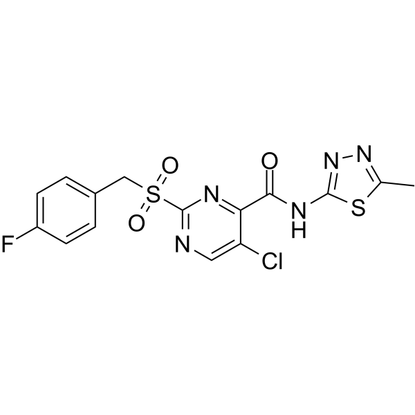
PK11007 is a mild thiol alkylator with anticancer activity. PK11007 stabilizes p53 via selective alkylation of two surface-exposed cysteines without compromising its DNA binding activity. PK11007 induces mutant p53 cancer cell death by increasing reactive oxygen species (ROS) levels.
For research use only. We do not sell to patients.
- Purity : 98.73%
- CAS No.: 874146-69-7
- Formula: C15H11ClFN5O3S2
- Molecular Weight:427.86
-
Storage:Powder -20°C, 3 years , 4°C, 2 years ; In solvent -80°C, 2 years , -20°C, 1 year

To place orders, for customer services and technical support, please contact: MedChemExpress USA
Tel: 609-228-6898 E-mail: [email protected] [email protected]


-
 Biological Activity
-
Chemical Information
-
Solvent & Solubility
- Protocols
- Purity & Documentation
- References
-
 Help & FAQs


-
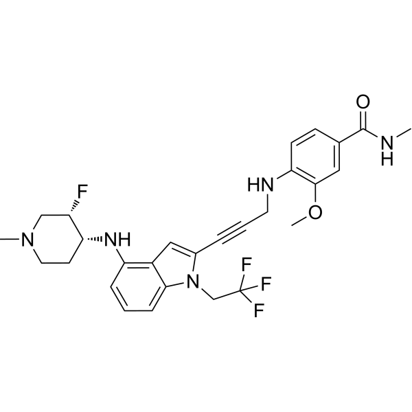
HY-156633
RezatapoptP53 Ligand -
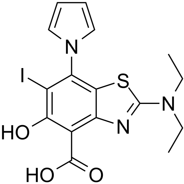
HY-120373
MB710p53-Y220C Stabilizer -
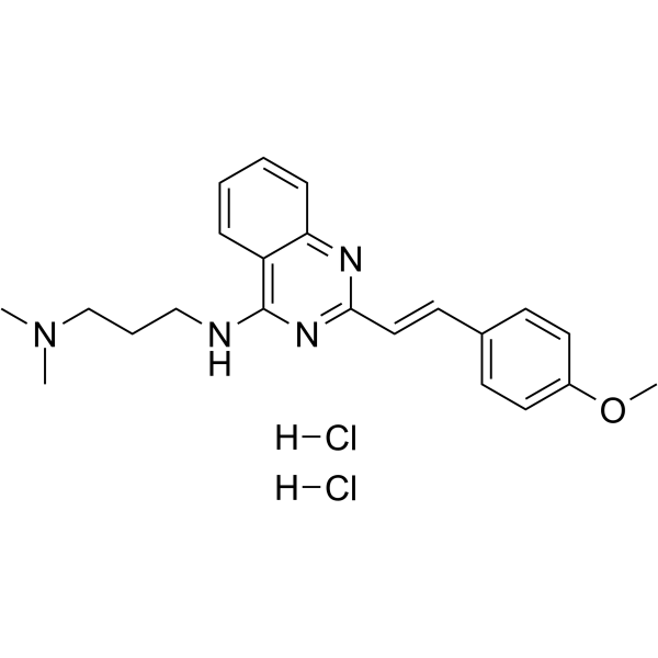
HY-18343A
CP-31398 dihydrochloridep53 Stabilizer -
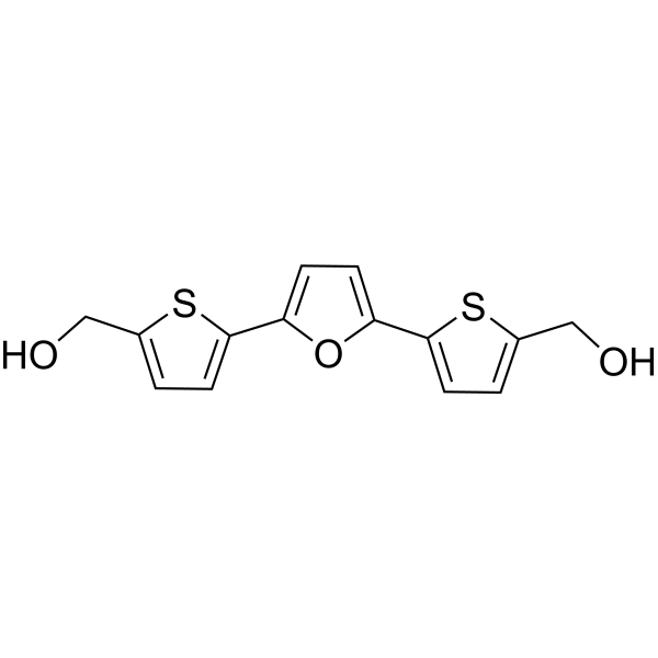
HY-13424
RITAp53-HDM-2 Interaction Inhibitor -
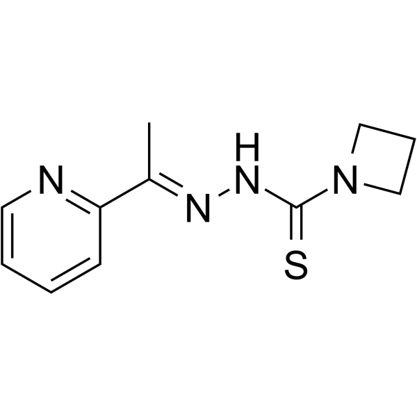
HY-18634
NSC319726MDM-2/p53 p53 Activator -
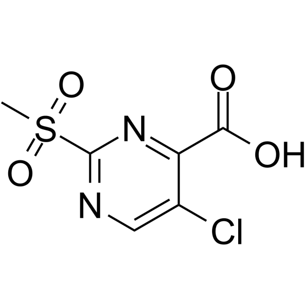
HY-U00447
PK11000p53 Activator -
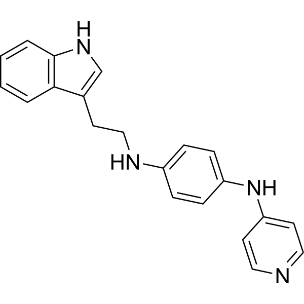
HY-12025
SerdemetanAnticancer Aagent -
HY-16664
SJ-172550MDM-2/p53 MDM2 Inhibitor -
HY-10029
Nutlin-3aMDM2 Antagonist -
HY-16702A
Pifithrin-β hydrobromideMDM-2/p53 p53 Inhibitor -
HY-15484
Pifithrin-α hydrobromidep53 Inhibitor -

HY-149461
KB-05Electrophilic Fragment Molecule


Biological Activity
Description
In Vitro
PK11007 (0-120 μM; 24 hours; four p53 wild-type cell lines and fours p53 mutant cell lines) treatment results in a large viability reduction in mutant p53 cell lines MKN1 (V143A), HUH-7 (Y220C), NUGC-3 (Y220C), and SW480 (R273H/P309S) at concentrations ranging from 15 to 30 μM. PK11007 induces mainly caspase-independent cell death[1].
PK11007 (0-60 μM; 3 hours or 6 hours; NUGC-4, NUGC-3, MKN1, HUH-6, and HUH-7 cancer cells) treatment up-regulates protein levels of the p53 target genes p21, MDM2, and PUMA in a mostly concentration-dependent manner in NUGC-3 (p53-Y220C), HUH-7 (p53-Y220C) and MKN1 (p53-V143A) cells, suggesting partial restoration of transcriptional activity to destabilized p53 mutants. PK11007 also increases p53 activity in HUH-6 and NUGC-4 cells, as indicated by the increase of MDM2, PUMA, and p21 protein levels[1].
PK11007 (15-20 μM; 4.5 hours or 6 hours; MKN1, HUH-7, NUGC-3, HUH-6 cells) treatment increases transcription of p53 target genes in three mutant p53 cell lines after 6-h treatment. PUMA and p21 mRNA levels are up-regulated by a factor of 2 upon treatment of NUGC-3, MKN, and HUH-7 cells, as well as NOXA for the latter two. MDM2 levels are halved in MKN1 and NUGC-3 cells[1].
PK11007 viability reduction is potentiated by glutathione depletion. To test whether PK11007 also increases ROS levels, NUGC-3, NUGC-4, HUH-6, HUH-7, and MKN1 cells with PK11007 are incubated for 2 h. There are elevated ROS levels in all cell lines after 2 h. In the mutant p53 cells MKN1, HUH-7, and NUGC-3, however, the ROS increase is higher at 60 μM PK11007 than in NUGC-4 and HUH-6 cells, suggesting that the higher PK11007 sensitivity of the mutant p53 cell lines is mediated by a stronger ROS induction. Basal and PK11007-induced ROS levels in MKN1 cells are at least twofold higher than in other cell lines[1].
PK11007 inhibits cell proliferation, induces apoptosis and alters genes involved in cell death are all consistent with the ability of PK11007 to reactivate mutant p53[2].
MedChemExpress (MCE) has not independently confirmed the accuracy of these methods. They are for reference only. Further protocols information, click here.
-
Cell Line:p53 wild-type cell lines (WI-38, HUH-6, NUGC-4, SJSA-1) and p53 mutant cell lines (HUH-7, NUGC-3, SW480, MKN1)
-
Concentration:0 μM, 20 μM, 40 μM, 60 µM, 80 µM, 100 µM and 120 µM
-
Incubation Time:24 hours
-
Result:There was a large viability reduction in mutant p53 cell lines MKN1 (V143A), HUH-7 (Y220C), NUGC-3 (Y220C), and SW480 (R273H/P309S) and in p53 WT cell line SJSA-1 at concentrations ranging from 15 to 30 µM. The p53 WT cancer cell lines HUH-6, NUGC-4 and WI-38 were less sensitive with reduced cell viability only at high concentrations of compound (60 and 120 µM).
-
Cell Line:NUGC-4, NUGC-3, MKN1, HUH-6, and HUH-7 cancer cells
-
Concentration:0 μM, 15 μM, 30 μM, 60 µM
-
Incubation Time:3 hours or 6 hours
-
Result:Up-regulated protein levels of the p53 target genes p21, MDM2, and PUMA in a mostly concentration-dependent manner in NUGC-3 (p53-Y220C), HUH-7 (p53-Y220C) and MKN1 (p53-V143A) cells. Also increased p53 activity in HUH-6 and NUGC-4 cells, as indicated by the increase of MDM2, PUMA, and p21 protein levels.
-
Cell Line:MKN1, HUH-7, NUGC-3, HUH-6 cells
-
Concentration:15 μM, 20 μM
-
Incubation Time:4.5 hours or 6 hours
-
Result:Increased transcription of p53 target genes in three mutant p53 cell lines after 6-h treatment. PUMA and p21 mRNA levels were up-regulated by a factor of 2 upon treatment of NUGC-3, MKN, and HUH-7 cells, as well as NOXA for the latter two. MDM2 levels were halved in MKN1 and NUGC-3 cells.


Chemical Information
-
CAS No. 874146-69-7
-
Appearance Solid
-
Molecular Weight 427.86
-
Formula C15H11ClFN5O3S2
-
Color White to off-white
-
SMILES
O=C(C1=NC(S(=O)(CC2=CC=C(F)C=C2)=O)=NC=C1Cl)NC3=NN=C(C)S3
-
Shipping
Room temperature in continental US; may vary elsewhere.
-
Storage
Powder -20°C 3 years 4°C 2 years In solvent -80°C 2 years -20°C 1 year


Solvent & Solubility
In Vitro:
DMSO : 250 mg/mL (584.30 mM; Need ultrasonic; Hygroscopic DMSO has a significant impact on the solubility of product, please use newly opened DMSO)
Please refer to the solubility information to select the appropriate solvent. Once prepared, please aliquot and store the solution to prevent product inactivation from repeated freeze-thaw cycles.
Storage method and period of stock solution: -80°C, 2 years; -20°C, 1 year. When stored at -80°C, please use it within 2 years. When stored at -20°C, please use it within 1 year.
Please refer to the solubility information to select the appropriate solvent. Once prepared, please aliquot and store the solution to prevent product inactivation from repeated freeze-thaw cycles.
Storage method and period of stock solution: -80°C, 2 years; -20°C, 1 year. When stored at -80°C, please use it within 2 years. When stored at -20°C, please use it within 1 year.
Concentration (start) × Volume (start) = Concentration (final) × Volume (final)
In Vivo:
Select the appropriate dissolution method based on your experimental animal and administration route.
- For the following dissolution methods, please ensure to first prepare a clear stock solution using an In Vitro approach and then sequentially add co-solvents:
- To ensure reliable experimental results, the clarified stock solution can be appropriately stored based on storage conditions. As for the working solution for In Vivo experiments, it is recommended to prepare freshly and use it on the same day.
- The percentages shown for the solvents indicate their volumetric ratio in the final prepared solution. If precipitation or phase separation occurs during preparation, heat and/or sonication can be used to aid dissolution.
Add each solvent one by one: 10% DMSO 40% PEG300 5% Tween-80 45% Saline
Solubility: ≥ 2.08 mg/mL (4.86 mM); Clear solution
This protocol yields a clear solution of ≥ 2.08 mg/mL (saturation unknown).
Taking 1 mL working solution as an example, add 100 μL DMSO stock solution (20.8 mg/mL) to 400 μL PEG300, and mix evenly; then add 50 μL Tween-80 and mix evenly; then add 450 μL Saline to adjust the volume to 1 mL.
Preparation of Saline: Dissolve 0.9 g sodium chloride in ddH₂O and dilute to 100 mL to obtain a clear Saline solution.
In Vivo Dissolution Calculator
Please enter the basic information of animal experiments:
-
-
-
-
Recommended: Prepare an additional quantity of animals to account for potential losses during experiments.
Please enter your animal formula composition:
-
%DMSO +
Recommended: Keep the proportion of DMSO in working solution below 2% if your animal is weak.
-
%+
-
+%Tween-80 + +
-
%Saline +
The co-solvents required include: DMSO, . All of co-solvents are available by MedChemExpress (MCE). , Tween 80. All of co-solvents are available by MedChemExpress (MCE).
Working solution concentration: 0.22 mg/mL
Method for preparing stock solution: mg drug dissolved in μL DMSO. Stock solution concentration: mg/mL.
1. Take μL DMSO stock solution;
2. Add μL .
μL , mix evenly;
3. Then add μL Tween 80, mix evenly;
4. Then add μL
Please ensure that the stock solution in the first step is dissolved to a clear state, and add co-solvents in sequence. You can use ultrasonic heating (ultrasonic cleaner, recommended frequency 20-40 kHz), vortexing, etc. to assist dissolution.

Protocols
-
Protocol for Electrophoretic Mobility Shift Assay (EMSA)
Electrophoretic mobility shift assay detects protein-nucleic acid binding by incubating a labeled DNA or RNA probe with purified protein or cell extract, then separating free probe from slower-migrating protein-probe complexes on a native gel. For cancer cells, primary neurons, mouse tumor samples, intestinal organoids, inflammatory macrophages, or drug-treated samples, EMSA can measure transcription-factor DNA binding or RNA-binding protein activity in extracts, but it does not directly measure transcription, protein expression, or chromatin occupancy in intact cells. Specificity is judged by competition with unlabeled wild-type probe, failure of mutated or unrelated competitors to compete, and antibody supershift or disruption when the binding protein identity must be confirmed.
-
Cell Cytotoxicity Assay
Cytotoxicity assays are usually based on the assessment of cell membrane damage, which can also be indirectly detected by measuring cell viability. Detection methods include MTT assay, CKK-8 assay, LDH assay and ATP assay, etc.
-
Mammalian live/dead viability and cytotoxicity staining
Live/dead viability and cytotoxicity staining assays are based on the simultaneous detection of intracellular esterase activity in metabolically active (viable) cells and membrane integrity loss in non-viable cells. In commonly used dual-staining approaches, membrane-permeant fluorogenic substrates are converted by intracellular esterases into fluorescent products in live cells, while impermeant DNA-binding dyes selectively enter cells with compromised plasma membranes and label nucleic acids in dead or dying cells, enabling discrimination between viable and non-viable populations by fluorescence microscopy or flow cytometry.
-
ROS/oxidative-stress fluorescent staining
ROS/oxidative-stress fluorescent staining uses cell-permeant fluorogenic probes that become fluorescent after oxidation inside cells or tissues; commonly used examples include DCFH-DA/DCFDA for broad cellular oxidant detection, DHE for superoxide-related signal detection, MitoSOX for mitochondrial superoxide-related signal detection, and CellROX probes for oxidative-stress-associated fluorescence readouts. The assay detects probe oxidation rather than a single ROS species unless the probe and analysis method have been chemically validated for that species. DCFH-DA enters cells, is deacetylated by intracellular esterases to DCFH, and produces fluorescent DCF after oxidation, so the readout is used as an operational measure of total cellular oxidative stress rather than a species-specific ROS measurement. DHE and MitoSOX can report superoxide-related oxidation, but red fluorescence alone can include non-specific ethidium-like oxidation products; HPLC or optimized spectral approaches are
-
Apoptosis Solutions
Apoptosis is a regulated, generally non-lytic cell-death pathway that removes unwanted, damaged, infected, or abnormal cells through coordinated morphological changes, caspase activation, DNA fragmentation, and membrane remodeling. The intrinsic apoptosis pathway is controlled mainly by mitochondrial outer membrane permeabilization, BCL-2 family proteins, cytochrome c release, apoptosome formation, caspase-9 activation, and downstream executioner caspase-3/7 activation. The extrinsic apoptosis pathway is initiated by death receptors such as Fas, TNFR, and TRAIL receptors, which recruit adaptor proteins and activate caspase-8 before engaging executioner caspases or mitochondrial amplification through BID cleavage. Apoptosis is linked to many phenotypes, including cancer cell killing, tissue homeostasis, immune regulation, neurodegeneration, infection response, and treatment-induced cytotoxicity; unresolved questions include how apoptosis interacts with necroptosis, pyroptosis, ferroptos


Purity & Documentation
-
Data Sheet (277 KB)
-
SDS (393 KB)
- English - EN (393 KB)
- Français - FR (393 KB)
- Deutsch - DE (393 KB)
- Norwegian - NO (393 KB)
- Español - ES (393 KB)
- Swedish - SV (393 KB)
- Italian - IT (393 KB)
- Korean - KR (393 KB)
- Portuguese - PT (393 KB)
-
Handling Instructions (2659 KB)
References
[1]. Bauer MR, et al. 2-Sulfonylpyrimidines: Mild alkylating agents with anticancer activity toward p53-compromised cells. Proc Natl Acad Sci U S A. 2016 Sep 6;113(36):E5271-80. [Content Brief]
[2]. Synnott NC, et al. Mutant p53 as a therapeutic target for the treatment of triple-negative breast cancer: Preclinical investigation with the anti-p53 drug, PK11007. Cancer Lett. 2018 Feb 1;414:99-106 [Content Brief]


Complete Stock Solution Preparation Table
Please refer to the solubility information to select the appropriate solvent. Once prepared, please aliquot and store the solution to prevent product inactivation from repeated freeze-thaw cycles.
Storage method and period of stock solution: -80°C, 2 years; -20°C, 1 year. When stored at -80°C, please use it within 2 years. When stored at -20°C, please use it within 1 year.
| Optional Solvent | Concentration Solvent Mass | 1 mg | 5 mg | 10 mg | 25 mg |
|---|---|---|---|---|---|
| DMSO | 1 mM | 2.3372 mL | 11.6861 mL | 23.3721 mL | 58.4303 mL |
| 5 mM | 0.4674 mL | 2.3372 mL | 4.6744 mL | 11.6861 mL | |
| 10 mM | 0.2337 mL | 1.1686 mL | 2.3372 mL | 5.8430 mL | |
| 15 mM | 0.1558 mL | 0.7791 mL | 1.5581 mL | 3.8954 mL | |
| 20 mM | 0.1169 mL | 0.5843 mL | 1.1686 mL | 2.9215 mL | |
| 25 mM | 0.0935 mL | 0.4674 mL | 0.9349 mL | 2.3372 mL | |
| 30 mM | 0.0779 mL | 0.3895 mL | 0.7791 mL | 1.9477 mL | |
| 40 mM | 0.0584 mL | 0.2922 mL | 0.5843 mL | 1.4608 mL | |
| 50 mM | 0.0467 mL | 0.2337 mL | 0.4674 mL | 1.1686 mL | |
| 60 mM | 0.0390 mL | 0.1948 mL | 0.3895 mL | 0.9738 mL | |
| 80 mM | 0.0292 mL | 0.1461 mL | 0.2922 mL | 0.7304 mL | |
| 100 mM | 0.0234 mL | 0.1169 mL | 0.2337 mL | 0.5843 mL |


PK11007 Related Classifications
HY-128784 Related Classifications
 Powered by Bioz