



Precocene II

Based on 1 Customer Validation
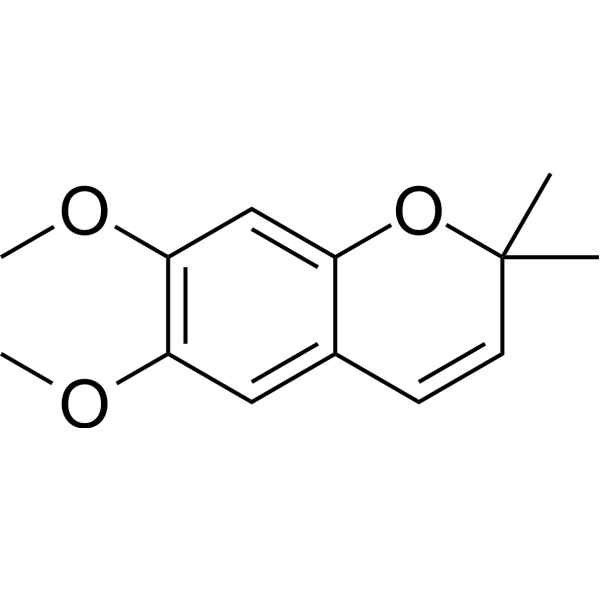
Precocene II (Ageratochromene) is a plant larva hormone antagonists that inhibits the biosynthesis of juvenile hormone. Precocene II inhibits corpora allata function and downregulates juvenile hormone levels, while exerting multiple effects including inducing precocious metamorphosis, forming macropterous morphs, inhibiting ovarian growth, producing cytotoxicity, disrupting insect development, and causing sterility and kidney damage. Precocene II blocks normal nymphal development, causes renal tubular damage and increases blood urea nitrogen levels, and also blocks juvenile hormone biosynthesis in vitro. Precocene II undergoes oxidative metabolism catalyzed by NADPH-dependent monooxygenase to generate a variety of metabolites. Precocene II can be applied in studies related to insect growth regulation and nephrotoxicity.
For research use only. We do not sell to patients.
- Purity : 99.98%
- CAS No.: 644-06-4
- Formula: C13H16O3
- Molecular Weight:220.26
-
Storage:
4°C, protect from light
* In solvent : -80°C, 6 months; -20°C, 1 month (protect from light)

To place orders, for customer services and technical support, please contact: MedChemExpress USA
Tel: 609-228-6898 E-mail: [email protected] [email protected]


-
 Biological Activity
-
Chemical Information
-
Solvent & Solubility
- Protocols
- Purity & Documentation
- References
-
 Help & FAQs


-

HY-122940R
Precocene II (Standard)Plant Larva Hormone Antagonists -
HY-14648A
Dexamethasone acetateCorticosteroid Ester -
HY-14648AR
Dexamethasone acetate (Standard)Analytical Standard -
HY-106019
LiarozoleRAMBA -
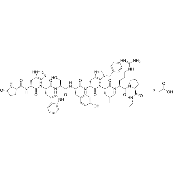
HY-P0056A
Histrelin acetateGnRH Receptor Agonist -

HY-117168
1,2-Dilinoleoyl-sn-glycerolDiacylglycerol -
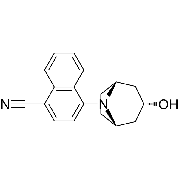
HY-122025
AC-262536Androgen Receptor Agonist -

HY-112527
7(Z),11(Z)-HeptacosadieneEster Product -

HY-121149B
(E/Z)-DroloxifeneER Modulator -

HY-106019R
Liarozole (Standard)RAMBA -

HY-W673589
Hexacosane, 2-Methyl- -

HY-133984
(7Z)-7-TricosenePheromone
All Endogenous Metabolite Isoforms
More

Biological Activity
Description
In Vitro
Precocene II (1-1000 ng) exhibits instar, dose, and sex-dependent effects on the brown planthopper Nilaparvata lugens: it induces precocious metamorphosis in both short-winged and long-winged nymphs of different instars; 1 ng is sufficient to induce differentiation of 2-3 instar short-winged nymphs into long-winged nymphs, with males being more sensitive. A single treatment of 100 ng in nymphs of all instars does not affect ovarian development, while a second administration 6 hours before adult emergence significantly inhibits ovarian development in 4-5 instar individuals treated for the first time[1].
Precocene II (2 × 10⁻⁵ M) can be metabolizes in vitro by the fat body homogenate of Cephalotaxus fortunei. The metabolic rate of the 1975 strain was significantly faster than that of the 1976 strain. The metabolic pathways of the two strains are consistent, mainly including epoxidation and O-demethylation, with trans-diol being the main product of the epoxidation pathway[2].
Precocene II (10-10000 μM; 30 min preincubation, 3 h incubation) inhibits the synthesis of C16 juvenile hormone in the pharyngeal lateral body of female American cockroaches in vitro. Compared to highly active glands, less active glands showed slightly lower sensitivity to Precocene II[4].
Precocene II (0.25-0.5 mM; 0-6 h preincubation, 2 h incubation) shows time-dependent inhibitory effect on the spontaneous C16JH biosynthesis of adult female American cockroaches in vitro[4].
MedChemExpress (MCE) has not independently confirmed the accuracy of these methods. They are for reference only. Further protocols information, click here.
In Vivo
Precocene II (1-1000 ng; local administration; single application; dissolved in 0.1 μl acetone on the abdomen) induces dose-dependent precocial metamorphosis in 3rd or 4th instar nymphs of the potentially long-winged brown planthopper without altering wing shape[1].
Precocene II (100-300 mg/kg; intraperitoneal injection; single administration; once daily for 5 consecutive days) induces nephrotoxicity in male Sprague-Dawley rats, characterized by histopathological damage to renal tissues[3].
MedChemExpress (MCE) has not independently confirmed the accuracy of these methods. They are for reference only.
-
Animal Model:presumptive brachypterous strain (yellowish brown brachypters, selected over 70 generations to produce ~100% short-winged adults across densities)[1]
-
Dosage:1 ng, 10 ng, 100 ng, 1000 ng (single topical application); 100 ng (two topical applications)
-
Administration:topical; single application (abdomen in 0.1 μl acetone); topical; two applications (first in 2nd/3rd/4th/5th stadium, second ~6h before adult emergence)
-
Result:Prolonged developmental duration, eliminated the 5th stadium and induced precocious metamorphosis in 2nd and early 3rd instar nymphs, while such effects were absent in older nymphs even at high doses.
Induced dose-dependent macroptery exclusively in 2nd and 3rd instar nymphs, with higher susceptibility observed in male individuals.
Exerted no impact on ovarian growth after single dosing.
Two administrations markedly inhibited ovarian growth when the first treatment was applied at late instars, and only generated mild suppression for early-instar initial treatment.
-
Animal Model:presumptive macropterous strain (blackish macropters, selected over 70 generations to produce ~100% long-winged adults across densities)[1]
-
Dosage:1 ng, 10 ng, 100 ng, 1000 ng (single topical application); 100 ng (two topical applications)
-
Administration:topical; single application (abdomen in 0.1 μl acetone); topical; two applications (first in 3rd/4th/5th stadium, second ~6h before adult emergence)
-
Result:Prolonged the 3rd stadium and altered late development in 3rd instar nymphs, and induced precocious metamorphosis in 3rd and 4th instar nymphs in a dose- and age-dependent manner.
No developmental responses were found in 2nd instar individuals.
No wing-form shift occurred in all treated nymphs, and precocious adults only presented smaller body size.
Single 100 ng dosing exerted no influence on ovarian growth.
Two rounds of 100 ng treatment caused severe ovarian growth inhibition when the first dose was applied at late instars, and mild inhibition for initial treatment at the 3rd instar.
-
Animal Model:Sprague-Dawley (male, 200 g)[3]
-
Dosage:100 mg/kg (daily x5); 200 mg/kg (single dose); 300 mg/kg (single dose)
-
Administration:i.p.; single dose; daily for 5 consecutive days
-
Result:Induced glomerular blood congestion, proximal convoluted tubule and collecting tubule epithelial degeneration, sloughing of tubular cell nuclei into lumens, and tubular epithelial cell regeneration within 24 h of dosing.
Increased average serum BUN level to 23 mg/100 mL in rats treated with a single 200 mg/kg dose.


Chemical Information
-
CAS No. 644-06-4
-
Appearance Solid
-
Molecular Weight 220.26
-
Formula C13H16O3
-
Color White to light yellow
-
SMILES
CC1(C)C=CC2=CC(OC)=C(OC)C=C2O1
-
Synonyms
Ageratochromene
-
Structure Classification
-
Initial Source
Insects
-
Shipping
Room temperature in continental US; may vary elsewhere.
-
Storage
4°C, protect from light
* In solvent : -80°C, 6 months; -20°C, 1 month (protect from light)


Solvent & Solubility
In Vitro:
DMSO : 100 mg/mL (454.01 mM; ultrasonic and warming and heat to 80°C; Hygroscopic DMSO has a significant impact on the solubility of product, please use newly opened DMSO)
Please refer to the solubility information to select the appropriate solvent. Once prepared, please aliquot and store the solution to prevent product inactivation from repeated freeze-thaw cycles.
Storage method and period of stock solution: -80°C, 6 months; -20°C, 1 month (protect from light). When stored at -80°C, please use it within 6 months. When stored at -20°C, please use it within 1 month.
Please refer to the solubility information to select the appropriate solvent. Once prepared, please aliquot and store the solution to prevent product inactivation from repeated freeze-thaw cycles.
Storage method and period of stock solution: -80°C, 6 months; -20°C, 1 month (protect from light). When stored at -80°C, please use it within 6 months. When stored at -20°C, please use it within 1 month.
Concentration (start) × Volume (start) = Concentration (final) × Volume (final)

Protocols
-
Nephrotoxicity Study
This protocol assesses nephrotoxicity by combining functional kidney injury readouts, urinary/tissue injury biomarkers, and renal histopathology. Serum creatinine and BUN reflect impaired kidney function, while KIM-1, NGAL, clusterin, osteopontin, IL-18, cystatin C, nephrin, Oat5, urinary protein, glucose, and alkaline phosphatase have been used to detect tubular injury in cisplatin-, gentamicin-, and acetaminophen-induced nephrotoxicity models.
-
Cell Cytotoxicity Assay
Cytotoxicity assays are usually based on the assessment of cell membrane damage, which can also be indirectly detected by measuring cell viability. Detection methods include MTT assay, CKK-8 assay, LDH assay and ATP assay, etc.
-
Mammalian live/dead viability and cytotoxicity staining
Live/dead viability and cytotoxicity staining assays are based on the simultaneous detection of intracellular esterase activity in metabolically active (viable) cells and membrane integrity loss in non-viable cells. In commonly used dual-staining approaches, membrane-permeant fluorogenic substrates are converted by intracellular esterases into fluorescent products in live cells, while impermeant DNA-binding dyes selectively enter cells with compromised plasma membranes and label nucleic acids in dead or dying cells, enabling discrimination between viable and non-viable populations by fluorescence microscopy or flow cytometry.
-
Research Protocol for Endocrine Diseases
Endocrine diseases often arise from disrupted hormone production, hormone signaling, or target-tissue responsiveness; for diabetes-focused endocrine disease models, insulin signaling regulates glucose uptake, hepatic glucose output, lipid metabolism, and β-cell compensation. Type 2 diabetes develops through interacting defects in insulin resistance, β-cell dysfunction, adipose inflammation, hepatic glucose overproduction, altered incretin signaling, and ectopic lipid metabolism. A major unresolved question is whether endocrine dysfunction is driven primarily by target-tissue insulin resistance, intrinsic β-cell failure, immune/inflammatory stress, or combined multi-organ failure that differs by disease stage.
-
Cell Viability Determination by MTT Colorimetric Assay
The following protocol uses the MTT colorimetric assay as a classic literature-established method for assessing cell viability/metabolic activity in cultured mammalian cells. MTT[3-(4,5-dimethylthiazol-2-yl)-2,5-diphenyltetrazolium bromide] is reduced by metabolically active cells to a colored formazan product; the amount of formazan is quantified spectrophotometrically and provides an indirect measure of metabolically active viable cells. Importantly, MTT reduction reflects cellular oxidoreductase/metabolic activity rather than an absolute direct count of living cells, so changes in cellular metabolism can alter the signal independently of cell number.


Purity & Documentation
-
Data Sheet (280 KB)
-
SDS (419 KB)
- English - EN (419 KB)
- Français - FR (419 KB)
- Deutsch - DE (419 KB)
- Norwegian - NO (419 KB)
- Español - ES (419 KB)
- Swedish - SV (419 KB)
- Italian - IT (419 KB)
- Korean - KR (419 KB)
- Portuguese - PT (419 KB)
-
Handling Instructions (2659 KB)
References


Complete Stock Solution Preparation Table
Please refer to the solubility information to select the appropriate solvent. Once prepared, please aliquot and store the solution to prevent product inactivation from repeated freeze-thaw cycles.
Storage method and period of stock solution: -80°C, 6 months; -20°C, 1 month (protect from light). When stored at -80°C, please use it within 6 months. When stored at -20°C, please use it within 1 month.
| Optional Solvent | Concentration Solvent Mass | 1 mg | 5 mg | 10 mg | 25 mg |
|---|---|---|---|---|---|
| DMSO | 1 mM | 4.5401 mL | 22.7004 mL | 45.4009 mL | 113.5022 mL |
| 5 mM | 0.9080 mL | 4.5401 mL | 9.0802 mL | 22.7004 mL | |
| 10 mM | 0.4540 mL | 2.2700 mL | 4.5401 mL | 11.3502 mL | |
| 15 mM | 0.3027 mL | 1.5134 mL | 3.0267 mL | 7.5668 mL | |
| 20 mM | 0.2270 mL | 1.1350 mL | 2.2700 mL | 5.6751 mL | |
| 25 mM | 0.1816 mL | 0.9080 mL | 1.8160 mL | 4.5401 mL | |
| 30 mM | 0.1513 mL | 0.7567 mL | 1.5134 mL | 3.7834 mL | |
| 40 mM | 0.1135 mL | 0.5675 mL | 1.1350 mL | 2.8376 mL | |
| 50 mM | 0.0908 mL | 0.4540 mL | 0.9080 mL | 2.2700 mL | |
| 60 mM | 0.0757 mL | 0.3783 mL | 0.7567 mL | 1.8917 mL | |
| 80 mM | 0.0568 mL | 0.2838 mL | 0.5675 mL | 1.4188 mL | |
| 100 mM | 0.0454 mL | 0.2270 mL | 0.4540 mL | 1.1350 mL |


Precocene II Related Classifications
HY-122940 Related Classifications
 Powered by Bioz