
PRO-905
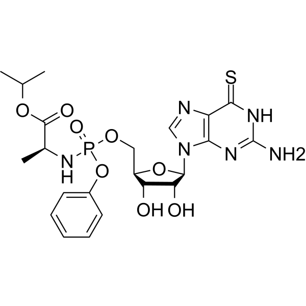
Pro-905 is a phosphite peptide with antitumor activity. Pro-905 delivers the active nucleotide antimetabolite thioguanosine monophosphate (TGMP) to the tumor. Pro-905 effectively prevents incorporation of purine salvage substrates into nucleic acids and inhibits colony formation in human malignant peripheral nerve sheath tumors (MPNST) cells. Pro-905 inhibits purine salvage incorporation to nucleic acids and prevents cell growth. Pro-905 inhibits the growth of MPNST and enhances the anti-tumor efficacy of JHU395 (HY-124778) .
For research use only. We do not sell to patients.
- CAS No.: 2762209-68-5
- Formula: C22H29N6O8PS
- Molecular Weight:568.54
-
Storage:
Please store the product under the recommended conditions in the Certificate of Analysis.
Biological Activity
Description
In Vitro
Pro-905 (10 µM, 120 h) inhibits cell proliferation and guanine incorporation to newly synthesized DNA and RNA using JH-2-002 cells with JHU395 (HY-124778) 1 µM, 120 h[1].
Pro-905 (10 µM, 120 h) inhibits colony formation in sNF96.2 and JH-2-002 human MPNST cell with JHU395 (1 µM, 120 h)[1].
Pro-905 (10 µM, 6 h) inhibits purine salvage-dependent 3H-hypoxanthine incorporation to DNA and RNA in sNF96.2 cells[1]
MedChemExpress (MCE) has not independently confirmed the accuracy of these methods. They are for reference only. Further protocols information, click here.
-
Cell Line:sNF96.2 and JH-2-002 human MPNST cells
-
Concentration:10 µM (in combination with JHU395 (HY-124778)(1µM))
-
Incubation Time:120 h
-
Result:Inhibited cell proliferation with effects on nucleic acid and induced DNA damage (γH2AX).
In Vivo
Pro-905 (10 mg/kg, i.p, 5 d/week) in B6 mice with flank MPNST inhibits tumor nucleotide metabolism and abrogates MPNST proliferation with JHU395 (1.2 mg/kg, p.o.) [1].
MedChemExpress (MCE) has not independently confirmed the accuracy of these methods. They are for reference only.
-
Animal Model:B6 mice with flank MPNST[1]
-
Dosage:10 mg/kg (in combination with JHU3955 (1.2 mg/kg, p.o.))
-
Administration:Intraperitoneal injection
-
Result:Prevented growth of established tumors to a greater extent.
Induced tumors significantly decrease, mean percent proliferation, JHU395 0.82%, Pro-905 1.25%, combination 0.18%.
-
Animal Model:Nod scid gamma (NSG) mice with JH-2-031 MPNST [1]
-
Dosage:20 mg/kg
-
Administration:Intraperitoneal injection
-
Result:Caused animal body weights remained within 10% of starting weights over two weeks of dosing.
Chemical Information
-
CAS No. 2762209-68-5
-
Molecular Weight 568.54
-
Formula C22H29N6O8PS
-
SMILES
O[C@H]1[C@@H](O)[C@H](N2C(N=C(N)NC3=S)=C3N=C2)O[C@@H]1COP(N[C@H](C(OC(C)C)=O)C)(OC4=CC=CC=C4)=O
-
Synonyms
MK-905
-
Shipping
Room temperature in continental US; may vary elsewhere.
-
Storage
Please store the product under the recommended conditions in the Certificate of Analysis.
Protocols
-
Protocol for Cell Counting and Cell Density Analysis
Cell counting and cell-density analysis estimate the number of cells in a known volume or field area. Manual hemocytometer counting uses a chamber of defined geometry to convert counted cells into cells/mL, while automated counters and image-analysis workflows detect cell objects from optical, brightfield, fluorescence, impedance, or digital-image features. Trypan blue viability counting is based on dye exclusion: viable cells with intact membranes exclude dye, while non-viable cells with compromised membranes stain blue. The readout is total cell density, viable-cell density, dead-cell density, and percent viability. Cell density can also be estimated from microscopy images by counting objects per image area, from flow cytometry using calibrated volume or reference particles, or from in situ microscopy in bioreactors after calibration against reference methods such as hemocytometer or flow cytometry.
-
Somatic Cell Culture
A method of simulating the in vivo environment in vitro to maintain the cell growth, differentation and main functions.
-
CCK-8/WST-8 Cell Proliferation Assay
The CCK-8/WST-8 assay is based on the reduction of the water-soluble tetrazolium salt WST-8 to a water-soluble formazan product by cellular dehydrogenases in metabolically active cells, where the generated formazan amount is proportional to the number of living cells and is quantified by measuring absorbance in the visible range, providing a colorimetric readout for cell viability and proliferation assessment. This class of tetrazolium-based assays improves upon earlier MTT-based systems by producing a water-soluble formazan, eliminating the need for organic solubilization steps and enabling direct spectrophotometric measurement in culture medium.
-
Soft Agar Colony Formation Assay
Soft agar colony formation assay measures anchorage-independent growth, in which transformed or tumorigenic cells proliferate as colonies in a semisolid agar matrix while many non-transformed adherent cells fail to proliferate without attachment; classic studies showed that growth in semisolid medium correlates with tumorigenicity in nude mice, and later protocol papers describe the method as a stringent in vitro assay for malignant transformation. The readout is the number, size, morphology, or signal intensity of colonies formed within agar after incubation; published formats include manual colony counting after staining, 96-well or 384-well quantitative formats, DNA-binding dye detection, MTT/tetrazolium-based detection, digital image analysis, and PCR-based marker detection from soft agar cultures.
-
Cell Counting-Based Growth Curve Assay
Cell counting-based growth curve assays quantify cell proliferation by directly measuring changes in viable cell number over time using manual or automated counting methods such as hemocytometer-based counting or instrument-assisted cell enumeration, enabling construction of growth curves that reflect population expansion dynamics in response to culture conditions. A widely used approach is trypan blue exclusion with hemocytometer counting, where membrane-compromised (non-viable) cells take up the dye, allowing discrimination between viable and non-viable cells while simultaneously enabling total cell number quantification. Repeated sampling across time points allows estimation of proliferation rate, growth phases, and comparative growth kinetics between experimental conditions.
-
Subcutaneous Cell-Line-Derived Xenograft
Subcutaneous cell-line-derived xenograft (CDX) models are established by implanting cultured human cancer cell lines into immunodeficient mice, where the injected cells form localized tumors that can be monitored in vivo as a measure of tumorigenic potential, growth kinetics, and treatment response. These models are widely used in oncology research because they allow reproducible tumor formation and enable comparative assessment of tumor growth between different cell lines or genetic manipulations in a controlled in vivo microenvironment. Subcutaneous implantation of cancer cells in immunodeficient mice is a standard approach for evaluating tumor growth behavior and therapeutic response across multiple cancer types, including prostate, esophageal, pancreatic, and colon cancer models.
-
Colony Formation (Clonogenic) Assay
The clonogenic (colony formation) assay measures the ability of a single cell to retain reproductive viability and form a macroscopic colony, typically defined as a cluster derived from one progenitor cell after a defined growth period. This assay is widely used to evaluate cell survival after exposure to ionizing radiation or cytotoxic treatments and is considered a standard method in radiation biology for generating dose-response relationships of reproductive cell death. Colony formation reflects long-term proliferative capacity rather than short-term metabolic activity, and survival is quantified by comparing treated versus untreated conditions based on colony number and derived survival fractions.
-
MTT Cell Proliferation Assay
The MTT assay is a colorimetric endpoint assay for estimating viable cell number, cell growth, cytotoxicity, or cell activation in cultured mammalian cells. Living cells reduce the yellow tetrazolium salt MTT into purple/blue formazan, while dead cells do not generate the same signal; the resulting color can be quantified with a multiwell spectrophotometer. MTT reduction is commonly interpreted as a readout of metabolic activity that often correlates with viable cell number, but it should not be treated as a direct cell-counting method unless the assay is optimized for the cell type and experimental condition. Studies show that MTT reduction can involve mitochondrial and non-mitochondrial reducing systems, and formazan may accumulate in intracellular lipid droplets rather than simply marking mitochondria.
-
Cell Viability Determination by MTT Colorimetric Assay
The following protocol uses the MTT colorimetric assay as a classic literature-established method for assessing cell viability/metabolic activity in cultured mammalian cells. MTT[3-(4,5-dimethylthiazol-2-yl)-2,5-diphenyltetrazolium bromide] is reduced by metabolically active cells to a colored formazan product; the amount of formazan is quantified spectrophotometrically and provides an indirect measure of metabolically active viable cells. Importantly, MTT reduction reflects cellular oxidoreductase/metabolic activity rather than an absolute direct count of living cells, so changes in cellular metabolism can alter the signal independently of cell number.
Purity & Documentation
References
Calculators
Concentration (start) × Volume (start) = Concentration (final) × Volume (final)

