



PSDalpha
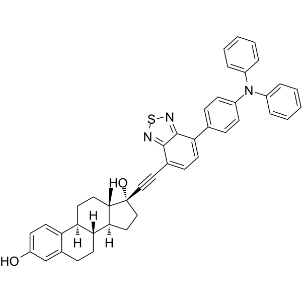
PSDalpha is a photoactivatable PS-Degron targeting estrogen receptor α (ERα/ESR1), with a Ki of 72.8 nM for ERα. PSDalpha consists of an ERα-targeting estradiol moiety conjugated via an alkyne bond to triphenylamine-benzothiadiazole (TB), an aggregation-induced emission (AIE) photosensitizer. Upon visible light irradiation, PSDalpha generates singlet oxygen (1O2) and induces controllable ERα degradation. PSDalpha also induces G1 phase arrest and apoptosis. PSDalpha can be used in studies related to ERα-positive breast cancer and light-controlled targeted protein degradation.
For research use only. We do not sell to patients.
- CAS No.: 2906869-08-5
- Formula: C44H39N3O2S
- Molecular Weight:673.86
-
Storage:
Please store the product under the recommended conditions in the Certificate of Analysis.

To place orders, for customer services and technical support, please contact: MedChemExpress USA
Tel: 609-228-6898 E-mail: [email protected] [email protected]


-
 Biological Activity
-
Chemical Information
- Protocols
- Purity & Documentation
- References
-
 Help & FAQs




Biological Activity
Description
IC50 & Target
[1]|
ERα 72.8 nM (Ki) |
Cellular Effect
|
Cell Line

|
Type | Value | Description | References |
|---|---|---|---|---|
| MCF7 | EC50 |
17 nM
|
ERα degradation in human MCF-7 breast cancer cells incubated for 6 h, irradiated for 30 min, harvested 4 h post-irradiation, measured via immunoblotting.
ERα degradation in human MCF-7 breast cancer cells incubated for 6 h, irradiated for 30 min, harvested 4 h post-irradiation, measured via immunoblotting.
|
35164509 |
| MCF7 | EC50 |
0.5 nM
|
ERα degradation in human MCF-7 breast cancer cells incubated for 6 h, irradiated for 45 min, harvested 4 h post-irradiation, measured via immunoblotting.
ERα degradation in human MCF-7 breast cancer cells incubated for 6 h, irradiated for 45 min, harvested 4 h post-irradiation, measured via immunoblotting.
|
35164509 |
| MCF7 | IC50 |
8.9 μM
|
Cell viability inhibition in human MCF-7 breast cancer cells incubated for 6 h, single irradiated for 15 min, viability measured 72 h post-irradiation by MTT assay.
Cell viability inhibition in human MCF-7 breast cancer cells incubated for 6 h, single irradiated for 15 min, viability measured 72 h post-irradiation by MTT assay.
|
35164509 |
| MCF7 | IC50 |
6.1 μM
|
Cell viability inhibition in human MCF-7 breast cancer cells incubated for 6 h, single irradiated for 30 min, viability measured 72 h post-irradiation by MTT assay.
Cell viability inhibition in human MCF-7 breast cancer cells incubated for 6 h, single irradiated for 30 min, viability measured 72 h post-irradiation by MTT assay.
|
35164509 |
| MCF7 | IC50 |
5.9 μM
|
Cell viability inhibition in human MCF-7 breast cancer cells incubated for 6 h, single irradiated for 45 min, viability measured 72 h post-irradiation by MTT assay.
Cell viability inhibition in human MCF-7 breast cancer cells incubated for 6 h, single irradiated for 45 min, viability measured 72 h post-irradiation by MTT assay.
|
35164509 |
| MCF7 | IC50 |
0.024 μM
|
Cell viability inhibition in human MCF-7 breast cancer cells incubated for 6 h, daily irradiated for 45 min over 3 days, viability measured 72 h post-initial incubation by MTT assay.
Cell viability inhibition in human MCF-7 breast cancer cells incubated for 6 h, daily irradiated for 45 min over 3 days, viability measured 72 h post-initial incubation by MTT assay.
|
35164509 |
In Vitro
PSDalpha binds to the ligand-binding domain of human ERα with a Ki of 72.8 nM; isothermal titration calorimetry (ITC) shows that PSDalpha interacts with ERα in an exothermic manner, with a binding molar ratio of approximately 1:1[1].
PSDalpha (1 μM) reduces the level of purified ERα after 15 min of illumination, and ERα disappears completely when illumination is extended to 30-45 min; PSDalpha remains inert under dark conditions[1].
The ERα degradation induced by PSDalpha in MCF-7 cells is light-duration dependent, with EC50 values of 17 nM and 0.5 nM upon 460 nm light irradiation for 30 min and 45 min, respectively[1].
PSDalpha inhibits the viability of MCF-7 cells after a single irradiation, with IC50 values of 8.9, 6.1, and 5.9 μM following 15, 30, and 45 min of irradiation, respectively; PSDalpha shows essentially no obvious toxicity to MCF-7 cells under dark conditions[1].
MedChemExpress (MCE) has not independently confirmed the accuracy of these methods. They are for reference only.
-
Cell Line:MCF-7
-
Concentration:0.1 or 1 μM
-
Incubation Time:6 h; 30 min irradiation; 2-24 h post-irradiation
-
Result:Induced time-dependent ERα degradation; complete depletion occurred at 8 h with 0.1 μM.
-
Cell Line:MCF-7
-
Concentration:0.1 nM-10 μM
-
Incubation Time:6 h; 30 or 45 min irradiation; 4 h post-irradiation
-
Result:Induced ERα degradation with EC50 values of 17 nM and 0.5 nM after 30- and 45-min irradiation, respectively.
-
Cell Line:MCF-7
-
Concentration:0.01 μM
-
Incubation Time:6 h; 0-45 min irradiation
-
Result:Increased cytochrome c release as irradiation time increased.
-
Cell Line:MCF-7
-
Concentration:0.01, 0.1, 1 μM
-
Incubation Time:30 min irradiation; assessed at 3 d
-
Result:Increased late apoptosis, with a stronger response under daily irradiation.


Chemical Information
-
CAS No. 2906869-08-5
-
Molecular Weight 673.86
-
Formula C44H39N3O2S
-
SMILES
C[C@@]12[C@](CC[C@]2(O)C#CC3=CC=C(C4=CC=C(C=C4)N(C5=CC=CC=C5)C6=CC=CC=C6)C7=NSN=C37)([H])[C@@]8([H])[C@@](CC1)([H])C9=CC=C(O)C=C9CC8
-
Shipping
Room temperature in continental US; may vary elsewhere.
-
Storage
Please store the product under the recommended conditions in the Certificate of Analysis.


Purity & Documentation
References

Calculators
Concentration (start) × Volume (start) = Concentration (final) × Volume (final)


PSDalpha Related Classifications
HY-144314 Related Classifications
 Powered by Bioz