



Pyraclostrobin
Based on 3 publication(s) in Google Scholar
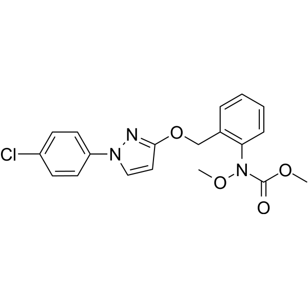
Pyraclostrobin is a highly effective and broad-spectrum strobilurin fungicide. Pyraclostrobin can induce oxidative DNA damage, mitochondrial dysfunction and autophagy through the activation of AMPK/mTOR signaling. Pyraclostrobin can be used to control crop diseases.
For research use only. We do not sell to patients.
- Purity : 99.64%
- CAS No.: 175013-18-0
- Formula: C19H18ClN3O4
- Molecular Weight:387.82
-
Storage:
4°C, protect from light
* In solvent : -80°C, 6 months; -20°C, 1 month (protect from light)

To place orders, for customer services and technical support, please contact: MedChemExpress USA
Tel: 609-228-6898 E-mail: [email protected] [email protected]


-
 Biological Activity
-
Chemical Information
-
Solvent & Solubility
- Protocols
- Purity & Documentation
- References
-
 Help & FAQs


-
HY-B0849
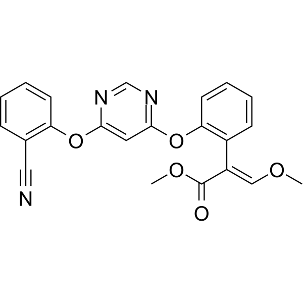
AzoxystrobinFungicide -
HY-B0824
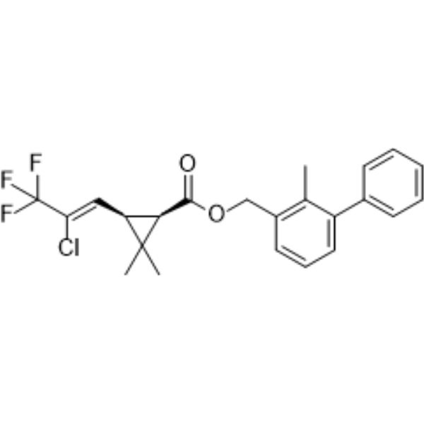
BifenthrinSodium Channel Activator -
HY-B0850
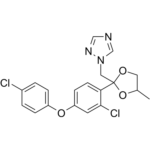
DifenoconazoleTriazole Fungicide -
HY-16658B
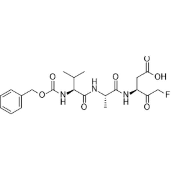
Z-VAD-FMKPan-Caspase Inhibitor -
HY-B0815
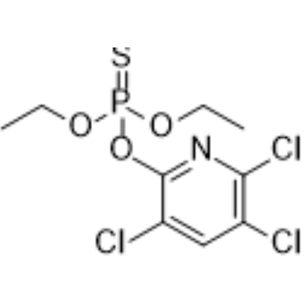
ChlorpyrifosMuscarinic Antagonist -
HY-B0827
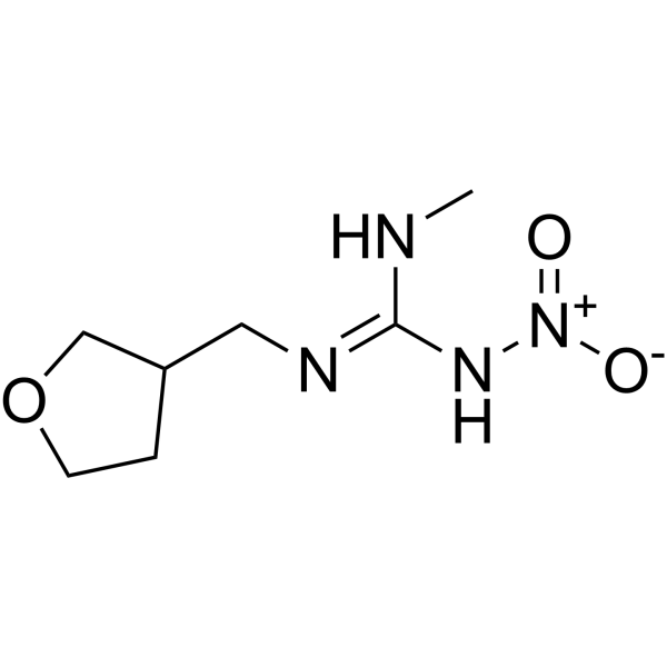
DinotefuranNicotinic acetylcholine receptors Inhibitor -
HY-B0817
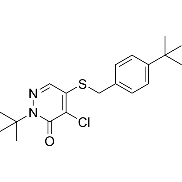
PyridabenMETI Acaricide -
HY-B0845
ProchlorazAntifungal -

HY-W036120
Benzophenone-2UV Filter -
HY-117386
MethoxyfenozideInsecticide -
HY-N6626R
Pyraclostrobin (Standard)Fungicide -
HY-B1978
IprodioneFungicide
Publications Citing Use of MedChemExpress (MCE) Pyraclostrobin
MoreAll AMPK Isoforms
More

Biological Activity
Description
IC50 & Target
|
Bax |
Bcl-2 |
In Vitro
Pyraclostrobin (10-80 μmol/L, 24 h) exhibits toxic effects in HepG2 cells[1].
Pyraclostrobin (10-80 μmol/L, 6 h) induced DNA damage and mitochondrial dysfunction in HepG2 cells[1].
MedChemExpress (MCE) has not independently confirmed the accuracy of these methods. They are for reference only.
-
Cell Line:HepG2 cells
-
Concentration:10 μmol/L, 20 μmol/L, 40 μmol/L, 80 μmol/L
-
Incubation Time:24 h
-
Result:Inhibited cell survival in concentration-dependent manner with IC50 value of 30.22 μmol/L.
-
Cell Line:HepG2 cells
-
Concentration:10 μmol/L, 20 μmol/L, 40 μmol/L, 80 μmol/L
-
Incubation Time:6 h
-
Result:Increased the content of cytochrome c (Cyt c) in the cytoplasm in a concentration-dependent way.
Increased pro-apoptotic protein Bax expression and down-regulated anti-apoptosis protein Bcl-2 expression.
In Vivo
Pyraclostrobin (0.001-0.02 mg/L, Fish were assigned to a vessel containing 20 L of water dissolved Pyraclostrobin for 7-28 days) has the toxic effects on DNA damage and antioxidant enzymatic activities in the zebrafish liver[2].
Pyraclostrobin (33-48 μg/L, Zebrafish larvae at 4 days post fertilization (dpf) were transferred into 24-well plates and subjected to pyraclostrobin until 8 dpf) has toxic effects on zebrafish larvae[3].
MedChemExpress (MCE) has not independently confirmed the accuracy of these methods. They are for reference only.
-
Animal Model:Zebrafish [2]
-
Dosage:0.001 mg/L, 0.01 mg/L, 0.02 mg/L
-
Administration:Fish were assigned to a vessel containing 20 L of water dissolved Pyraclostrobin
-
Result:Showed higher ROS levels in zebrafish livers.
Decreased SOD levels and increased MDA levels.
Increased the olive tail moments as the dose increased on days 7, 14, 21, and 28.
-
Animal Model:Zebrafish larvae [3]
-
Dosage:33 mg/L, 36 mg/L, 40 mg/L, 44 mg/L, 48 mg/L
-
Administration:Zebrafish larvae at 4 days post fertilization (dpf) were transferred into 24-well plates and subjected to pyraclostrobin until 8 dpf
-
Result:Damaged histological and subcellular structure of larval heart and brain.
Changed the expression level of cardiac muscle contraction pathway- and neural-related genes and proteins.
Impaired larval cardiac function and locomotor behavior.


Chemical Information
-
CAS No. 175013-18-0
-
Appearance Solid
-
Molecular Weight 387.82
-
Formula C19H18ClN3O4
-
Color Off-white to light yellow
-
SMILES
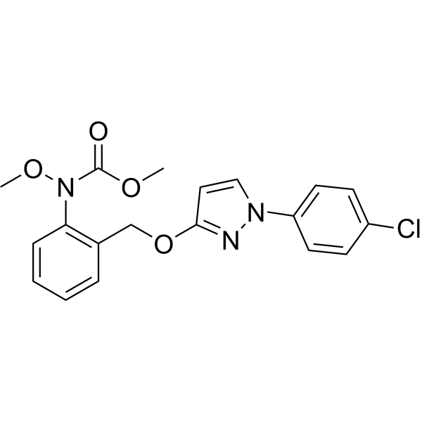
COC(N(C1=C(C=CC=C1)COC2=NN(C3=CC=C(Cl)C=C3)C=C2)OC)=O
-
Shipping
Room temperature in continental US; may vary elsewhere.
-
Storage
4°C, protect from light
* In solvent : -80°C, 6 months; -20°C, 1 month (protect from light)


Publications (3)
-
Journal Impact Factor
-
Most Recent
-
Food Res Int
Analysis of pesticide residues in açaí-based food products: an approach using polymeric ionic liquid-based solid-phase microextraction coupled to gas chromatography-mass spectrometry. [Abstract]2025 Dec;222(Pt 2):117720. PMID: 41271335 -
-
Pestic Biochem Physiol
The function of the alternative oxidase gene in the tolerance of Fusarium graminearum to azoxystrobin. [Abstract]2026 Feb:217:106853. PMID: 41461432


Solvent & Solubility
In Vitro:
DMSO : 100 mg/mL (257.85 mM; Need ultrasonic; Hygroscopic DMSO has a significant impact on the solubility of product, please use newly opened DMSO)
Please refer to the solubility information to select the appropriate solvent. Once prepared, please aliquot and store the solution to prevent product inactivation from repeated freeze-thaw cycles.
Storage method and period of stock solution: -80°C, 6 months; -20°C, 1 month (protect from light). When stored at -80°C, please use it within 6 months. When stored at -20°C, please use it within 1 month.
Please refer to the solubility information to select the appropriate solvent. Once prepared, please aliquot and store the solution to prevent product inactivation from repeated freeze-thaw cycles.
Storage method and period of stock solution: -80°C, 6 months; -20°C, 1 month (protect from light). When stored at -80°C, please use it within 6 months. When stored at -20°C, please use it within 1 month.
Concentration (start) × Volume (start) = Concentration (final) × Volume (final)
In Vivo:
Select the appropriate dissolution method based on your experimental animal and administration route.
- For the following dissolution methods, please ensure to first prepare a clear stock solution using an In Vitro approach and then sequentially add co-solvents:
- To ensure reliable experimental results, the clarified stock solution can be appropriately stored based on storage conditions. As for the working solution for In Vivo experiments, it is recommended to prepare freshly and use it on the same day.
- The percentages shown for the solvents indicate their volumetric ratio in the final prepared solution. If precipitation or phase separation occurs during preparation, heat and/or sonication can be used to aid dissolution.
Add each solvent one by one: 10% DMSO 40% PEG300 5% Tween-80 45% Saline
Solubility: ≥ 2.08 mg/mL (5.36 mM); Clear solution
This protocol yields a clear solution of ≥ 2.08 mg/mL (saturation unknown).
Taking 1 mL working solution as an example, add 100 μL DMSO stock solution (20.8 mg/mL) to 400 μL PEG300, and mix evenly; then add 50 μL Tween-80 and mix evenly; then add 450 μL Saline to adjust the volume to 1 mL.
Preparation of Saline: Dissolve 0.9 g sodium chloride in ddH₂O and dilute to 100 mL to obtain a clear Saline solution.
In Vivo Dissolution Calculator
Please enter the basic information of animal experiments:
-
-
-
-
Recommended: Prepare an additional quantity of animals to account for potential losses during experiments.
Please enter your animal formula composition:
-
%DMSO +
Recommended: Keep the proportion of DMSO in working solution below 2% if your animal is weak.
-
%+
-
+%Tween-80 + +
-
%Saline +
The co-solvents required include: DMSO, . All of co-solvents are available by MedChemExpress (MCE). , Tween 80. All of co-solvents are available by MedChemExpress (MCE).
Working solution concentration: 0.22 mg/mL
Method for preparing stock solution: mg drug dissolved in μL DMSO. Stock solution concentration: mg/mL. * In solvent : -80°C, 6 months; -20°C, 1 month (protect from light)
1. Take μL DMSO stock solution;
2. Add μL .
μL , mix evenly;
3. Then add μL Tween 80, mix evenly;
4. Then add μL
Please ensure that the stock solution in the first step is dissolved to a clear state, and add co-solvents in sequence. You can use ultrasonic heating (ultrasonic cleaner, recommended frequency 20-40 kHz), vortexing, etc. to assist dissolution.


Purity & Documentation
-
Data Sheet (277 KB)
-
SDS (624 KB)
- English - EN (624 KB)
- Français - FR (624 KB)
- Deutsch - DE (624 KB)
- Norwegian - NO (624 KB)
- Español - ES (624 KB)
- Swedish - SV (624 KB)
- Italian - IT (624 KB)
- Korean - KR (624 KB)
- Portuguese - PT (624 KB)
-
Handling Instructions (2659 KB)
References
[1]. Wu M, et al. Characterization of hepatotoxic effects induced by pyraclostrobin in human HepG2 cells and zebrafish larvae [J]. Chemosphere, 2023, 340: 139732. [Content Brief]
[2]. Zhang C, et al. Acute and subchronic toxicity of pyraclostrobin in zebrafish (Danio rerio) [J]. Chemosphere, 2017, 188: 510-516. [Content Brief]
[3]. Li H, et al. Mitochondrial dysfunction-based cardiotoxicity and neurotoxicity induced by pyraclostrobin in zebrafish larvae [J]. Environmental Pollution, 2019, 251: 203-211. [Content Brief]


Complete Stock Solution Preparation Table
Please refer to the solubility information to select the appropriate solvent. Once prepared, please aliquot and store the solution to prevent product inactivation from repeated freeze-thaw cycles.
Storage method and period of stock solution: -80°C, 6 months; -20°C, 1 month (protect from light). When stored at -80°C, please use it within 6 months. When stored at -20°C, please use it within 1 month.
| Optional Solvent | Concentration Solvent Mass | 1 mg | 5 mg | 10 mg | 25 mg |
|---|---|---|---|---|---|
| DMSO | 1 mM | 2.5785 mL | 12.8926 mL | 25.7852 mL | 64.4629 mL |
| 5 mM | 0.5157 mL | 2.5785 mL | 5.1570 mL | 12.8926 mL | |
| 10 mM | 0.2579 mL | 1.2893 mL | 2.5785 mL | 6.4463 mL | |
| 15 mM | 0.1719 mL | 0.8595 mL | 1.7190 mL | 4.2975 mL | |
| 20 mM | 0.1289 mL | 0.6446 mL | 1.2893 mL | 3.2231 mL | |
| 25 mM | 0.1031 mL | 0.5157 mL | 1.0314 mL | 2.5785 mL | |
| 30 mM | 0.0860 mL | 0.4298 mL | 0.8595 mL | 2.1488 mL | |
| 40 mM | 0.0645 mL | 0.3223 mL | 0.6446 mL | 1.6116 mL | |
| 50 mM | 0.0516 mL | 0.2579 mL | 0.5157 mL | 1.2893 mL | |
| 60 mM | 0.0430 mL | 0.2149 mL | 0.4298 mL | 1.0744 mL | |
| 80 mM | 0.0322 mL | 0.1612 mL | 0.3223 mL | 0.8058 mL | |
| 100 mM | 0.0258 mL | 0.1289 mL | 0.2579 mL | 0.6446 mL |


Pyraclostrobin Related Classifications
HY-N6626 Related Classifications
 Powered by Bioz