



FGF basic/bFGF Protein, Human (145a.a)
Based on 4 publication(s) in Google Scholar
FGF-2 is a member of the fibroblast family involved in bone healing, cartilage repair, bone repair, and nerve regeneration. FGF-2 is also a mitotic promoter that accelerates cell proliferation. FGF-2 regulates immune processes by specifically targeting tyrosine kinase receptors and activating the FGF/FGFR signaling pathway. For example, FGF-2 is involved in the JAK-STAT signaling pathway to regulate cartilage metabolism and also activates ERK signaling to promote cartilage regeneration. FGF-2 combined with FGFR1/3 promoted degeneration and repair of articular cartilage, respectively. FGF-2 is also a known carcinogen in GBM, which contributes to glioma growth and vascularization.FGF basic/bFGF Protein, Human (145a.a), consists of 145 amino acids, produced by E.coli with tag free.
- Species: Human
- Source: E. coli
-
Storage:Stored at -20°C for 2 years from date of receipt. After reconstitution, it is stable at 4°C for 1 week or -20°C for longer (with carrier protein). It is recommended to freeze aliquots at -20°C or -80°C for extended storage.


To place orders, for customer services and technical support, please contact: MedChemExpress USA
Tel: 609-228-6898 E-mail: [email protected] [email protected]


-
 Biological Activity
-
Technical Parameters
-
Product Properties
- Documentation
- References
- Help & FAQs


-

HY-P7109
EGF Protein, Human -

HY-10182
LaduviglusibGSK-3 Inhibitor -

HY-10431
SB-431542TGF-β Receptor Kinase Inhibitor -
HY-P7007
BMP-4 Protein, Human -
HY-P70311
Activin A Protein, Human/Mouse/Rat (HEK293) -
HY-P7048
KGF-2/FGF-10 Protein, Human (169a.a) -

HY-10071
Y-27632ROCK Inhibitor -
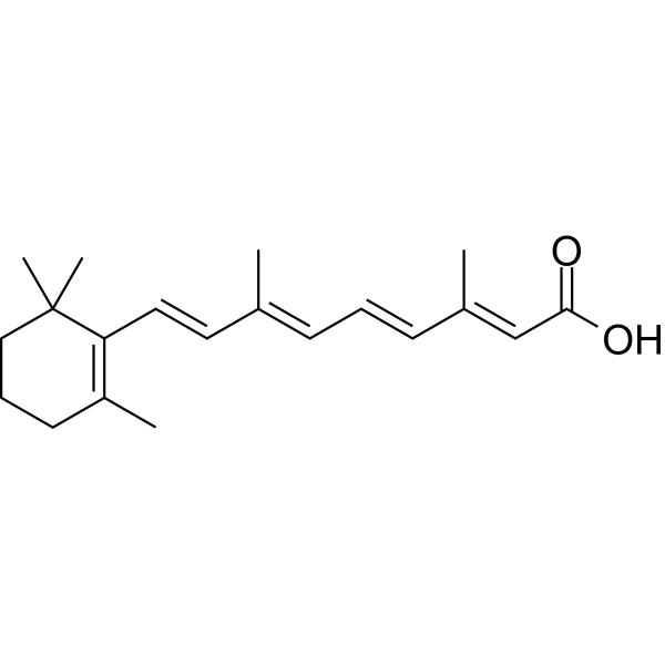
HY-14649
Retinoic acidRAR Agonist -

HY-10583
Y-27632 dihydrochlorideROCK Inhibitor -
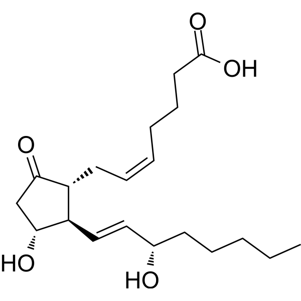
HY-101952
Prostaglandin E2Prostaglandin Receptor Agonist -
HY-P0035
Insulin (human)Recombinant Human Insulin -
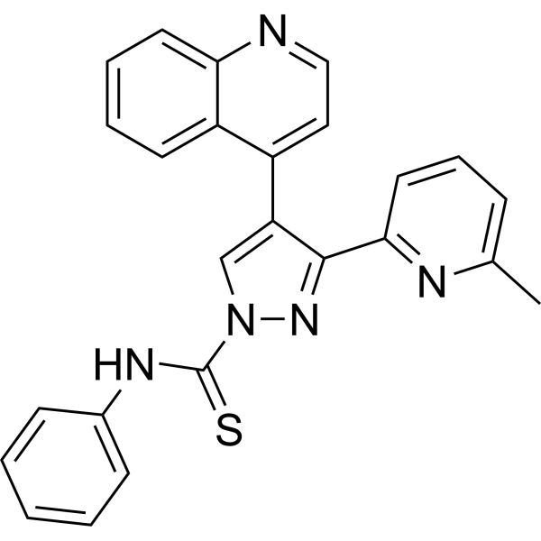
HY-10432
A 83-01ALK4/5/7 Inhibitor


Biological Activity
Description
FGF-2 is a member of the fibroblast family involved in bone healing, cartilage repair, bone repair, and nerve regeneration. FGF-2 is also a mitotic promoter that accelerates cell proliferation. FGF-2 regulates immune processes by specifically targeting tyrosine kinase receptors and activating the FGF/FGFR signaling pathway. For example, FGF-2 is involved in the JAK-STAT signaling pathway to regulate cartilage metabolism and also activates ERK signaling to promote cartilage regeneration. FGF-2 combined with FGFR1/3 promoted degeneration and repair of articular cartilage, respectively[1]. FGF-2 is also a known carcinogen in GBM, which contributes to glioma growth and vascularization[2].FGF basic/bFGF Protein, Human (145a.a), consists of 145 amino acids, produced by E.coli with tag free.
Background
FGF-2/bFGF is a member of the fibroblast family and has a high affinity for heparin. FGF-2 plays an important role in tendon to bone healing, cartilage repair, bone repair, and nerve regeneration. FGF-2 specifically binds to tyrosine kinase receptors and activates the FGF/FGFR signaling pathway. Subsequently, FGF-2 influences cell proliferation, differentiation and apoptosis, as well as immune regulation by transducing other classical pathways. For example, FGF-2 regulates the JAK-STAT signaling pathway to regulate cartilage metabolism. FGF-2 also acts as a mitotic promoter to accelerate cell proliferation. Therefore, (1) FGF-2 is an important growth factor in the healing process of ligament/tendon injury. In vitro experiments, low-dose FGF-2 can stimulate the proliferation and differentiation of bone marrow mesenchymal stem cells, and up-regulate the mRNA expression of type I/III collagen and fibronectin. However, high doses of FGF-2 did not stimulate extracellular matrix (ECM) protein proliferation and gene expression. (2) FGF-2 is also an endogenous and intrinsic growth factor in cartilage repair. FGF-2 binds to heparan sulfate proteoglycan and is stored in the ECM of articular cartilage. When cartilage is damaged or degenerated, ECM rapidly releases FGF-2 and activates ERK signaling pathways to promote cartilage regeneration. FGF-2 exhibits a biphasic effect in combination with its specific receptor. FGF-2 combined with FGFR3 promoted the repair of articular cartilage. FGF-2 combined with FGFR1 promoted the degeneration of articular cartilage[1]. FGF-2 is expressed in granulosa cells and colliculus cells, as well as hepatocellular cancer cells, but not in non-cancerous liver tissues. This reveals the role of FGF-2 in brain tumors, particularly glioblastoma. According to studies, FGF-2 is a known carcinogenic factor in GBM. FGF-2 increases the self-renewal of glioblastoma stem cells and contributes to the growth and vascularization of glioma[2]. FGF-2 protein is highly conserved in some species, and the similarity rate of human FGF-2 protein sequence to rat, mouse, and bovine was 97.4%, 95.45%, and 98.71%, respectively.
In Vitro
FGF-2 (human; 3 ng/mL, 30 ng/mL; 7-28 d) triggers the biphasic bone marrow stromal cell (BMSC) response at 3 ng/mL, promotes cell proliferation to peak on day 7, and significantly enhances mRNA expression of type I collagen, type III collagen, fibronectin, and alpha-smooth muscle actin on day 14 and 28. But there is no obvious effect at 30 ng/mL[3].
Verified Bioactivity
Measured in a cell proliferation assay using NIH-3T3 mouse embryonic fibroblasts. The ED50 for this effect is 0.1-0.8 ng/mL.
MCE Validation Data
-
Purity - SDS-PAGE
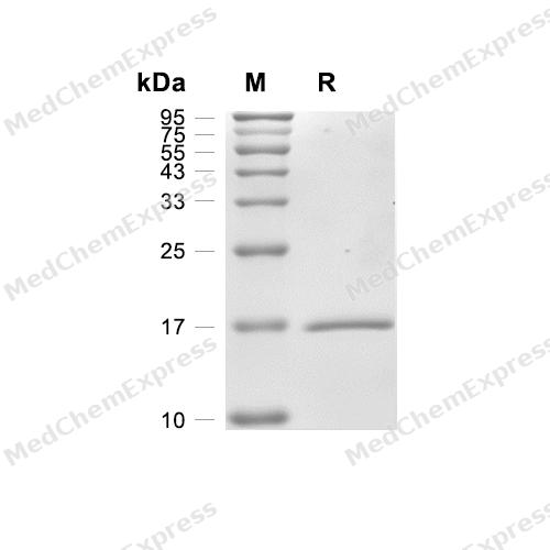
Purity - SDS-PAGE
-
Bioactivity - Cell-Based Assay
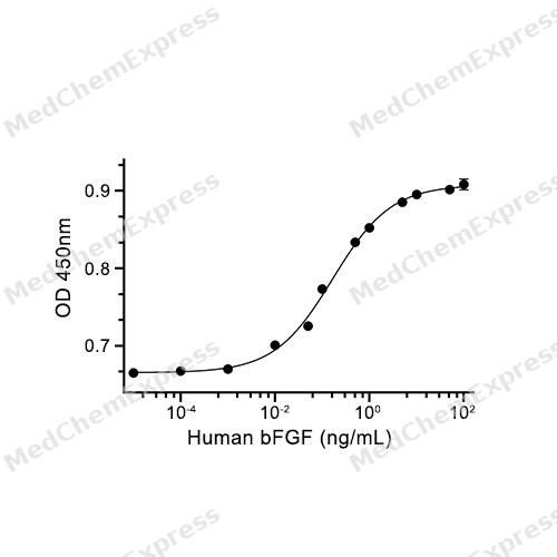
Bioactivity - Cell-Based Assay


Publications (4)
-
Journal Impact Factor
-
Most Recent
-
Adv Sci (Weinh)
Globotriaosylceramide Gb3 Influences Wound Healing and Scar Formation by Orchestrating Fibroblast Heterogeneity. [Abstract]2025 Aug 14:e09733. PMID: 40810720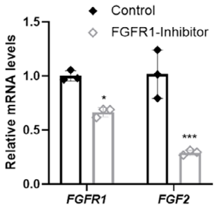
FGF basic/bFGF Protein, Human (145a.a) purchased from MedChemExpress. Usage Cited in: Adv Sci (Weinh). 2025 Aug 14:e09733. [Abstract]
After 24 h of serum‐free starvation culture, dHFBs were treated with FGFR1‐specific tyrosine kinase inhibitor FGF basic/bFGF Protein (FGF2, 1 nM, HY‐P7330) for 48 h. The expression of FGFR1 target gene mRNA was detected by qRT‐PCR.
-
Cell Rep Med
A human patient-derived organoid biobank to model tumor heterogeneity and therapeutic vulnerability for oral squamous cell carcinoma. [Abstract]2026 Feb 17;7(2):102622. PMID: 41707653 -
-


Technical Parameters
-
Species Human
-
Source E. coli
-
Tag Tag Free
-
Accession
P09038-4 (A144-S288)
-
Molecular Construction
-
N-term
-
bFGF (A144-S288)
Accession # P09038-4 -
C-term
-
-
Protein Length
Full Length of Fibroblast growth factor 2 Chain
-
Synonyms
FGF2; BFGF; Prev. FGFB; Basic Fibroblast Growth Factor; Fibroblast Growth Factor 2 (Basic); Fibroblast Growth Factor; Heparin-Binding Growth Factor 2; Prostatropin; HBGF-2; FGF2A; FGF-2; FGF; Fibroblast Growth Factor 2; Basic Fibroblast Growth Factor BFGF
-
AA Sequence
ALPEDGGSGAFPPGHFKDPKRLYCKNGGFFLRIHPDGRVDGVREKSDPHIKLQLQAEERGVVSIKGVCANRYLAMKEDGRLLASKCVTDECFFFERLESNNYNTYRSRKYTSWYVALKRTGQYKLGSKTGPGQKAILFLPMSAKS
-
Predicted Molecular Mass
16.3 kDa
-
Molecular Weight
Approximately 16 kDa, based on SDS-PAGE under reducing conditions.
-
Purity
≥ 95%, as determined by reducing SDS-PAGE.


Product Properties
Lyophilized powder
1.Lyophilized from a 0.22 μm filtered solution of PBS.
2.Lyophilized from a 0.22 μm filtered solution of PBS, pH 7.4.
3.Lyophilized from a 0.22 μm filtered solution of PBS, pH 7.4, 8% trehalose.
Please refer to the lot-specific COA for specific buffer information.
<0.2 EU/μg, determined by LAL method.
It is not recommended to reconstitute to a concentration less than 100 μg/mL in ddH2O. For long term storage it is recommended to add a carrier protein (0.1% BSA, 5% HSA, 10% FBS or 5% Trehalose).
Stored at -20°C for 2 years from date of receipt. After reconstitution, it is stable at 4°C for 1 week or -20°C for longer (with carrier protein). It is recommended to freeze aliquots at -20°C or -80°C for extended storage.
Room temperature in continental US; may vary elsewhere.


Documentation
-
Data Sheet (264 KB)
-
SDS (252 KB)
- English - EN (252 KB)
- Français - FR (252 KB)
- Deutsch - DE (252 KB)
- Norwegian - NO (252 KB)
- Español - ES (252 KB)
- Swedish - SV (252 KB)
- Italian - IT (252 KB)
- Korean - KR (252 KB)
- Portuguese - PT (252 KB)
-
Handling Instructions (2659 KB)


References
[1]. Zhang J, et al. FGF2: a key regulator augmenting tendon-to-bone healing and cartilage repair. Regen Med. 2020 Sep;15(9):2129-2142. [Content Brief]
[2]. Westermann R, et al. Basic fibroblast growth factor (bFGF), a multifunctional growth factor for neuroectodermal cells. J Cell Sci Suppl. 1990;13:97-117. [Content Brief]
[3]. Jimenez-Pascual A, et al. FGF2: a novel druggable target for glioblastoma? Expert Opin Ther Targets. 2020 Apr;24(4):311-318. [Content Brief]
[4]. Rusnati M, et al. Interaction of angiogenic basic fibroblast growth factor with endothelial cell heparan sulfate proteoglycans. Biological implications in neovascularization. Int J Clin Lab Res. 1996;26(1):15-23. [Content Brief]
[5]. Hankemeier S, et al. Modulation of proliferation and differentiation of human bone marrow stromal cells by fibroblast growth factor 2: potential implications for tissue engineering of tendons and ligaments. Tissue Eng. 2005 Jan-Feb;11(1-2):41-9. [Content Brief]

Calculators
Concentration (start) × Volume (start) = Concentration (final) × Volume (final)


 Powered by Bioz

