



IL-10R alpha Protein, Mouse (HEK293, Fc)

Based on 1 Customer Validation
IL-10R alpha protein is an IL10 cytokine surface receptor that is involved in IL10-mediated inflammation and immune regulation and inhibits the synthesis of pro-inflammatory cytokines. IL-10R alpha Protein, Mouse (HEK293, Fc) is expressed by HEK 293cells with a hFc tag at the C-terminus.
- Species: Mouse
- Source: HEK293
-
Storage:Stored at -80°C for 1 year from date of receipt. It is stable at -20°C for 3 months after opening. It is recommended to freeze aliquots at -80°C for extended storage. Avoid repeated freeze-thaw cycles.


To place orders, for customer services and technical support, please contact: MedChemExpress USA
Tel: 609-228-6898 E-mail: [email protected] [email protected]


-
 Biological Activity
-
Technical Parameters
-
Product Properties
- Documentation
- References
- Help & FAQs




Biological Activity
Description
IL-10R alpha protein is an IL10 cytokine surface receptor that is involved in IL10-mediated inflammation and immune regulation and inhibits the synthesis of pro-inflammatory cytokines. IL-10R alpha Protein, Mouse (HEK293, Fc) is expressed by HEK 293cells with a hFc tag at the C-terminus[1].
Background
IL-10R alpha is a ligand-binding subunit of the type II cytokine receptor consisting of 2 alpha and 2 beta subunits and is expressed primarily in hematopoietic cells such as B cells, T cells, NK cells, monocytes and macrophages, but not in non-hematopoietic cells such as fibroblasts or endothelial cells[1].
IL-10R alpha binds to the ligand and leads to a conformational change in the beta subunit, which results in the beta subunit also binding IL-10, forming a heterotetramer that leads to activation of the signalling complex of JAK1 and TYK2 kinases. In this case, JAK1 binds to the alpha subunit and TYK2 binds to the beta subunit, phosphorylating specific tyrosine residues in the intracellular structural domain of IL10R alpha. This further leads to phosphorylation and activation of the transcription factor STAT3, which dimerises STAT3 monomers into the nucleus and induces transcriptional expression of the target gene[1].
IL-10R alpha is involved in suppressing inflammatory responses and Th 1 cell-mediated immune responses, and also regulates neutrophil functional responses. In addition, IL10R alpha-mediated activation of STAT3 also inhibits starvation-induced autophagy[2].
In Vitro
IL-1R alpha expression levels is low in naive (CD45RBhi), memory/effector (Treg cell-depleted CD45RBlo), and nTreg CD4 T cells, but IL-1Rα expression levels can be upregulated by in vitro stimulation with anti-CD3 and CD28 antibodies[3].
In Vivo
IL1R alpha expression is low in naive CD4+ T cells but is efficiently induced in small intestinal Th17 cells of anti-CD3-treated mice[4].
IL-1R alpha can be induced by IFN-γ in a mouse model of colitis DSS, while deletion of IL-1R alpha in an epithelial-specific IL-1R alpha knockout (KO) mouse model leads to increased colitis, suggesting an important role for epithelial IL-1R alpha in mucosal integrity and possibly barrier function[5].
MCE Validation Data
-
Purity - SDS-PAGE
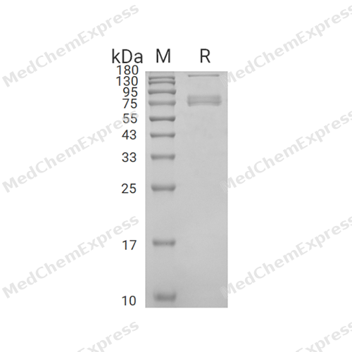
Purity - SDS-PAGE


Technical Parameters
-
Species Mouse
-
Source HEK293
-
Tag C-hFc
-
Accession
Q61727/NP_032374.1 (L17-N238)
-
Molecular Construction
-
N-term
-
IL-10Rα (L17-N238)
Accession # Q61727/NP_032374.1 -
hFc
-
C-term
-
-
Protein Length
Extracellular Domain
-
Synonyms
IL10RA; Interleukin 10 Receptor, Alpha; Prev. IL10R; IL-10R Subunit Alpha; Interleukin-10 Receptor Subunit Alpha; IL-10R Subunit 1; Interleukin-10 Receptor Subunit 1; IL-10R1; HIL-10R; CDw210a; CDW210A; Interleukin-10 Receptor Alpha Chain; CD210a; CD210 A
-
AA Sequence
LEFIAYGTELPSPSYVWFEARFFQHILHWKPIPNQSESTYYEVALKQYGNSTWNDIHICRKAQALSCDLTTFTLDLYHRSYGYRARVRAVDNSQYSNWTTTETRFTVDEVILTVDSVTLKAMDGIIYGTIHPPRPTITPAGDEYEQVFKDLRVYKISIRKFSELKNATKRVKQETFTLTVPIGVRKFCVKVLPRLESRINKAEWSEEQCLLITTEQYFTVTN
-
Predicted Molecular Mass
52.7 kDa
-
Glycosylation
Yes
-
Purity
≥ 95%, as determined by reducing SDS-PAGE.


Product Properties
Solution
Supplied as a 0.22 μm filtered solution of PBS, pH 7.4.
<1 EU/μg, determined by LAL method.
Stored at -80°C for 1 year from date of receipt. It is stable at -20°C for 3 months after opening. It is recommended to freeze aliquots at -80°C for extended storage. Avoid repeated freeze-thaw cycles.
Shipping with dry ice.


Documentation
-
Data Sheet (264 KB)
-
SDS (251 KB)
- English - EN (251 KB)
- Français - FR (251 KB)
- Deutsch - DE (251 KB)
- Norwegian - NO (251 KB)
- Español - ES (251 KB)
- Swedish - SV (251 KB)
- Italian - IT (251 KB)
- Korean - KR (251 KB)
- Portuguese - PT (251 KB)
-
Handling Instructions (2659 KB)


References
[1]. Dror S Shouval, et al. Interleukin 10 receptor signaling: master regulator of intestinal mucosal homeostasis in mice and humans. Adv Immunol. 2014;122:177-210. [Content Brief]
[2]. J Shi, et al. IL10 inhibits starvation-induced autophagy in hypertrophic scar fibroblasts via cross talk between the IL10-IL10R-STAT3 and IL10-AKT-mTOR pathways. Cell Death Dis. 2016 Mar 10;7(3):e2133. [Content Brief]
[3]. Masahito Kamanaka, et al. Memory/effector (CD45RB(lo)) CD4 T cells are controlled directly by IL-10 and cause IL-22-dependent intestinal pathology. J Exp Med. 2011 May 9;208(5):1027-40. [Content Brief]
[4]. Samuel Huber, et al. Th17 cells express interleukin-10 receptor and are controlled by Foxp3⁻ and Foxp3+ regulatory CD4+ T cells in an interleukin-10-dependent manner. Immunity. 2011 Apr 22;34(4):554-65. [Content Brief]
[5]. Douglas J Kominsky, et al. IFN-γ-mediated induction of an apical IL-10 receptor on polarized intestinal epithelia. J Immunol. 2014 Feb 1;192(3):1267-76. [Content Brief]

Calculators
Concentration (start) × Volume (start) = Concentration (final) × Volume (final)


 Powered by Bioz

