



MIP-3 beta/CCL19 Protein, Mouse
Based on 1 Customer Validation
MIP-3 beta/CCL19 Protein, Mouse is a CC chemokine that is strongly chemotactic for CD4 T cells and CD8 T cells and acts as a ligand that binds specifically to the chemokine receptor CCR7 to mediate tissue immunity, inflammatory responses, and antiviral infections. MIP-3 beta/CCL19 Protein, Mouse is a recombinant mouse MIP-3 beta/CCL19 (G26-S108) protein expressed by E. coli.
- Species: Mouse
- Source: E. coli
-
Storage:Stored at -20°C for 2 years from date of receipt. After reconstitution, it is stable at 4°C for 1 week or -20°C for longer (with carrier protein). It is recommended to freeze aliquots at -20°C or -80°C for extended storage.


To place orders, for customer services and technical support, please contact: MedChemExpress USA
Tel: 609-228-6898 E-mail: [email protected] [email protected]


-
 Biological Activity
-
Technical Parameters
-
Product Properties
- Documentation
- References
- Help & FAQs


-

HY-P72756
6Ckine/CCL21A Protein, Mouse -
HY-P7063
IL-6 Protein, Mouse -
HY-P72782
SDF-1 beta/CXCL12 Protein, Mouse -
HY-P7044
IL-6 Protein, Human -
HY-P7167
6Ckine/CCL21B Protein, Mouse -
HY-P7285
SDF-1 alpha/CXCL12 Protein, Mouse -
HY-P7227
IP-10/CRG-2/CXCL10 Protein, Mouse -
HY-133073
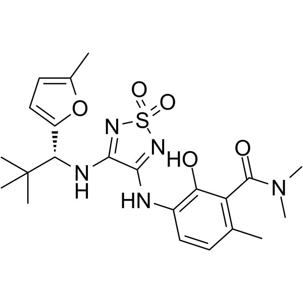
CCR7 Ligand 1 -
HY-P77911
BCA-1/CXCL13 Protein, Mouse (HEK293, His) -
HY-P76651
SPARCL1 Protein, Mouse (HEK293, His) -
HY-P72966
Cystatin C/CST3 Protein, Mouse (HEK293, His) -
HY-P702526
MIP-3 beta/CCL19 Protein, Human


Biological Activity
Description
MIP-3 beta/CCL19 Protein, Mouse is a CC chemokine that is strongly chemotactic for CD4 T cells and CD8 T cells and acts as a ligand that binds specifically to the chemokine receptor CCR7 to mediate tissue immunity, inflammatory responses, and antiviral infections. MIP-3 beta/CCL19 Protein, Mouse is a recombinant mouse MIP-3 beta/CCL19 (G26-S108) protein expressed by E. coli[1][2].
Background
CCL19, also known as MIP-3 beta, is an immunostable chemokine that is located on chromosome 9 in the human genome. CCL19 is abundantly expressed in the thymus and lymph nodes, moderately expressed in the trachea and colon, and less expressed in the stomach, small intestine, lung, kidney, and spleen. CCL19 binds to and functions as a chemokine receptor, CCR7. CCR7 is the first CCR7 is the first lymphocyte-specific G protein-coupled receptor (GPCR) identified with seven transmembrane alpha helices. CCR7 is expressed on both double-negative and single-positive thymocytes, including naive T cells, central memory T cells, regulatory T cells, naive B cells, semimature/mature DCs and NK cells, as well as a few tumor cells, where it serves as a key regulator for directing steady-state lymphocytes to secondary lymphoid organs. In contrast, CCL19 is the only chemokine known to effectively stimulate β-arrestin-mediated phosphorylation and internalization of CCR7, leading to receptor desensitization and migration of antigen (Ag)-presenting DCs, thereby affecting T cell responses. The CCL19-CCR7-based signaling pathway plays an important role in tissue immunity and inflammatory response memory. In addition, it also plays a role in vaccine-based protection against a variety of viruses, such as HIV-1 infection, hepatitis C virus (HCV), and herpes simplex virus 1 (HSV-1), etc. The interaction of CCL19 and CCR7 also contributes to the release of antiviral-associated cytokines (e.g., IFN-γ and IL-4) by immune cells, thereby promoting T cell proliferation and DC uptake of antigens[1][2].
In Vitro
CCL19 (1 μg/mL, 6 h) induces T cell proliferation in immature mouse dendritic cells (DC)-T cell co-culture systems, induces upregulation of costimulatory molecules and cytokine production in DCs, and maturation of DC to induce the Th1 response in DC isolated from spleens and mesenteric lymph nodes of naïve BALB/c mice[3].
In Vivo
CCL19 (5 mg/mouse by osmotic pump) significantly increases IL-10 production in plt (plt) mice,and after treatment with 100 mg acetylcholine, the number of lymphocytes at bronchoalveolar lavage fluid (BALF) and enhanced pause (Penh) levels is significantly lower in CCL19-treated mice than in untreated mice[2].
CCL19 can lead to rapid clearance of intrahepatic HBV likely through increased intrahepatic CD8+ T-cell proportion, decreased frequency of PD-1+ CD8+ T cells in blood and compromised suppression of hepatic APCs, with lymphocytes producing a significantly high level of Ag-responsive TNF-α and IFN-γ from CD8+ T cells in C57BL/6 mice transfected with the mouse CCL19-encoding plasmid[4].
Verified Bioactivity
1.Full biological activity determined by a chemotaxis bioassay using human mature dendritic cells is in a concentration range of 10-100 ng/mL.
2.Biological activity was determined by its ability to chemoattract Jurkat human T cells using a concentration of 10-50 ng/mL.
3.Measured by its ability to chemoattract BaF3 mouse pro-B cells transfected with human CCR7. The ED50 for this effect is 3-15 ng/mL.
MCE Validation Data
-
Purity - SDS-PAGE
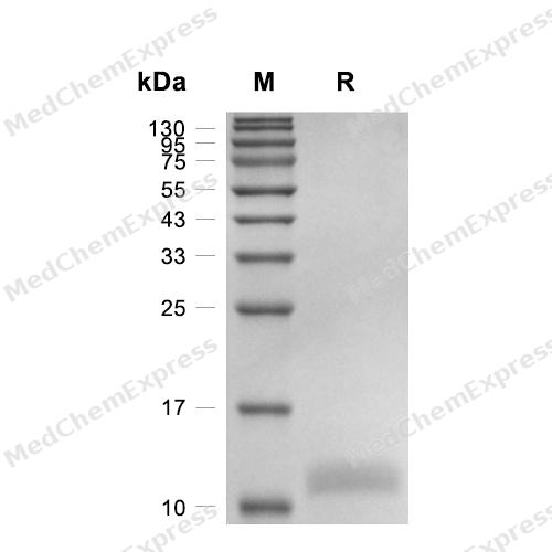
Purity - SDS-PAGE
-
Bioactivity - Cell-Based Assay
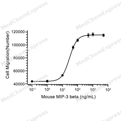
Bioactivity - Cell-Based Assay


Technical Parameters
-
Species Mouse
-
Source E. coli
-
Tag Tag Free
-
Accession
O70460 (G26-S108)
-
Molecular Construction
-
N-term
-
CCL19 (G26-S108)
Accession # O70460 -
C-term
-
-
Protein Length
Full Length of Mature Protein
-
Synonyms
CCL19; Small-Inducible Cytokine A19; Prev. SCYA19; CC Chemokine Ligand 19; ELC; C-C Motif Chemokine 19; CK Beta-11; EBI1-Ligand Chemokine; Exodus-3; MIP-3-Beta; MIP-3b; MIP3B; CKb11; Macrophage Inflammatory Protein 3 Beta; Small Inducible Cytokine Subfamily A (Cys-Cys), Member 19; Beta-Chemokine Exodus-3; Epstein-Barr Virus-Induced Molecule 1 Ligand Chemokine; EBI1 Ligand Chemokine; Macrophage Inflammatory Protein 3-Beta; C-C Motif Chemokine; Chemokine (C-C Motif) Ligand 19; C-C Motif Chemokine Ligand 19; Beta Chemokine Exodus-3
-
AA Sequence
GANDAEDCCLSVTQRPIPGNIVKAFRYLLNEDGCRVPAVVFTTLRGYQLCAPPDQPWVDRIIRRLKKSSAKNKGNSTRRSPVS
-
Predicted Molecular Mass
9.2 kDa
-
Molecular Weight
Approximately 12 kDa, based on SDS-PAGE under reducing conditions.
-
Purity
≥ 95%, as determined by reducing SDS-PAGE.


Product Properties
Lyophilized powder
1.Lyophilized from a 0.22 μm filtered solution of 0.1% TFA, pH 2.5.
2.Lyophilized from a 0.22 μm filtered solution of PBS, pH 7.4.
Please refer to the lot-specific COA for specific buffer information.
<1 EU/μg, determined by LAL method.
It is not recommended to reconstitute to a concentration less than 100 μg/mL in ddH2O. For long term storage it is recommended to add a carrier protein (0.1% BSA, 5% HSA, 10% FBS or 5% Trehalose).
Stored at -20°C for 2 years from date of receipt. After reconstitution, it is stable at 4°C for 1 week or -20°C for longer (with carrier protein). It is recommended to freeze aliquots at -20°C or -80°C for extended storage.
Room temperature in continental US; may vary elsewhere.


Documentation
-
Data Sheet (264 KB)
-
SDS (252 KB)
- English - EN (252 KB)
- Français - FR (252 KB)
- Deutsch - DE (252 KB)
- Norwegian - NO (252 KB)
- Español - ES (252 KB)
- Swedish - SV (252 KB)
- Italian - IT (252 KB)
- Korean - KR (252 KB)
- Portuguese - PT (252 KB)
-
Handling Instructions (2659 KB)


References
[1]. Yan Yan, et al. CCL19 and CCR7 Expression, Signaling Pathways, and Adjuvant Functions in Viral Infection and Prevention. Front Cell Dev Biol. 2019 Oct 1;7:212. [Content Brief]
[2]. Zhang X, et al. Increased CCL19 expression is associated with progression in cervical cancer. Oncotarget. 2017 May 18;8(43):73817-73825. [Content Brief]
[3]. Naomi Yamashita, et al. Role of CCL21 and CCL19 in allergic inflammation in the ovalbumin-specific murine asthmatic model. J Allergy Clin Immunol. 2006 May;117(5):1040-6 [Content Brief]
[4]. Benjamin J Marsland, et al. CCL19 and CCL21 induce a potent proinflammatory differentiation program in licensed dendritic cells. Immunity. 2005 Apr;22(4):493-505. [Content Brief]
[5]. Yan Yan, et al. CCL19 enhances CD8+ T-cell responses and accelerates HBV clearance. J Gastroenterol. 2021 Aug;56(8):769-785. [Content Brief]

Calculators
Concentration (start) × Volume (start) = Concentration (final) × Volume (final)


 Powered by Bioz

