



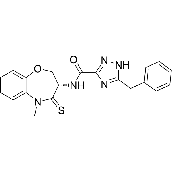
RIPK1-IN-16
RIPK1-IN-16 is an orally active and potent inhibitor of RIPK1. RIPK1-IN-16 inhibits excessive inflammation by blocking RIPK1-mediated necroptosis in vivo. RIPK1-IN-16 protects mouse from TNF-induced systemic inflammatory response syndrome and sepsis.
Nur für Forschungszwecke. Wir verkaufen nicht an Patienten.
- CAS. Nr.: 2561431-77-2
- Formel: C20H19N5O2S
- Molecular Weight:393.46
-
Speicherung:
Please store the product under the recommended conditions in the Certificate of Analysis.

To place orders, for customer services and technical support, please contact: MedChemExpress USA
Tel: 609-228-6898 E-mail: [email protected] [email protected]


-
 Biologische Aktivität
-
Chemical Information
- Protokoll
- Reinheit & Dokumentation
- Verweise
-
 Help & FAQs




Biologische Aktivität
Beschreibung
Cellular Effect
|
Cell Line

|
Type | Value | Description | References |
|---|---|---|---|---|
| HT-29 | CC50 |
>100 μM
Compound: 1
|
Cytotoxicity against human HT-29 cells assessed as reduction in cell viability measured for 16 to 24 hrs by Cell-titer-glo assay
Cytotoxicity against human HT-29 cells assessed as reduction in cell viability measured for 16 to 24 hrs by Cell-titer-glo assay
|
[PMID: 36724216] |
| HT-29 | CC50 |
>50 μM
Compound: 11
|
Cytotoxicity against human HT-29 cells assessed as reduction in cell viability measured after 8 to 10 hrs by celltiter-glo luminescent cell viability assay
Cytotoxicity against human HT-29 cells assessed as reduction in cell viability measured after 8 to 10 hrs by celltiter-glo luminescent cell viability assay
|
[PMID: 33930803] |
| HT-29 | CC50 |
>50 μM
Compound: 1
|
Cytotoxicity against human HT-29 cells assessed as reduction in cell viability
Cytotoxicity against human HT-29 cells assessed as reduction in cell viability
|
[PMID: 35398729] |
| HT-29 | EC50 |
0.0028 μM
Compound: 11
|
Anti-necroptotic activity in human HT-29 cells assessed as inhibition of hTNFalpha/Z-VAD-fmk (TSZ)-induced necroptosis by measuring increase in cell viability measured after 8 to 10 hrs by celltiter-glo luminescent cell viability assay
Anti-necroptotic activity in human HT-29 cells assessed as inhibition of hTNFalpha/Z-VAD-fmk (TSZ)-induced necroptosis by measuring increase in cell viability measured after 8 to 10 hrs by celltiter-glo luminescent cell viability assay
|
[PMID: 33930803] |
| HT-29 | EC50 |
2.8 nM
Compound: 1
|
Anti-necroptosis activity against human HT-29 cells assessed as inhibition of TSZ-induced necroptosis preincubated for 30 mins followed by compound addition and measured for 16 hrs by Cell-titer-glo assay
Anti-necroptosis activity against human HT-29 cells assessed as inhibition of TSZ-induced necroptosis preincubated for 30 mins followed by compound addition and measured for 16 hrs by Cell-titer-glo assay
|
[PMID: 36724216] |


Chemical Information
-
CAS. Nr. 2561431-77-2
-
Molecular Weight 393.46
-
Formel C20H19N5O2S
-
SMILES
CN1C2=CC=CC=C2OC[C@H](NC(C3=NNC(CC4=CC=CC=C4)=N3)=O)C1=S
-
Versand
Room temperature in continental US; may vary elsewhere.
-
Speicherung
Please store the product under the recommended conditions in the Certificate of Analysis.


Reinheit & Dokumentation
Verweise

Calculators
Konzentration (Stammlösung) × Volumen (Stammlösung) = Konzentration (Ziellösung) × Volumen (Ziellösung)


RIPK1-IN-16 Related Classifications
HY-156367 Related Classifications
 Powered by Bioz