



SC13
Based on 1 publication(s) in Google Scholar
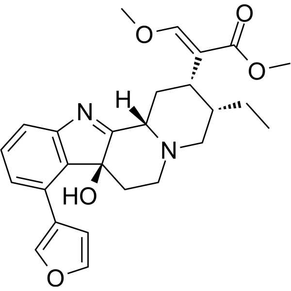
SC13 is an orally active, selective Flap structure-specific endonuclease 1 (FEN1) inhibitor and mu opioid receptor (MOR) activator. SC13 impairs DNA damage repair and induces apoptosis in cancer cells. SC13 activates cGAS-STING signaling, increases chemokine secretion, and promotes CAR-T cell infiltration at solid tumour sites. SC13 can be used for the research of solid tumours and pain.
For research use only. We do not sell to patients.
- CAS No.: 2839142-69-5
- Formula: C26H30N2O5
- Molecular Weight:450.53
-
Storage:
Please store the product under the recommended conditions in the Certificate of Analysis.

To place orders, for customer services and technical support, please contact: MedChemExpress USA
Tel: 609-228-6898 E-mail: [email protected] [email protected]


-
 Biological Activity
-
Chemical Information
- Protocols
- Purity & Documentation
- References
-
 Help & FAQs


Publications Citing Use of MedChemExpress (MCE) SC13
MoreAll Opioid Receptor Isoforms
More

Biological Activity
Description
IC50 & Target
[2]|
μ Opioid Receptor/MOR |
In Vitro
SC13 (20 μM; 48 h) induces DNA double-strand break accumulation and cytoplasmic dsDNA leakage in HeLa, MCF-7, and SKOV-3 human cancer cells[1].
SC13 (20 μM; 48 h) activates the cGAS-STING pathway and increases chemokine (CCL5, CXCL10) and IFN-β expression in HeLa, MCF-7, and SKOV-3 human cancer cells, dependent on STING signalling[1].
SC13 (20 μM; 48 h) induces HeLa, MCF-7, and SKOV-3 cells to secrete chemokines that promote MSLN CAR-T cell migration via a TBK1-dependent mechanism[1].
SC13 (20 μM; 48 h) induces mild DNA damage but does not impair proliferation, CAR expression, or cytotoxic function of MSLN CAR-T cells[1].
SC13 potently inhibits cAMP production in human mu-opioid receptor-expressing HEK-T cells with an EC50 of 7.25 nM and maximum efficacy[2].
SC13 recruits β-arrestin2 in human mu-opioid receptor-expressing HTLA cells with low potency (EC50 = 11000 nM) and moderate efficacy[2].
SC13 acts as a partial agonist for Gi-1 G-protein activation in human mu-opioid receptor-expressing cells, with high selectivity for MOR over DOR and KOR[2].
MedChemExpress (MCE) has not independently confirmed the accuracy of these methods. They are for reference only.
-
Cell Line:HeLa, MCF-7, SKOV-3 human cancer cell lines
-
Concentration:20 μM
-
Incubation Time:48 h
-
Result:Upregulated γ-H2AX (a marker of DNA double-strand breaks) in HeLa, MCF-7, and SKOV-3 cells.
Increased cytoplasmic dsDNA leakage in these cell lines.
-
Cell Line:HeLa, MCF-7, SKOV-3 human cancer cell lines
-
Concentration:20 μM
-
Incubation Time:48 h
-
Result:Activated the cGAS-STING pathway and increased chemokine (CCL5, CXCL10) and IFN-β expression, dependent on STING signalling.
-
Cell Line:MSLN CAR-T cells, HeLa, MCF-7, SKOV-3 human cancer cell lines
-
Concentration:20 μM
-
Incubation Time:48 h
-
Result:Significantly increased the number of migrating MSLN CAR-T cells compared to medium from vehicle-treated tumour cells; this effect was blocked by pre-treatment of tumour cells with the TBK1 inhibitor Amlexanox.
Increased secretion of CCL5 and CXCL10 in the supernatant of SC13-treated tumour cells.
In Vivo
SC13 (1-15 mg/kg; s.c. or p.o.; single dose; 10 mg/kg) produces MOR-dependent antinociception in mice with an ED50 of 3.05 mg/kg s.c., and maintains efficacy via oral administration[2].
SC13 (15 mg/kg; s.c.; daily; 2 days) does not induce reward or dysphoria in Mus musculus[2].
SC13 (15 mg/kg; s.c.; single dose) does not inhibit gastrointestinal transit in mice[2].
SC13 (45 mg/kg; s.c.; single dose) does not induce respiratory depression or hyperlocomotion in mice[2].
MedChemExpress (MCE) has not independently confirmed the accuracy of these methods. They are for reference only.
-
Animal Model:B-NDG (6 weeks old; injected 5 × 10⁶ HeLa cells subcutaneously into the mice)[1]
-
Dosage:5 mg/kg
-
Administration:i.p.; every other day for 32 days
-
Result:Reduced mean tumour weight to ~0.2 g compared to ~0.5 g in CAR-T alone group.
Increased intratumoural CD8+ T-cell infiltration to 18.65% of tumour-infiltrating cells compared to 6.95% in CAR-T alone group.
Increased CD8 signal intensity ~4-fold compared to CAR-T alone group.
Elevated CCL5 fluorescence intensity ~3-fold compared to CAR-T alone group.
Elevated CXCL10 fluorescence intensity ~2-fold compared to CAR-T alone group.
Caused no significant change in mouse body weight.
Showed no exacerbated organ damage to heart, liver, spleen, lung, or kidney.
-
Animal Model:C57BL/6J mice (20-32 g)[2]
-
Dosage:1 mg/kg; 3 mg/kg; 10 mg/kg; 15 mg/kg
-
Administration:s.c. or p.o.; single dose
-
Result:Produced dose-dependent antinociception.
Reached a maximum of ~30% MPE at 20 minutes post-administration with 1 mg/kg s.c..
Reached ~30% MPE at 20 minutes post-administration with 3 mg/kg s.c..
Reached 100% MPE at 20 minutes post-administration with 10 mg/kg s.c..
Achieved an ED50 of 3.05 mg/kg s.c. for antinociception.
Showed significantly reduced antinociception in MOR knockout mice, but remained intact in KOR knockout and DOR knockout mice.
Reached a peak of 87% MPE at 30 minutes post-administration with 10 mg/kg p.o..
-
Animal Model:C57BL/6J mice (20-32 g)[2]
-
Dosage:15 mg/kg
-
Administration:s.c.; daily; 2 days
-
Result:Produced no significant conditioned place preference or aversion.
Showed no statistically significant difference in time spent in the drug-paired compartment compared to pre-conditioning.
-
Animal Model:C57BL/6J mice (20-32 g)[2]
-
Dosage:15 mg/kg
-
Administration:s.c.; single dose
-
Result:Had no significant effect on gastrointestinal transit.
Resulted in charcoal traveling 30.8 cm, a value indistinguishable from saline-treated mice.
-
Animal Model:C57BL/6J mice (20-32 g)[2]
-
Dosage:45 mg/kg
-
Administration:s.c.; single dose
-
Result:Showed no statistically significant respiratory depression compared to vehicle.
Had breath rate comparable to vehicle/saline controls at all time points except 180 and 200 minutes, where it showed a slight increase.
Produced no significant hyperlocomotion.
Had ambulation distance indistinguishable from vehicle/saline controls at all time points.


Chemical Information
-
CAS No. 2839142-69-5
-
Molecular Weight 450.53
-
Formula C26H30N2O5
-
SMILES
O=C(OC)/C([C@@H](C[C@]12[H])[C@H](CC)CN1CC[C@@]3(O)C2=NC4=C3C(C5=COC=C5)=CC=C4)=C/OC
-
Shipping
Room temperature in continental US; may vary elsewhere.
-
Storage
Please store the product under the recommended conditions in the Certificate of Analysis.


Publications (1)
-
Journal Impact Factor
-
Most Recent
-
Nat Commun
2025 Mar 10;16(1):2401. PMID: 40064914


Purity & Documentation
References
[1]. Dong Y, et al. FEN1 inhibitor SC13 promotes CAR-T cells infiltration into solid tumours through cGAS-STING signalling pathway. Immunology. 2023;170(3):388-400. [Content Brief]
[2]. Chakraborty S, et al. A Novel Mitragynine Analog with Low-Efficacy Mu Opioid Receptor Agonism Displays Antinociception with Attenuated Adverse Effects. J Med Chem. 2021;64(18):13873-13892. [Content Brief]

Calculators
Concentration (start) × Volume (start) = Concentration (final) × Volume (final)


SC13 Related Classifications
HY-139678 Related Classifications
 Powered by Bioz