1316 Results for "

image

" in MedChemExpress (MCE) Product Catalog:
Products (1316)

1316 Results for "image" in MCE Product Catalog:

Cat. No.: HY-D0077
CAS No.: 195136-58-4
Synonyms: Oregon green 488
Target:  

Fluorescent Dye

Research Areas:  

Others

2',7'-Difluorofluorescein (Oregon green 488) is a fluorescein derivative and a pH-sensitive fluorescent probe (pKa ~4.7). Upon excitation at 488 nm, 2',7'-Difluorofluorescein exhibits pH-sensitive fluorescence intensity through the formation of dianions, while its pH sensitivity decreases under excitation at 450 nm, allowing its use in ratiometric pH analysis. 2',7'-Difluorofluorescein can be used for the quantitative analysis of pH values in the range of 2-7 in submicron aerosol particles. 2',7'-Difluorofluorescein undergoes buffer-mediated and buffer-free excited-state proton transfer between different protonated forms, and its cationic form undergoes rapid excited-state deprotonation. 2',7'-Difluorofluorescein is resistant to photodegradation, maintains stable absorption and fluorescence properties within the physiological pH range, and serves as a fluorescent protein label, a component of Ca 2+ indicators, a fluorescent imaging agent, and an anisotropy probe .
loading...
    loading...
Cat. No.: HY-D1850
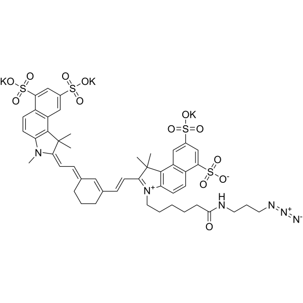
Sulfo-Cy7.5 azide is a Cyanine 7.5 (Cy7.5) (HY-D0926) dye derivative with azide and sulfonate functional groups. The sulfonate ion increases the water solubility of the compound, making it suitable for use in aqueous solutions. Cy7.5 is a near-infrared fluorescent dye commonly used for biolabeling and cell imaging. The azide group of Sulfo-Cy7.5 azide can react chemically with molecules containing alkyne functionality, such as alkyne or cyclooctyne, to form covalent bonds. Therefore, Sulfo-Cy7.5 azide can bind to biomolecules such as proteins and antibodies to track their location and dynamic changes in biological samples. It contains an azide group and can undergo copper-catalyzed azide-alkyne cycloaddition (CuAAc) with molecules containing alkyne groups. It can also undergo ring strain-promoted alkyne-azide cycloaddition (SPAAC) with molecules containing DBCO or BCN groups.
loading...
    loading...
Cat. No.: HY-D3074
Research Areas:  

Others

TCM-4COOH is an AIE-active fluorescent probe for calcium ions. TCM-4COOH relies on aggregation-induced emission activity, and its carboxyl chelating group can bind to Ca 2+ and Mg 2+ to form insoluble aggregates, triggering fluorescence enhancement via restricting intramolecular motion (Ex/Em = 477/603 nm). TCM-4COOH shows pH independence in physiological environments (pH 4-9), can be strongly activated at Ca 2+ concentrations ranging from low nM to mM, and has a stronger binding ability to Ca 2+ than to Mg 2+. TCM-4COOH, as its acetoxymethyl ester derivative TCM-AM, can penetrate cell membranes for intracellular detection. TCM-4COOH can be used for imaging of cellular calcium overload, detection of bone microcracks, and studies related to calcium-rich biological environments .
loading...
    loading...
Cat. No.: HY-D3088
Research Areas:  

Others

CQPP is a Fluorescent probe designed for monitoring polarity changes in the cellular microenvironment, including polarity tracking of lipid droplets/nuclei during ferroptosis. CQPP exhibits ratiometric fluorescence emission and fluorescence lifetime variations in response to polarity changes; a nonpolar environment stimulates fluorescence from the locally excited (LE) state, while a polar environment drives solvation relaxation to the intramolecular charge transfer (ICT) state, which attenuates the LE emission at ~470 nm and simultaneously enhances the ICT emission at ~670 nm. CQPP binds to intranuclear DNA through electrostatic interactions and hydrogen bonds in the DNA minor groove, which enhances its emission at ~670 nm and prolongs its fluorescence lifetime. CQPP can simultaneously target lipid droplets (green LE fluorescence) and the nucleus (red ICT fluorescence). The detection wavelengths of CQPP are Ex/Em = 405/470 nm and Ex/Em = 405/670 nm, and it also supports fluorescence lifetime imaging via 810 nm two-photon excitation .
loading...
    loading...
Cat. No.: HY-D3106
CAS No.: 2659241-66-2
Target:  

Fluorescent Dye

Research Areas:  

Others

Lys-VBOD is a Fluorescent probe for lysosomal viscosity monitoring and live cell imaging. Its detection mechanism depends on viscosity: increased viscosity restricts the twisting of the double bond between the indole and BODIPY fluorophore, as well as the rotation of the single bond between quinoline and BODIPY, which reduces non-radiative decay and leads to enhanced fluorescence intensity in high viscosity environments; it also features a morpholine group that provides lysosome-targeting properties, allowing it to localize to lysosomes for viscosity detection. The probe has excitation/emission wavelengths of Ex/Em = 602/637 nm, and its fluorescence intensity shows a good linear relationship with log-transformed viscosity values. Lys-VBOD is stable across a wide pH range of 5.35 to 10.74, is not affected by macromolecules and proteins like BSA, has a fluorescence lifetime that increases with viscosity, and can visualize real-time lysosomal viscosity changes in live cells under Dexamethasone (HY-14648) stimulation[1].
loading...
    loading...
Cat. No.: HY-Y1804R
CAS No.: 7274-88-6
D-Lysine monohydrochloride (Standard) is the analytical standard of D-Lysine monohydrochloride (HY-Y1804). This product is intended for research and analytical applications. D-Lysine monohydrochloride is the D-enantiomer of L-Lysine (HY-N0469). D-Lysine monohydrochloride is metabolically inert and not utilized for protein synthesis by mammalian ribosomes. D-Lysine monohydrochloride blocks renal uptake of 111In/ 90Y-Octreotide (HY-P0036)-based probes without inhibiting uptake by tumor/receptor tissues, and thus acts as a renoprotective agent in diagnostic imaging and peptide receptor radionuclide therapy (PRRT). D-Lysine monohydrochloride specifically inhibits the early steps of non-enzymatic glycation by competing with glucose via its free amino group, theoretically, it can serve as a glycation competitor that "does not interfere with protein synthesis" under chronic hyperglycemia in diabetes. D-Lysine monohydrochloride can be used in research related to cancer and diabetes .
loading...
    loading...
Cat. No.: HY-D3120
CAS No.: 2246946-53-0
Target:  

Fluorescent Dye

Research Areas:  

Others

LysoAIE2 is a Fluorescent probe for lysosomal viscosity detection and live-cell imaging. Its detection mechanism relies on the aggregation-induced emission effect: it exhibits only weak fluorescence in non-viscous media; in viscous environments, restricted intramolecular motion inhibits non-radiative energy dissipation pathways, thereby significantly enhancing fluorescence intensity. It achieves specific targeting of lysosomes through the proton acceptor property of its indole ring structure, while its hydroxyl group endows it with excellent water solubility. This probe is basically unaffected by microenvironmental polarity and pH within the range of pH 4.0 to pH 8.0, which avoids interference from these factors in viscosity measurement. LysoAIE2 has an emission wavelength of 570 nm. It can be used to monitor lysosomal viscosity changes during processes such as Dexamethasone (HY-14648)-induced lysosomal migration and starvation-induced mitophagy in live cells; at concentrations up to 40 μM, cell viability remains above 80%, demonstrating excellent biocompatibility .
loading...
    loading...
Cat. No.: HY-D3187
CAS No.: 2484831-02-7
HMRef-αMan is a substrate-based green fluorescent probe (Ex/Em=465 nm/515 nm) targeting MAN2C1 (α-mannosidase). HMRef-αMan can be specifically cleaved by MAN2C1 to generate a highly fluorescent product, which thus gets activated to produce green fluorescence in malignant breast tissues, benign lesions and living cancer cells. The signal intensity of HMRef-αMan is directly correlated with MAN2C1 activity, and it can effectively detect tiny breast cancer lesions with a diameter of less than 1 mm. When used in combination with the red-emitting γ-glutamyl transpeptidase (GGT) probe gGlu-2OMe SiR600 (HY-D3188), HMRef-αMan enables precise optical differentiation of breast tissue types via a dual-color imaging strategy. HMRef-αMan has been widely used in the research of breast diseases such as breast cancer, fibroadenoma, phyllodes tumor and various types of papilloma .
loading...
    loading...
Cat. No.: HY-D3416
Synonyms: TAMRA-2,4-dinitroaniline
Target:  

Fluorescent Dye

Research Areas:  

Others

TMR-DN (TAMRA-2,4-dinitroaniline) is a fluorescent probe for RNA imaging that consists of a covalently coupled fluorophore (5-carboxy tetramethylrhodamine (TMR)) and quencher (dinitroaniline (DN)). The Kd value of TMR-DN with SRB-2 is 35 nM, and its Kd value with RhoBAST is approximately 30 nM. TMR-DN has a contact quenching function and forms a non-fluorescent intramolecular ground-state dimer in the free state; the excitation/emission wavelengths of the free probe are Ex/Em = 557/579 nm, those of the complex formed by binding to SRB-2 are Ex/Em = 561/587 nm, and those of the probe bound to the DNB aptamer are Ex/Em = 555/582 nm. TMR-DN reduces non-specific binding to genomic nucleic acids via negatively charged carboxyl-functionalized aromatic rings. Fluorescence dequenching occurs when TMR-DN specifically binds to SRB-2, RhoBAST or the DNB aptamer, among which the binding of RhoBAST requires Mg 2+ for proper folding .
loading...
    loading...
Cat. No.: HY-100138R
CAS No.: 173308-19-5
2-Aminoethyl-mono-amide-DOTA-tris(tBu ester) (Standard) is the analytical standard of 2-Aminoethyl-mono-amide-DOTA-tris(tBu ester) (HY-100138). This product is intended for research and analytical applications. 2-Aminoethyl-mono-amide-DOTA-tris(tBu ester) is a metal chelator precursor containing a DOTA macrocyclic structure. DOTA can form highly stable complexes with metal ions (such as 68Ga, 177Lu) through four nitrogen atoms and four carboxylic acid groups to mediate targeted delivery of radionuclides. The tert-butyl ester group (tBu ester) of 2-Aminoethyl-mono-amide-DOTA-tris(tBu ester) also protects the carboxylic acid group during synthesis, and forms a free carboxyl group after deprotection reaction for coupling with targeting molecules (such as antibodies, peptides). 2-Aminoethyl-mono-amide-DOTA-tris(tBu ester) may be combined with tumor pre-targeting systems through bioorthogonal reactions (such as reverse electron demand Diels-Alder reaction) to study radioactive imaging or therapy of tumor tissues, and is mainly used in tumor pre-targeting research in the field of nuclear medicine .
loading...
    loading...
Cat. No.: HY-D3084
Target:  

Fluorescent Dye

Research Areas:  

Cancer

C-TPA is a fluorescent probe used for hypochlorite detection and bioimaging of lipid droplet polarity in cancer cells. For hypochlorite detection, when its derivative C-TPA-S is exposed to hypochlorite, an oxidative desulfurization reaction converts the thiocarbonyl group of C-TPA-S to the carbonyl group of C-TPA, thus restoring bright fluorescence from the almost non-fluorescent C-TPA-S. For bioimaging of lipid droplet polarity, the intramolecular charge transfer process between its Triphenylamine (HY-W011998) donor group and Coumarin (HY-N0709) acceptor group endows C-TPA with solvatochromism-it exhibits stronger fluorescence in less polar environments (such as lipid droplets in cancer cells) and weaker fluorescence in more polar environments (such as lipid droplets in normal cells), enabling the differentiation of cancer cells and cancer tissues from normal cells and normal tissues. The excitation wavelength of C-TPA is 405 nm, and the emission wavelengths used for cell and tissue imaging cover 425-475 nm and 500-550 nm; in solvents, its emission peak shifts from 494 nm in non-polar toluene to 528 nm in polar water .
loading...
    loading...
Cat. No.: HY-D3094
CAS No.: 2687198-42-9
Target:  

Fluorescent Dye

Research Areas:  

Others

TPE-SQ5 is a ratiometric Fluorescent probe for hypochlorite (ClO -) detection with a limit of detection of 5.6 nM. TPE-SQ5 exhibits high selectivity and high sensitivity towards ClO -. TPE-SQ5 can be oxidized by ClO - to form an epoxide, which quenches its deep red fluorescence; subsequently, the epoxide decomposes into a hemicyanine and a sulfonic acid-substituted oxindole, producing blue-green fluorescence to achieve a ratiometric response. TPE-SQ5 can form nanoparticles with Pluronic F-127 via the nanoprecipitation method, and these nanoparticles possess cell permeability. TPE-SQ5 has unique Ex/Em spectra in different solvents and nanoparticle forms, with emission peaks at 643 nm (380 nm excitation, 95% H2O/THF), 631 nm (564 nm excitation, DMSO), 652 nm (536 nm excitation, THF) and 630 nm (524 nm excitation, nanoparticles); the Ex/Em settings used for cell imaging are 488/550-650 nm (red channel) and 405/450-550 nm (green channel) .
loading...
    loading...
Cat. No.: HY-L105S
867 compounds

Peptides, composed of amino acids, serve as crucial building blocks for proteins and have gained significant attention in drug development over the past decade. The advancements in production, modification, and analytical technologies have led to a surge in the potential applications of peptides in medicine. Peptides offer a number of advantages over small molecule drugs, including: greater target specificity and efficacy, more predictable metabolic profiles, easier delivery to where they are needed in the body, and fewer side effects. Peptides are increasingly appearing in all branches of medicine as components of innovative drugs, imaging agents, diagnostic agents, and other complex drugs such as peptide-drug conjugates. To date, more than 80 peptide drugs have been approved to treat a variety of diseases, including microbial infections, obesity, anti-diabetes, and cancer, as well as to develop cell targeting platforms and improve cell penetration properties.

MCE designs a unique collection of 867 peptide compounds. HY-L105S is a peptide compound library that can be provided with solution form based on HY-L105, and can be applied to peptides-based drug development.

Cat. No.: HY-D2465
Target:  

Fluorescent Dye

Research Areas:  

Infection

CY5-Dextran (MW60-80k) is a fluorescent probe formed by covalent conjugation of the cyanine dye Cy5 to dextran. It is a pH-insensitive fluid-phase endocytosis tracer (the pH of endosomal lumen is precisely quantified by the fluorescence ratio of FITC/Cy5). CY5-Dextran (MW60-80k) is internalized by cells via a clathrin-independent fluid-phase pinocytosis pathway, and is sequentially evidenced by early endosomes, late endosomes are transported to lysosomes. CY5-D I cannot pass through the limited pore size formed by uncoating of human rhinovirus type 2 (HRV2), but can be released into the neutral cytoplasm upon adenovirus-mediated endosomal rupture, thus distinguishing between the two viral modes of "pore-forming escape" and "lysis escape". CY5-Dextran (MW60) -80k) has an excitation wavelength of 635 nm, and it can also be used for time-lapse imaging of endocytosis dynamics of stromal cells in tissues such as rat salivary glands under in vivo two-photon microscopy .
loading...
    loading...
Cat. No.: HY-W594061
CAS No.: 1811541-14-6
Target:  

Fluorescent Dye

Research Areas:  

Others

JF635-HTL is a Fluorescent dye for spatiotemporally controlled live cell imaging and single-molecule localization microscopy SMLM. Its detection mechanism depends on the conformational change of a photoswitchable HaloTag psHaloTag, which integrates the light-responsive AsLOV2 domain: in the dark state, the folded Jα helix of AsLOV2 maintains the dye in a predominantly closed, non-fluorescent form; upon 450 nm illumination, a metastable photo-adduct forms between a cysteine side chain and the FMN cofactor of AsLOV2, causing undocking and unfolding of the Jα helix, which propagates a conformational change to the HaloTag near the dye binding site, shifting the dye's equilibrium to the open, fluorescent form; this process is fully reversible in the dark as the Jα helix refolds spontaneously, returning the dye to the closed, non-fluorescent state. For psHaloTag1a labeled with JF635-HTL, the excitation/emission wavelengths for the ON state are Ex/Em = 642/655 nm, while for psHaloTag1b labeled with JF635-HTL, the wavelengths are Ex/Em = 639/655 nm; when bound to wild-type HaloTag, the wavelengths are Ex/Em = 640/656 nm .
loading...
    loading...
Cat. No.: HY-D3105
Target:  

Fluorescent Dye

Research Areas:  

Others

DCA is a Fluorescent probe for visualization of phase behavior in ER membranes. DCA is an ER-targeting, polarity-responsive NIR ratiometric probe, with its p-toluenesulfonamide group responsible for ER localization; its sensitivity to polarity relies on its donor-π-acceptor (D-π-A) structure, where aniline acts as the donor and dicyanomethylene acts as the acceptor, driving an intramolecular charge transfer (ICT) process upon excitation. In environments with low polarity, such as the closely packed, low water content ERₒ phase of ER membranes, DCA emits at a shorter wavelength, while in high polarity environments like the loosely packed, higher water content ERd phase, ICT leads to a red-shifted emission, allowing discrimination of the two phases via dual NIR emission colors and ratiometric imaging. Ex/Em = 488/570–620 nm and 488/665–735 nm; additional excitation/emission pairs include Ex/Em = 488/631 nm in low polarity 1,4-dioxane and Ex/Em = 488/677 nm in 1,4-dioxane with 30% water, the higher polarity condition. It shows a large Stokes shift of ~170 nm, and pH, viscosity, and biologically relevant species including Cys, GSH, H₂O₂, and metal ions do not exert marked interference on its fluorescence spectra[1].
loading...
    loading...

