16 Results for "

lipid droplet imaging

" in MedChemExpress (MCE) Product Catalog:
Products (16)

16 Results for "lipid droplet imaging" in MCE Product Catalog:

Cat. No.: HY-D1581
CAS No.: 878674-84-1
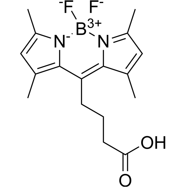
BODIPY 505/515-8-C3-COOH is a green fluorescing derivative, as a fluorescent dye for imaging lipid droplets in nannochloropsis. BODIPY 505/515-8-C3-COOH can be used for the research of flow cytometric high-throughput screening and cell sorting .
loading...
    loading...
Cat. No.: HY-152073
CAS No.: 2924598-24-1
Purity:  98.76%
Target:  

Fluorescent Dye

Research Areas:  

Others

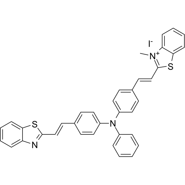
BETA-1 is the first twisted intramolecular charge transfer (TICT)-aggregation-induced emission (AIE) integration molecule. BETA-1 emits cyan fluorescence in lipid droplets (LDs) and red fluorescence in mitochondria. BETA-1 can be used for the simultaneous and dual-color imaging of LDs and mitochondria in vivo and in vitro .
loading...
    loading...
Cat. No.: HY-139337
CAS No.: 2417507-09-4
Purity:  ≥95.0%
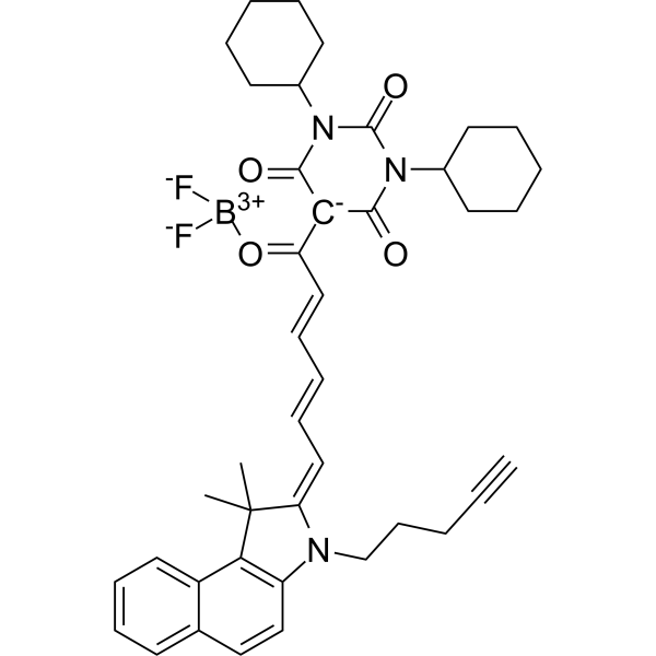
SMCy5.5 is a SMCy dye (ex. 638 nm; em. 705 nm). The high brightness of SMCy5.5 allows for effective tracking and imaging of lipid droplet exchange between adipocytes. SMCy5.5 can be used for lipid droplet labeling studies .
loading...
    loading...
Cat. No.: HY-134096
CAS No.: 78323-98-5
Synonyms: DNS-M
Target:  

Fluorescent Dye

Research Areas:  

Others

Dansyl-morpholine (DNS-M) is a Fluorescent probe for lipid droplet imaging, cancer cell discrimination, and real-time tracking of lipid droplet dynamics. As a solvatochromic probe with a donor-π-acceptor structure, it relies on hydrophobic interaction for its mechanism of action: its good lipophilicity, confirmed by an oil-water partition coefficient LogP = 2.35, allows it to rapidly penetrate cell membranes, and it specifically localizes to the hydrophobic core of lipid droplets; its fluorescence is strongly enhanced in the nonpolar environment of lipid droplets, while it emits very weak fluorescence in polar environments like PBS buffer, and it exhibits a bathochromic shift in emission wavelength with increasing solvent polarity. It has negligible cytotoxicity, with cell viability remaining over 95% after 24-hour incubation with 100 μM of the probe, and it possesses excellent photostability, retaining over 97% of initial fluorescence intensity after 60 continuous laser scans. For cell imaging applications, its excitation/emission wavelengths for lipid droplet labeling are Ex/Em = 405/480−540 nm, and in a simulative lipid environment O/W emulsion, it has an excitation wavelength of ~346 nm and emission wavelength of ~500 nm, giving a Stokes shift of 154 nm[1].
loading...
    loading...
Cat. No.: HY-D3368
Target:  

Fluorescent Dye

Research Areas:  

Inflammation/Immunology

DMA-SiRd is a fluorescent probe that can be used for crosstalk-free dual-channel imaging of lipid droplets and mitochondria, mapping of lipid microenvironment heterogeneity, and imaging of lipid droplet-mitochondria interactions. DMA-SiRd has two imaging channels: Ex/Em = 633/650-750 nm and Ex/Em = 546/570-620 nm, with two-photon excitation wavelengths of 1040 nm and 1200 nm for brain imaging. DMA-SiRd can be applied to the research of atherosclerosis .
loading...
    loading...
Cat. No.: HY-D3075
CAS No.: 2490504-23-7
Target:  

Fluorescent Dye

Research Areas:  

Others

TPE-2E N-oxide is a fluorescent probe used for hypoxia detection and tumor hypoxia imaging. Due to its active intramolecular motion, TPE-2E N-oxide shows no fluorescence in aqueous solution. In hypoxic environments, its N-oxide group undergoes two-electron reduction by CYP450 reductase and other heme protein reductases expressed under hypoxia, forming hydrophobic aggregates that trigger aggregation-induced emission through the restriction of intramolecular motion. TPE-2E N-oxide exhibits selective lipid droplet localization in cells. The absorption wavelength of TPE-2E N-oxide in dichloromethane is 313 nm; in a 99% hexane/dichloromethane mixed system, its excitation wavelength is 330 nm and emission wavelength is 460 nm. For in vitro cell imaging, excitation is performed at 405 nm, with an emission wavelength range of 430-560 nm .
loading...
    loading...
Cat. No.: HY-D3071
CAS No.: 1510814-96-6
Target:  

Fluorescent Dye

Research Areas:  

Inflammation/Immunology

LW-1 is a fluorescent probe targeting polarity detection of lipid droplets (LD). LW-1 produces a strong solvatochromic effect via the intramolecular charge transfer (ICT) mechanism: as environmental polarity increases, the emission wavelength red-shifts and the fluorescence intensity decreases significantly (emission at 557 nm in toluene and 642 nm in acetonitrile, with an 85 nm red-shift); fluorescence enhances in low-polarity lipid droplets. LW-1 can be used for lipid droplet polarity detection, differentiation between cancer cells and normal cells, monitoring of lipid droplet polarity changes under different physiological conditions such as starvation, oleic acid treatment and oxidative stress, as well as ex vivo imaging of polarity changes in lung tissues of asthmatic mice. The detection wavelength for cell imaging with LW-1 is Ex/Em = 405/500-550 nm (green channel) .
loading...
    loading...
Cat. No.: HY-D3090
CAS No.: 2757682-04-3
Target:  

Fluorescent Dye

Research Areas:  

Others

NRLD is a lipid droplet-targeting solvatochromic fluorescent probe developed based on Nile Red (HY-D0718), which can target lipid droplets and sense the non-polar lipid microenvironment inside lipid droplets. NRLD exhibits superior lipid droplet-targeting selectivity compared to the parent Nile Red dye in HeLa cell imaging. NRLD achieves detection relying on the solvatochromic effect generated by excited-state charge transfer, and its emission wavelength shifts according to the local polarity changes of the microenvironment it locates in: the more compact and ordered the lipid packing and the lower the environmental polarity, the more blue-shifted the emission; the looser the lipid packing or the stronger the hydration and the higher the environmental polarity, the more red-shifted the emission. NRLD shows blue shift in the non-polar oil core of lipid droplets and red shift in high-polarity environments such as phosphate buffer .
loading...
    loading...
Cat. No.: HY-D3085
CAS No.: 2882924-23-2
SSR-LDs is a fluorescent probe for lipid droplet polarity detection and lipid droplet imaging. SSR-LDs exhibits polarity-dependent fluorescence: it produces intense short-wavelength emission in low-polarity environments, while generating weak long-wavelength emission in high-polarity environments. SSR-LDs enters cells via free diffusion, specifically targets lipid droplets, and is unaffected by intracellular viscosity or pH values ranging from 4.5 to 9.0. SSR-LDs shows variable Ex/Em wavelengths in different solvents, including 590/663 nm in THF, 600/660 nm in mouse tissue and in vivo imaging, and an excitation wavelength of 580 nm with an emission wavelength range of 610-750 nm in cell imaging. SSR-LDs can be used for research related to fatty liver, liver injury and hepatitis .
loading...
    loading...
Cat. No.: HY-D3068
CAS No.: 3077446-86-4
Target:  

Fluorescent Dye

Research Areas:  

Metabolic Disease

LDs-BCA is a BF2-chelated AIE fluorescent probe with a positive solvatochromic effect, which can specifically target intracellular lipid droplets (Ex/Em = 565/620-730 nm). LDs-BCA monitors the dynamic changes in the morphology, size and quantity of intracellular lipid droplets under oleic acid induction and starvation stress. LDs-BCA, after intraperitoneal injection, accumulates in the gastric fat of living obese mice, allowing continuous monitoring via its fluorescent signal. LDs-BCA is applicable to studies related to intracellular lipid droplet polarity imaging and in vivo gastric fat visualization .
loading...
    loading...
Cat. No.: HY-D3007
LD-CK is a fluorescent probe based on a coumarin-chromone structure, specifically designed to visualize lipid droplet polarity changes and endowed with multimodal imaging capability. LD-CK contains a high-performance lipophilic coumarin moiety, which enables it to specifically target lipid droplets upon cellular entry while minimizing background fluorescence to the greatest extent. LD-CK has an excitation wavelength (Ex) of 488 nm and an emission wavelength (Em) of 540 nm (in low-polarity environments such as toluene) or 640 nm (in high-polarity environments such as glycerol), with its emission peak undergoing a redshift as solvent polarity increases .
loading...
    loading...
Cat. No.: HY-D3084
Target:  

Fluorescent Dye

Research Areas:  

Cancer

C-TPA is a fluorescent probe used for hypochlorite detection and bioimaging of lipid droplet polarity in cancer cells. For hypochlorite detection, when its derivative C-TPA-S is exposed to hypochlorite, an oxidative desulfurization reaction converts the thiocarbonyl group of C-TPA-S to the carbonyl group of C-TPA, thus restoring bright fluorescence from the almost non-fluorescent C-TPA-S. For bioimaging of lipid droplet polarity, the intramolecular charge transfer process between its Triphenylamine (HY-W011998) donor group and Coumarin (HY-N0709) acceptor group endows C-TPA with solvatochromism-it exhibits stronger fluorescence in less polar environments (such as lipid droplets in cancer cells) and weaker fluorescence in more polar environments (such as lipid droplets in normal cells), enabling the differentiation of cancer cells and cancer tissues from normal cells and normal tissues. The excitation wavelength of C-TPA is 405 nm, and the emission wavelengths used for cell and tissue imaging cover 425-475 nm and 500-550 nm; in solvents, its emission peak shifts from 494 nm in non-polar toluene to 528 nm in polar water .
loading...
    loading...
Cat. No.: HY-D3073
CAS No.: 2477607-64-8
Target:  

Fluorescent Dye

Research Areas:  

Others

DXB-NIR is a push-pull dioxaboron fluorescent probe with solvatochromic properties, featuring high fluorescence brightness, photostability, and a large two-photon absorption cross-section. DXB-NIR exhibits Ex/Em of 550/620–780 nm (with two-photon excitation at 930 nm). DXB-NIR quantifies local polarity via the intensity ratio I (>640)/I (<640) between the far-red channel (<640 nm) and the near-infrared channel (>640 nm), and it can simultaneously label the plasma membrane, endoplasmic reticulum, and lipid droplets in live cells. DXB-NIR monitors the increase in local polarity of various cell compartments under conditions of cholesterol depletion, starvation, and oxidative stress. DXB-NIR can be used for polarity imaging of biological membranes and lipid droplets, as well as studies related to cellular stress .
loading...
    loading...
Cat. No.: HY-D3088
Research Areas:  

Others

CQPP is a Fluorescent probe designed for monitoring polarity changes in the cellular microenvironment, including polarity tracking of lipid droplets/nuclei during ferroptosis. CQPP exhibits ratiometric fluorescence emission and fluorescence lifetime variations in response to polarity changes; a nonpolar environment stimulates fluorescence from the locally excited (LE) state, while a polar environment drives solvation relaxation to the intramolecular charge transfer (ICT) state, which attenuates the LE emission at ~470 nm and simultaneously enhances the ICT emission at ~670 nm. CQPP binds to intranuclear DNA through electrostatic interactions and hydrogen bonds in the DNA minor groove, which enhances its emission at ~670 nm and prolongs its fluorescence lifetime. CQPP can simultaneously target lipid droplets (green LE fluorescence) and the nucleus (red ICT fluorescence). The detection wavelengths of CQPP are Ex/Em = 405/470 nm and Ex/Em = 405/670 nm, and it also supports fluorescence lifetime imaging via 810 nm two-photon excitation .
loading...
    loading...
Cat. No.: HY-D3101
Target:  

Fluorescent Dye

Research Areas:  

Others

Nile-TPA is a fluorescent probe for polarity detection, which can be used for lipid droplet targeting, cell polarity analysis, in vivo polarity imaging, and blood polarity monitoring in circulating tumor mouse models. The detection mechanism of Nile-TPA relies on the dual intramolecular charge transfer (ICT) effect generated by the electron-donating triphenylamine and Nile Red moieties and the electron-withdrawing carbonyl group; this probe emits strong red fluorescence in a low-polarity environment, and as polarity increases, the fluorescence intensity decreases and a red shift occurs due to changes in the excited-state dipole moment. In spectral tests, its excitation wavelength is 540 nm, and the emission wavelength varies in the range of 550-800 nm depending on solvent polarity; for cell, zebrafish and mouse imaging, Ex/Em = 561/570-620 nm, and for in vivo mouse imaging, Ex/Em = 560/620 nm .
loading...
    loading...
Cat. No.: HY-D3113
Target:  

Fluorescent Dye

Research Areas:  

Cancer

BF6 is a blood-brain barrier permeable NIR probe. BF6 can diffuse from intracranial blood vessels into brain tissue, enabling visualization of brain structures and differentiation between glioblastoma (GBM) and normal tissues. The absorption peaks of BF6 in DMSO and FBS range from 727 to 859 nm, with a broad fluorescence emission band spanning 800 to 1400 nm; its emission wavelength falls within the NIR-II region (800-1400 nm) .
loading...
    loading...
  • 1

