AD-8007
Based on 1 Customer Validation
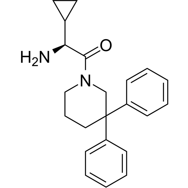
AD-8007 is a blood-brain barrier-permeable ACSS2 inhibitor with human IC50 of 0.89 μM and Kd of 116.6 μM. AD-8007 reduces colony-forming ability, lipid storage levels and FASN protein expression, and induces cancer cell death. AD-8007 shrinks established tumors, acts synergistically with radiotherapy to inhibit tumor growth, reduces tumor burden, causes no obvious toxicity to normal brain tissue, and prolongs survival. AD-8007 exhibits superior metabolic stability. AD-8007 is applicable to research related to breast cancer brain metastasis.
For research use only. We do not sell to patients.
- Purity : 99.36%
- CAS No.: 1497439-74-3
- Formula: C22H26N2O
- Molecular Weight:334.45
-
Storage:Powder -20°C, 3 years ; In solvent -80°C, 6 months , -20°C, 1 month
Biological Activity
Description
IC50 & Target
[1]|
ACSS2 0.89 μM (IC50) |
ACSS2 116.6 μM (Kd) |
In Vitro
AD-8007 (0.6-150 μM; 60 min) potently and specifically inhibits human ACSS2 (but not hACSS1), with an IC50 of 0.89 μM for its ATPase inhibitory activity[1].
AD-8007 exhibits high metabolic stability in human liver microsomes, and shows moderate blood-brain barrier permeability without inhibiting P-glycoprotein in MDCK cell-based assays[1].
AD-8007 (100 μM; 48 h) reduces the clonogenic survival rate of MDA-MB-231BR and 4T1BR breast cancer brain metastasis cells and induces their cell death in vitro[1].
AD-8007 (20 μM; 6 days) inhibits the growth of established MDA-MB-231BR tumors in an ex vivo brain slice model without affecting the viability of normal brain tissue; its combination with 6 Gy radiotherapy synergistically blocks tumor growth[1].
AD-8007 (100 μM; 24-48 h) reduces acetyl-CoA levels, lipid droplet content, and FASN protein levels in MDA-MB-231BR and 4T1BR breast cancer brain metastatic cells in vitro[1].
MedChemExpress (MCE) has not independently confirmed the accuracy of these methods. They are for reference only. Further protocols information, click here.
In Vivo
AD-8007 (50 mg/kg; i.p.; daily; 14 days) reduces breast cancer brain metastasis tumor burden, decreases tumor proliferation, and extends survival in BalbC mice without significant weight loss[1].
AD-8007 (50 mg/kg; i.p.; single dose) penetrates the blood-brain barrier in BalbC mice, with significantly higher brain-to-blood levels than control compound VY-3-135[1].
MedChemExpress (MCE) has not independently confirmed the accuracy of these methods. They are for reference only.
-
Animal Model:Nu/Nu (female, 4-6 weeks old, intracranial injection of luciferase-tagged MDA-MB-231BR breast cancer brain tropic cells)[1]
-
Dosage:50 mg/kg
-
Administration:i.p.; daily; 14 days
-
Result:Significantly reduced relative bioluminescence signal (tumor burden) compared to vehicle control.
Showed reduced tumor size and lower Ki-67 staining in brain sections compared to vehicle control.
Significantly extended mouse survival relative to vehicle control.
Caused no significant weight loss during treatment.
-
Animal Model:BalbC (female, 4-6 weeks old, intracranial injection of luciferase-tagged 4T1BR breast cancer brain tropic cells)[1]
-
Dosage:50 mg/kg
-
Administration:i.p.; daily
-
Result:Significantly reduced tumor burden compared to vehicle control.
Decreased Ki-67 staining in tumors compared to vehicle control.
Extended mouse survival compared to vehicle control.
Caused no significant weight loss compared to vehicle control.
Chemical Information
-
CAS No. 1497439-74-3
-
Appearance Solid
-
Molecular Weight 334.45
-
Formula C22H26N2O
-
Color White to off-white
-
SMILES
O=C([C@@H](N)C1CC1)N2CC(C3=CC=CC=C3)(C4=CC=CC=C4)CCC2
-
Shipping
Room temperature in continental US; may vary elsewhere.
-
Storage
Powder -20°C 3 years In solvent -80°C 6 months -20°C 1 month
Solvent & Solubility
In Vitro:
DMSO : 16.67 mg/mL (49.84 mM; ultrasonic and warming and heat to 60°C; Hygroscopic DMSO has a significant impact on the solubility of product, please use newly opened DMSO)
Please refer to the solubility information to select the appropriate solvent. Once prepared, please aliquot and store the solution to prevent product inactivation from repeated freeze-thaw cycles.
Storage method and period of stock solution: -80°C, 6 months; -20°C, 1 month. When stored at -80°C, please use it within 6 months. When stored at -20°C, please use it within 1 month.
Please refer to the solubility information to select the appropriate solvent. Once prepared, please aliquot and store the solution to prevent product inactivation from repeated freeze-thaw cycles.
Storage method and period of stock solution: -80°C, 6 months; -20°C, 1 month. When stored at -80°C, please use it within 6 months. When stored at -20°C, please use it within 1 month.
Concentration (start) × Volume (start) = Concentration (final) × Volume (final)
Protocols
-
RNA extraction experimental
By lysing cells, releasing RNA, and removing impurities such as proteins and DNA, high-purity RNA products are finally obtained. The commonly used traditional method is the guanidine isothiocyanate/phenol/chloroform method (Trizol), which is suitable for a variety of animal materials including animal tissues, microorganisms, cultured cells, etc., and most plant materials.
-
Western Blot
Western blotting (WB) is a commonly used experimental method in molecular biology, biochemistry, and immunogenetics for identifying and quantifying target proteins. It combines gel electrophoresis with immunoassay, enabling researchers to analyze protein expression, post-translational modifications, and molecular weight.
-
Cell Cytotoxicity Assay
Cytotoxicity assays are usually based on the assessment of cell membrane damage, which can also be indirectly detected by measuring cell viability. Detection methods include MTT assay, CKK-8 assay, LDH assay and ATP assay, etc.
-
Mammalian live/dead viability and cytotoxicity staining
Live/dead viability and cytotoxicity staining assays are based on the simultaneous detection of intracellular esterase activity in metabolically active (viable) cells and membrane integrity loss in non-viable cells. In commonly used dual-staining approaches, membrane-permeant fluorogenic substrates are converted by intracellular esterases into fluorescent products in live cells, while impermeant DNA-binding dyes selectively enter cells with compromised plasma membranes and label nucleic acids in dead or dying cells, enabling discrimination between viable and non-viable populations by fluorescence microscopy or flow cytometry.
-
Patient-Derived Xenograft (PDX)
Patient-derived xenograft (PDX) models are generated by engrafting primary human tumor tissue directly into immunodeficient mice, allowing in vivo propagation of patient tumor biology without initial in vitro adaptation. These models are used to preserve key histopathological and molecular characteristics of the original tumor and enable assessment of tumor growth dynamics and therapeutic response in a living organism. The biological readout is tumor engraftment and subsequent growth in the murine host, which reflects the ability of human tumor cells to survive, vascularize, and expand in an immunocompromised microenvironment.
-
Apoptosis Solutions
Apoptosis is a regulated, generally non-lytic cell-death pathway that removes unwanted, damaged, infected, or abnormal cells through coordinated morphological changes, caspase activation, DNA fragmentation, and membrane remodeling. The intrinsic apoptosis pathway is controlled mainly by mitochondrial outer membrane permeabilization, BCL-2 family proteins, cytochrome c release, apoptosome formation, caspase-9 activation, and downstream executioner caspase-3/7 activation. The extrinsic apoptosis pathway is initiated by death receptors such as Fas, TNFR, and TRAIL receptors, which recruit adaptor proteins and activate caspase-8 before engaging executioner caspases or mitochondrial amplification through BID cleavage. Apoptosis is linked to many phenotypes, including cancer cell killing, tissue homeostasis, immune regulation, neurodegeneration, infection response, and treatment-induced cytotoxicity; unresolved questions include how apoptosis interacts with necroptosis, pyroptosis, ferroptos
-
Patient-Derived Orthotopic Xenograft (PDOX)
Patient-derived orthotopic xenograft (PDOX) modeling implants fresh patient tumor tissue or patient-derived tumor cells into the anatomically corresponding organ or tissue site of immunodeficient mice, usually by surgical orthotopic implantation, to preserve patient tumor histology, local microenvironmental context, invasion, metastatic behavior, and treatment-response features better than subcutaneous implantation. PDOX readouts include tumor engraftment, orthotopic tumor growth, local invasion, metastasis, recurrence after resection, histologic similarity to the donor tumor, biomarker retention, molecular concordance, survival, and response or resistance to therapy. PDOX models are used for preclinical drug testing and individualized therapy evaluation, but engraftment success varies by tumor type and specimen quality.
-
Colony Formation (Clonogenic) Assay
The clonogenic (colony formation) assay measures the ability of a single cell to retain reproductive viability and form a macroscopic colony, typically defined as a cluster derived from one progenitor cell after a defined growth period. This assay is widely used to evaluate cell survival after exposure to ionizing radiation or cytotoxic treatments and is considered a standard method in radiation biology for generating dose-response relationships of reproductive cell death. Colony formation reflects long-term proliferative capacity rather than short-term metabolic activity, and survival is quantified by comparing treated versus untreated conditions based on colony number and derived survival fractions.
-
Subcutaneous Cell-Line-Derived Xenograft
Subcutaneous cell-line-derived xenograft (CDX) models are established by implanting cultured human cancer cell lines into immunodeficient mice, where the injected cells form localized tumors that can be monitored in vivo as a measure of tumorigenic potential, growth kinetics, and treatment response. These models are widely used in oncology research because they allow reproducible tumor formation and enable comparative assessment of tumor growth between different cell lines or genetic manipulations in a controlled in vivo microenvironment. Subcutaneous implantation of cancer cells in immunodeficient mice is a standard approach for evaluating tumor growth behavior and therapeutic response across multiple cancer types, including prostate, esophageal, pancreatic, and colon cancer models.
-
Breast Cancer Modeling
Breast cancer is a heterogeneous cancer, and it has been distinguished into four subtypes: luminal A, luminal B, HER2-positive and basal-like. Molecular mutations, epigenetic alterations, hormone exposure and immune microenvironment are related to the progression of breast cancer.
-
Orthotopic Cell-Line Xenograft
Orthotopic cell-line xenograft models involve implantation of human cancer cell lines into the anatomically corresponding organ of immunodeficient mice to reproduce tumor growth within a native microenvironment, enabling more clinically relevant tumor behavior compared with subcutaneous models. These models are widely used because orthotopic placement better recapitulates tumor progression, including invasion and metastatic spread, which are often underrepresented in heterotopic implantation systems. Compared with conventional xenografts, orthotopic implantation is described as more technically complex but provides improved simulation of tumor-microenvironment interactions and metastatic behavior, making it particularly valuable for translational oncology research. Surgical orthotopic implantation approaches have been emphasized as enabling faithful reproduction of clinical cancer features, including metastasis and disease progression patterns that align with the tumor’s organ of origi
-
Protocol For Protein Expression And Purification
Recombinant protein expression in Escherichia coli followed by purification of a His-tagged soluble protein by immobilized metal affinity chromatography (IMAC), with optional MBP fusion and TEV tag removal when the construct includes these elements. The biological readout is production of the encoded target protein, detected as an inducible band at the expected molecular mass by SDS-PAGE and quantified by total protein assay or chromatographic absorbance; the purification readout is enrichment of the target protein in elution fractions after selective binding of polyhistidine residues to immobilized Ni2+/metal-chelate resin and elution by imidazole-containing buffer. Expression is driven by an inducible bacterial expression system, commonly T7/lac-based, in which IPTG or lactose/auto-induction activates transcription and translation of the cloned gene; lower induction temperature, lower inducer concentration, induction timing, and solubility-enhancing fusion tags can influence the frac
Purity & Documentation
-
Data Sheet (287 KB)
-
SDS (251 KB)
- English - EN (251 KB)
- Français - FR (251 KB)
- Deutsch - DE (251 KB)
- Norwegian - NO (251 KB)
- Español - ES (251 KB)
- Swedish - SV (251 KB)
- Italian - IT (251 KB)
- Korean - KR (251 KB)
- Portuguese - PT (251 KB)
-
Handling Instructions (2659 KB)
References
Complete Stock Solution Preparation Table
Please refer to the solubility information to select the appropriate solvent. Once prepared, please aliquot and store the solution to prevent product inactivation from repeated freeze-thaw cycles.
Storage method and period of stock solution: -80°C, 6 months; -20°C, 1 month. When stored at -80°C, please use it within 6 months. When stored at -20°C, please use it within 1 month.
| Optional Solvent | Concentration Solvent Mass | 1 mg | 5 mg | 10 mg | 25 mg |
|---|---|---|---|---|---|
| DMSO | 1 mM | 2.9900 mL | 14.9499 mL | 29.8998 mL | 74.7496 mL |
| 5 mM | 0.5980 mL | 2.9900 mL | 5.9800 mL | 14.9499 mL | |
| 10 mM | 0.2990 mL | 1.4950 mL | 2.9900 mL | 7.4750 mL | |
| 15 mM | 0.1993 mL | 0.9967 mL | 1.9933 mL | 4.9833 mL | |
| 20 mM | 0.1495 mL | 0.7475 mL | 1.4950 mL | 3.7375 mL | |
| 25 mM | 0.1196 mL | 0.5980 mL | 1.1960 mL | 2.9900 mL | |
| 30 mM | 0.0997 mL | 0.4983 mL | 0.9967 mL | 2.4917 mL | |
| 40 mM | 0.0747 mL | 0.3737 mL | 0.7475 mL | 1.8687 mL |