



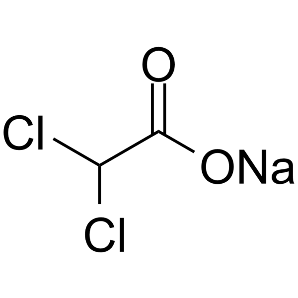
Sodium dichloroacetate
Based on 31 publication(s) in Google Scholar
Sodium dichloroacetate is an orally active pyruvate dehydrogenase kinase (PDK) inhibitor. Sodium dichloroacetate also stimulates pyruvate dehydrogenase (PDH) activity and works as a Na+-K+-2Cl− cotransporter (NKCC) inhibitor. Sodium dichloroacetate prevents the phosphorylation of the E1α subunit of PDC, promoting the entry of pyruvate into the mitochondria for oxidative metabolism, reducing lactate production, and simultaneously increasing the production of reactive oxygen species (ROS). Sodium dichloroacetate inhibits tumor cell proliferation and induces apoptosis. Sodium dichloroacetate is promising for research of cancers.
For research use only. We do not sell to patients.
- Purity : 99.78%
- CAS No.: 2156-56-1
- Formula: C2HCl2NaO2
- Molecular Weight:150.92
-
Storage:
4°C, sealed storage, away from moisture
* In solvent : -80°C, 6 months; -20°C, 1 month (sealed storage, away from moisture)

To place orders, for customer services and technical support, please contact: MedChemExpress USA
Tel: 609-228-6898 E-mail: [email protected] [email protected]


-
 Biological Activity
-
Chemical Information
-
Solvent & Solubility
- Protocols
- Purity & Documentation
- References
-
 Help & FAQs


-
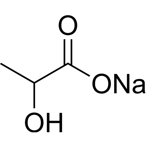
HY-B2227B
Lactic acid sodium (60% w/w in water)Glycolysis Production -
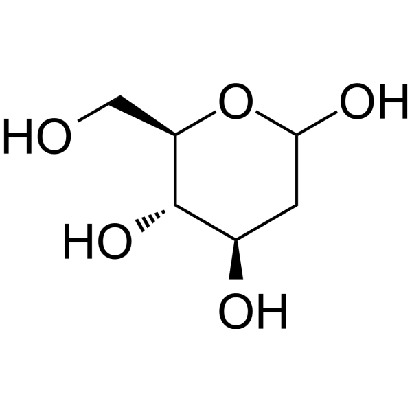
HY-13966
2-Deoxy-D-glucoseGlycolysis Inhibitor -
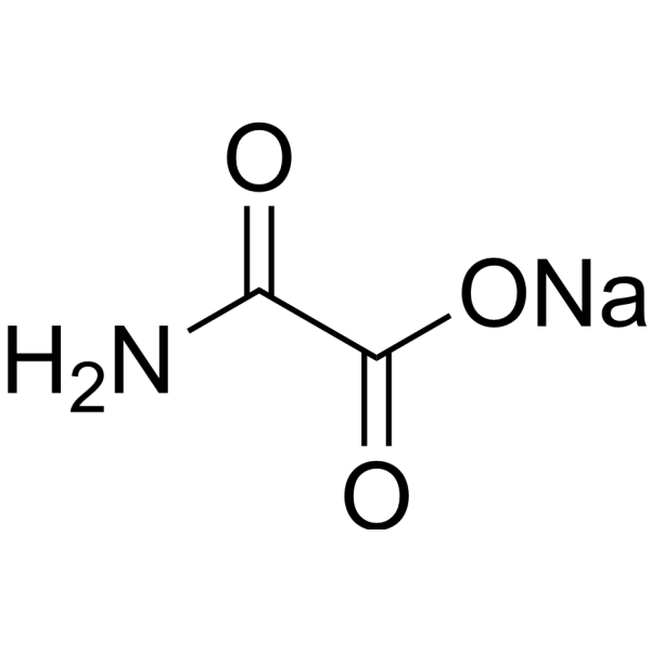
HY-W013032A
Oxamic acid sodiumLDH-A Inhibitor -
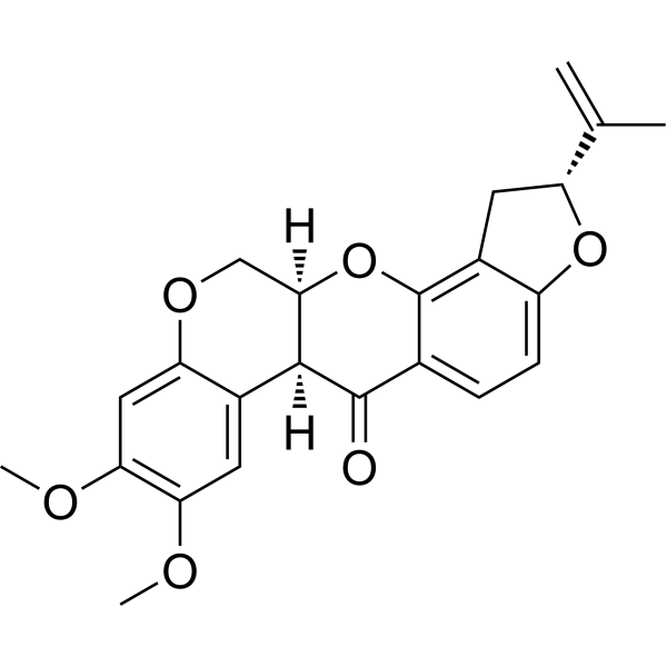
HY-B1756
RotenoneMitochondrial Complex I Inhibitor -
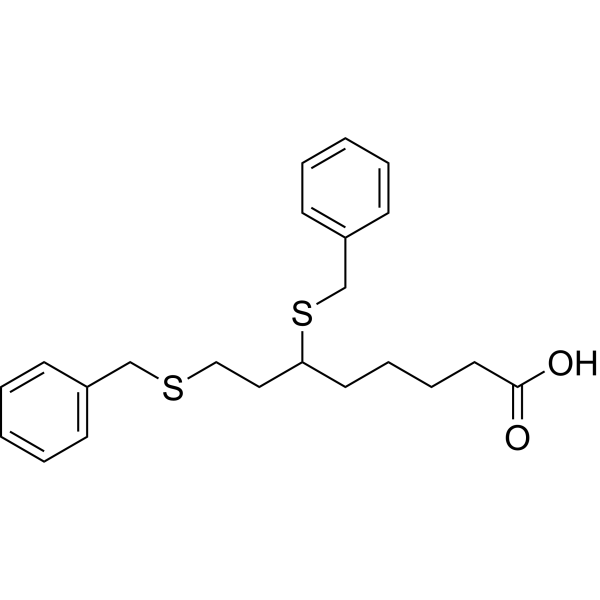
HY-15453
DevimistatMitochondrial Metabolism Inhibitor -
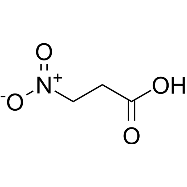
HY-W012875
3-Nitropropanoic acidSuccinate Dehydrogenase Inhibitor -
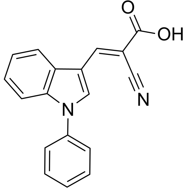
HY-15475
UK-5099Mitochondrial Pyruvate Carrier Inhibitor -

HY-B0215
AcetylcysteineMucolytic Agent -
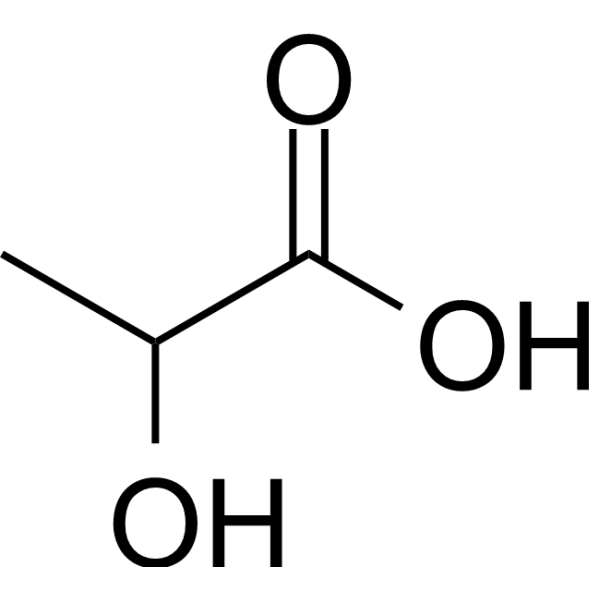
HY-B2227
Lactic acidGlycolysis Production -
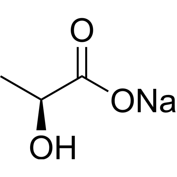
HY-W040233
L-Lactic acid sodiumBuildiing Block -
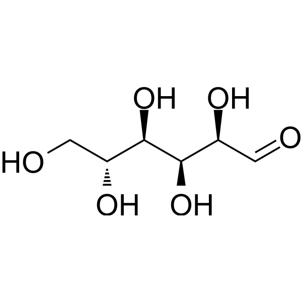
HY-B0389
D-GlucoseMonosaccharide -
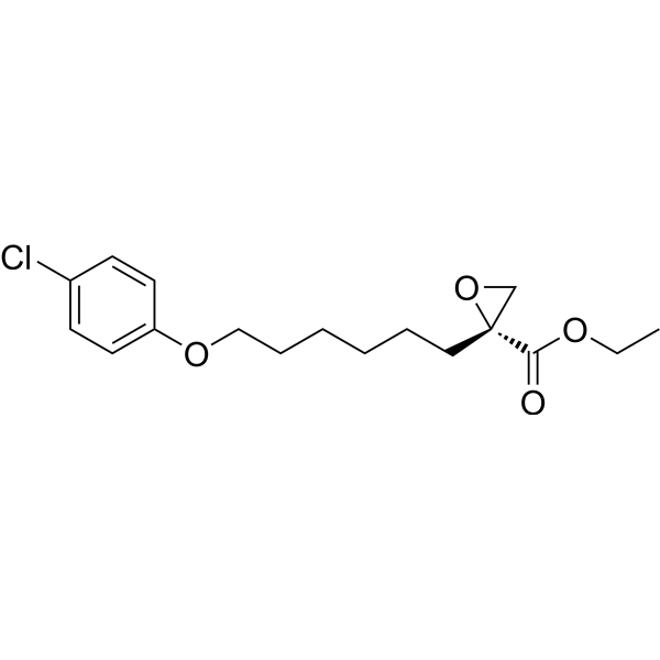
HY-50202
EtomoxirCPT1a Inhibitor
Publications Citing Use of MedChemExpress (MCE) Sodium dichloroacetate
More- Signal Transduct Target Ther. 2022 Sep 1;7(1):303. [Abstract]
- Redox Biol. 2025 Apr:81:103553. [Abstract]
- Adv Sci (Weinh). 2025 Jul 29:e05436. [Abstract]
- Adv Sci (Weinh). 2025 May;12(20):e2413709. [Abstract]
- Cell Death Differ. 2025 Mar;32(3):530-545. [Abstract]
- Cell Death Dis. 2023 Nov 7;14(11):722. [Abstract]
- Cell Death Dis. 2021 Sep 6;12(9):837. [Abstract]
- Clin Cancer Res. 2026 Apr 17. [Abstract]
- Proc Natl Acad Sci U S A. 2025 Mar 11;122(10):e2404899122. [Abstract]
- Cancer Drug Resist. 2020 Sep 4;3(4):947-958. [Abstract]
- Fundam Res. 2023 Mar 6;4(4):820-828. [Abstract]
- J Autoimmun. 2025 May:153:103411. [Abstract]
- J Ethnopharmacol. 2024 Aug 10:330:118235. [Abstract]
- Cell Mol Life Sci. 2025 Oct 28;82(1):370. [Abstract]
- Biochem Pharmacol. 2024 Jul:225:116294. [Abstract]
- Life Sci. 2023 Jan 1:312:121192. [Abstract]
- Inflammation. 2026 Jan 16;49(1):49. [Abstract]
- Cancers (Basel). 2022 Oct 10;14(19):4966. [Abstract]
- Cell Signal. 2026 Jun 27:112701. [Abstract]
- Cell Signal. 2023 Sep:109:110747. [Abstract]
- J Trace Elem Med Biol. 2023 Jul:78:127147. [Abstract]
- J Biol Chem. 2022 Mar 4:101775. [Abstract]
- J Proteome Res. 2025 Nov 7. [Abstract]
- Exp Cell Res. 2021 Sep 1;406(1):112755. [Abstract]
- Mol Reprod Dev. 2021 Jun;88(6):405-415. [Abstract]
- Biochem Biophys Res Commun. 2026 Sep 10:830:154296. [Abstract]
- bioRxiv. 2026 Jun 12:2026.06.11.731694. [Abstract]
- bioRxiv. 2026 May 21.
- Jacobs J Environ Sci. 2025 Sep 12.
- Oxid Med Cell Longev. 2021 Jun 4:2021:6633419. [Abstract]
- Oxid Med Cell Longev. 2021 Jun 27;2021:5549047. [Abstract]
 Customer Validation & Images

-
IF
-
WB
-
In Vivo Efficacy Study
-
Cell Proliferation/Viability Assay
-
WB


Biological Activity
Description
IC50 & Target
PDHK; Reactive oxygen species (ROS); Apoptosis; NKCC[1]
Cellular Effect
|
Cell Line

|
Type | Value | Description | References |
|---|---|---|---|---|
| 4T1 | IC50 |
>100 μM
Compound: Na-DCA
|
Anticancer activity against mouse 4T1 cells expressing PDK assessed as reduction in cell viability after 72 hrs by MTT assay
Anticancer activity against mouse 4T1 cells expressing PDK assessed as reduction in cell viability after 72 hrs by MTT assay
|
[PMID: 31509699] |
| A-375 | IC50 |
>100 μM
Compound: Na-DCA
|
Anticancer activity against human A375 cells expressing PDK assessed as reduction in cell viability after 72 hrs by MTT assay
Anticancer activity against human A375 cells expressing PDK assessed as reduction in cell viability after 72 hrs by MTT assay
|
[PMID: 31509699] |
| A-375 | IC50 |
20.1 mM
Compound: DCA
|
Antiproliferative activity against human A375 cells assessed as growth inhibition after 72 hrs by MTT assay
Antiproliferative activity against human A375 cells assessed as growth inhibition after 72 hrs by MTT assay
|
[PMID: 33550182] |
| A549 | IC50 |
1011 μM
Compound: DCA
|
Anticancer activity against human A549 cells after 72 hrs by MTT assay
Anticancer activity against human A549 cells after 72 hrs by MTT assay
|
[PMID: 20663593] |
| A549 | IC50 |
19.5 mM
Compound: DCA
|
Antiproliferative activity against human A549 cells assessed as growth inhibition after 72 hrs by MTT assay
Antiproliferative activity against human A549 cells assessed as growth inhibition after 72 hrs by MTT assay
|
[PMID: 33550182] |
| A549 | IC50 |
20.1 mM
Compound: DCA
|
Antiproliferative activity against human A549 cells assessed as reduction in cell viability after 72 hrs by MTT assay
Antiproliferative activity against human A549 cells assessed as reduction in cell viability after 72 hrs by MTT assay
|
[PMID: 30470491] |
| A549 | IC50 |
23.58 mM
Compound: DCA
|
Cytotoxicity against human A549 cells assessed as growth inhibition after 72 hrs by MTT assay
Cytotoxicity against human A549 cells assessed as growth inhibition after 72 hrs by MTT assay
|
[PMID: 27006991] |
| A549 | IC50 |
4286 μM
Compound: DCA
|
Cytotoxicity against human A549 cells after 68 hrs by MTT assay
Cytotoxicity against human A549 cells after 68 hrs by MTT assay
|
[PMID: 23067553] |
| Bel-7402 | IC50 |
860 μM
Compound: DCA
|
Anticancer activity against human Bel7402 cells after 72 hrs by MTT assay
Anticancer activity against human Bel7402 cells after 72 hrs by MTT assay
|
[PMID: 20663593] |
| BGC-823 | IC50 |
1051 μM
Compound: DCA
|
Anticancer activity against human BGC823 cells after 72 hrs by MTT assay
Anticancer activity against human BGC823 cells after 72 hrs by MTT assay
|
[PMID: 20663593] |
| CCD-841CoN | IC50 |
12.5 μM
Compound: SDA
|
Cytotoxicity against human CRL1790 cells assessed as inhibition of cell growth
Cytotoxicity against human CRL1790 cells assessed as inhibition of cell growth
|
[PMID: 31864797] |
| HCT-116 | IC50 |
>100 μM
Compound: Na-DCA
|
Anticancer activity against human HCT116 cells expressing PDK assessed as reduction in cell viability after 72 hrs by MTT assay
Anticancer activity against human HCT116 cells expressing PDK assessed as reduction in cell viability after 72 hrs by MTT assay
|
[PMID: 31509699] |
| HCT-116 | IC50 |
14.8 μM
Compound: SDA
|
Cytotoxicity against human HCT116 cells assessed as inhibition of cell growth
Cytotoxicity against human HCT116 cells assessed as inhibition of cell growth
|
[PMID: 31864797] |
| HCT-116 | IC50 |
9.8 mM
Compound: DCA
|
Antiproliferative activity against human HCT-116 cells assessed as growth inhibition after 72 hrs by MTT assay
Antiproliferative activity against human HCT-116 cells assessed as growth inhibition after 72 hrs by MTT assay
|
[PMID: 33550182] |
| KB | IC50 |
1424 μM
Compound: DCA
|
Anticancer activity against human KB cells after 72 hrs by MTT assay
Anticancer activity against human KB cells after 72 hrs by MTT assay
|
[PMID: 20663593] |
| KB 3-1 | IC50 |
5883 μM
Compound: DCA
|
Cytotoxicity against human KB-3-1 cells after 68 hrs by MTT assay
Cytotoxicity against human KB-3-1 cells after 68 hrs by MTT assay
|
[PMID: 23067553] |
| L02 | IC50 |
>100 μM
Compound: Na-DCA
|
Cytotoxicity against human LO2 cells assessed as reduction in cell viability after 72 hrs by MTT assay
Cytotoxicity against human LO2 cells assessed as reduction in cell viability after 72 hrs by MTT assay
|
[PMID: 31509699] |
| L02 | IC50 |
21.7 mM
Compound: DCA
|
Cytotoxicity against human L02 cells assessed as reduction in cell viability after 72 hrs by MTT assay
Cytotoxicity against human L02 cells assessed as reduction in cell viability after 72 hrs by MTT assay
|
[PMID: 33550182] |
| MCF7 | IC50 |
>100 μM
Compound: Na-DCA
|
Anticancer activity against human MCF7 cells overexpressing PDK assessed as reduction in cell viability after 72 hrs by MTT assay
Anticancer activity against human MCF7 cells overexpressing PDK assessed as reduction in cell viability after 72 hrs by MTT assay
|
[PMID: 31509699] |
| MCF7 | IC50 |
12.35 mM
Compound: DCA
|
Cytotoxicity against human MCF7 cells assessed as growth inhibition after 72 hrs by MTT assay
Cytotoxicity against human MCF7 cells assessed as growth inhibition after 72 hrs by MTT assay
|
[PMID: 27006991] |
| MCF7 | IC50 |
13.4 mM
Compound: DCA
|
Antiproliferative activity against human MCF7 cells assessed as growth inhibition after 72 hrs by MTT assay
Antiproliferative activity against human MCF7 cells assessed as growth inhibition after 72 hrs by MTT assay
|
[PMID: 33550182] |
| NCI-H1650 | IC50 |
16.3 mM
Compound: DCA
|
Antiproliferative activity against human NCI-H1650 cells assessed as reduction in cell viability after 72 hrs by MTT assay
Antiproliferative activity against human NCI-H1650 cells assessed as reduction in cell viability after 72 hrs by MTT assay
|
[PMID: 30470491] |
| NCI-H460 | IC50 |
3892 μM
Compound: DCA
|
Cytotoxicity against human H460 cells after 68 hrs by MTT assay
Cytotoxicity against human H460 cells after 68 hrs by MTT assay
|
[PMID: 23067553] |
| PC-3 | IC50 |
15.7 mM
Compound: DCA
|
Antiproliferative activity against human PC3 cells assessed as growth inhibition after 72 hrs by MTT assay
Antiproliferative activity against human PC3 cells assessed as growth inhibition after 72 hrs by MTT assay
|
[PMID: 33550182] |
| SH-SY5Y | IC50 |
17.4 mM
Compound: DCA
|
Antiproliferative activity against human SHSY-5Y cells assessed as growth inhibition after 72 hrs by MTT assay
Antiproliferative activity against human SHSY-5Y cells assessed as growth inhibition after 72 hrs by MTT assay
|
[PMID: 33550182] |
In Vitro
Sodium dichloroacetate increases ROS generation in mitochondria. Sodium dichloroacetate affects cell growth and viability through the ROS production increase derived from the promotion of oxidative metabolism. The effects of Sodium dichloroacetate on multiple myeloma cell viability, cell cycle arrest, and apoptotic cell death were associated with pyruvate dehydrogenase kinases (PDK) inhibition, restored pyruvate dehydrogenase (PDH) activity, and the promotion of oxidative metabolism in association with increased intracellular ROS production which depends on the Sodium dichloroacetate dose. The Sodium dichloroacetate effects cooperated with C I inhibition promoting the oxidative stress in rat VM-M3 glioblastoma cells. Increased ROS levels in Sodium dichloroacetate-treated cancer cells are related to the induction of apoptosis associated with the increased cytochrome c expression. Sodium dichloroacetate causes ROS-dependent T-cell differentiation[1].
MedChemExpress (MCE) has not independently confirmed the accuracy of these methods. They are for reference only. Further protocols information, click here.
In Vivo
A single Sodium dichloroacetate dose causes a significantly higher 24 h diuresis in Wistar male rats, and the increased diuresis is related to NKCC2 inhibition. The NKCC2 is more abundant in kidneys of intact females compared to intact males, with a greater transporter density in Sprague-Dawley female rats[1].
The oral Sodium dichloroacetate bioavailability in na ve male rats dosed 5, 20 and 100 mg/kg is significantly lower than in GSTζ-depleted ones (10%, 13%, 81% and 31%, 75%, 100%, respectively). The liver extraction of Sodium dichloroacetate in the GSTζ-depleted rats has linear kinetics, but it decreases with the metabolism saturation at higher doses[1].
MedChemExpress (MCE) has not independently confirmed the accuracy of these methods. They are for reference only.
Clinical Trial
| NCT Number | Sponsor | Condition | Start Date |
Phase
|
|---|---|---|---|---|
| NCT01329991 | Plexxikon| | 2011-05 | PHASE1 |


Chemical Information
-
CAS No. 2156-56-1
-
Appearance Solid
-
Molecular Weight 150.92
-
Formula C2HCl2NaO2
-
Color White to off-white
-
SMILES
O=C(O[Na])C(Cl)Cl
-
Shipping
Room temperature in continental US; may vary elsewhere.
-
Storage
4°C, sealed storage, away from moisture
* In solvent : -80°C, 6 months; -20°C, 1 month (sealed storage, away from moisture)


Publications (31)
-
Journal Impact Factor
-
Most Recent
-
Signal Transduct Target Ther
Suppression of PFKFB3-driven glycolysis restrains endothelial-to-mesenchymal transition and fibrotic response. [Abstract]2022 Sep 1;7(1):303. PMID: 36045132 -
Redox Biol
Histone lactylation drives liver cancer metastasis by facilitating NSF1-mediated ferroptosis resistance after microwave ablation. [Abstract]2025 Apr:81:103553. PMID: 39970777 -
Adv Sci (Weinh)
2025 Jul 29:e05436. PMID: 40726407 -
Adv Sci (Weinh)
UGDH Lactylation Aggravates Osteoarthritis by Suppressing Glycosaminoglycan Synthesis and Orchestrating Nucleocytoplasmic Transport to Activate MAPK Signaling. [Abstract]2025 May;12(20):e2413709. PMID: 40150862 -
Cell Death Differ
Tufm lactylation regulates neuronal apoptosis by modulating mitophagy in traumatic brain injury. [Abstract]2025 Mar;32(3):530-545. PMID: 39496783 -
Cell Death Dis
Pyruvate dehydrogenase kinase 1 protects against neuronal injury and memory loss in mouse models of diabetes. [Abstract]2023 Nov 7;14(11):722. PMID: 37935660 -
Cell Death Dis
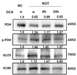
Mitochondrial dysfunction induces radioresistance in colorectal cancer by activating [Ca2+]m-PDP1-PDH-histone acetylation retrograde signaling. [Abstract]2021 Sep 6;12(9):837. PMID: 34489398
Sodium dichloroacetate purchased from MedChemExpress. Usage Cited in: Cell Death Dis. 2021 Sep 6;12(9):837. [Abstract]
Western blot analysis of PDH, H3K9ac, H3K56ac, and DNA repair proteins in NC cells and ROT cells with or without DCA (5 mM) for 6 h and 24 h in the ROT group.
-
Clin Cancer Res
Lactate-induced lactylation enhances BMAL1 transcription and nuclear translocation, promoting bevacizumab resistance in glioblastoma through VEGFA. [Abstract]2026 Apr 17. PMID: 41995718 -
Proc Natl Acad Sci U S A
2025 Mar 11;122(10):e2404899122. PMID: 40030031 -
Cancer Drug Resist
Dichloroacetate enhances the antitumor effect of pirarubicin via regulating the ROS-JNK signaling pathway in liver cancer cells. [Abstract]2020 Sep 4;3(4):947-958. PMID: 35582217 -
Fundam Res
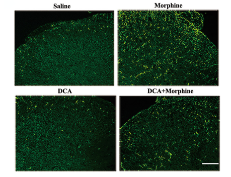
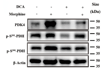
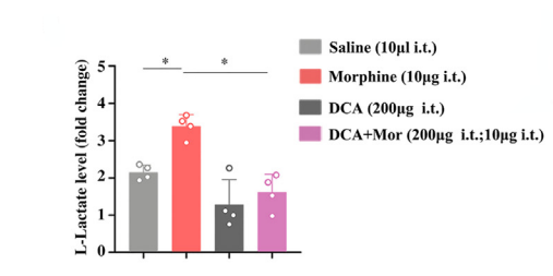
Astrocytic pyruvate dehydrogenase kinase-lactic acid axis involvement in glia-neuron crosstalk contributes to morphine-induced hyperalgesia in mice. [Abstract]2023 Mar 6;4(4):820-828. PMID: 39161415
Sodium dichloroacetate purchased from MedChemExpress. Usage Cited in: Fundam Res. 2023 Mar 6;4(4):820-828. [Abstract]
The PDK4 inhibitor DCA was diluted with saline to a concentration of 20 mg/ml, and injected intrathecally (i.t., 10 μl) 30 min before morphine. Immunostaining analysis of the activity of GFAP in SDHs was performed.
Sodium dichloroacetate purchased from MedChemExpress. Usage Cited in: Fundam Res. 2023 Mar 6;4(4):820-828. [Abstract]
The PDK4 inhibitor DCA was diluted with saline to a concentration of 20 mg/ml, and injected intrathecally (i.t., 10 μl) 30 min before morphine. Protein levels of PDK4, p-S293-PDH, and p-S300-PDH in SDHs were measured.
Sodium dichloroacetate purchased from MedChemExpress. Usage Cited in: Fundam Res. 2023 Mar 6;4(4):820-828. [Abstract]
The PDK4 inhibitor DCA was diluted with saline to a concentration of 20 mg/ml and injected intrathecally (i.t., 10 μl) 30 min before morphine. The accumulation of lactate in SDHs was detected with the lactate assay; n = 4
-
J Autoimmun
Neutrophil extracellular traps induce trophoblasts pyroptosis via enhancing NLRP3 lactylation in SLE pregnancies. [Abstract]2025 May:153:103411. PMID: 40179478 -
J Ethnopharmacol
Exploration of the underlying mechanism of Astragaloside III in attenuating immunosuppression via network pharmacology and vitro/vivo pharmacological validation. [Abstract]2024 Aug 10:330:118235. PMID: 38648891 -
Cell Mol Life Sci
The transcription factor RBPJ is required for inflammatory macrophage activation in thoracic aortic dissection by mediating mechanotransduction-induced glycolysis. [Abstract]2025 Oct 28;82(1):370. PMID: 41148245 -
Biochem Pharmacol
Dihydroartemisinin breaks the positive feedback loop of YAP1 and GLUT1-mediated aerobic glycolysis to boost the CD8+ effector T cells in hepatocellular carcinoma. [Abstract]2024 Jul:225:116294. PMID: 38754557 -
Life Sci
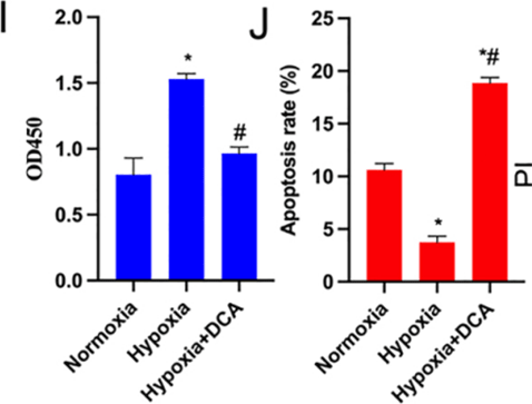
SIRT6 inhibits hypoxia-induced pulmonary arterial smooth muscle cells proliferation via HIF-1α/PDK4 signaling. [Abstract]2023 Jan 1:312:121192. PMID: 36396113
Sodium dichloroacetate purchased from MedChemExpress. Usage Cited in: Life Sci. 2023 Jan 1:312:121192. [Abstract]
Sodium dichloroacetate (DCA; 10 mM; 48 h) significantly inhibits hypoxia-induced proliferation of HPASMCs (Fig I) , and markedly increases the apoptosis rate (Fig J).
-
Inflammation
Narciclasine Alleviates Endothelial Inflammation and Atherosclerosis Initiation by Inhibiting Histone Lactylation-Mediated NF-κB Activation. [Abstract]2026 Jan 16;49(1):49. PMID: 41543766 -
Cancers (Basel)
2022 Oct 10;14(19):4966. PMID: 36230889 -
Cell Signal
PDK4-dependent lactate production and lactylation promote renal calcium oxalate crystal-induced EMT and mitochondrial dysfunction via the TGF-β/SMAD3/GPX4 axis. [Abstract]2026 Jun 27:112701. PMID: 42364861 -
Cell Signal
2023 Sep:109:110747. PMID: 37286120 -
J Trace Elem Med Biol
Sodium selenite inhibits proliferation of lung cancer cells by inhibiting NF-κB nuclear translocation and down-regulating PDK1 expression which is a key enzyme in energy metabolism expression. [Abstract]2023 Jul:78:127147. PMID: 36963369 -
J Biol Chem
Mitochondrial pyruvate carrier blockade results in decreased osteoclastogenesis and bone resorption via regulating mitochondrial energy production. [Abstract]2022 Mar 4:101775. PMID: 35257748 -
J Proteome Res
Regulating Energy Metabolism to Induce the Release of VOC Biomarkers in Lung Cancer Cells. [Abstract]2025 Nov 7. PMID: 41201462 -
Exp Cell Res
Dichloroacetate enhances the anti-tumor effect of sorafenib via modulating the ROS-JNK-Mcl-1 pathway in liver cancer cells. [Abstract]2021 Sep 1;406(1):112755. PMID: 34332981 -
Mol Reprod Dev
Dysfunction in Sertoli cells participates in glucocorticoid-induced impairment of spermatogenesis. [Abstract]2021 Jun;88(6):405-415. PMID: 34032349 -
Biochem Biophys Res Commun
Inhibition of mitochondrial complex I impedes zygotic genome activation via PDH-histone modification retrograde signaling. [Abstract]2026 Sep 10:830:154296. PMID: 42468378 -
bioRxiv
Citrate Compartmentalization Controls Calcium-Dependent Cytokine Production in Effector T Cells. [Abstract]2026 Jun 12:2026.06.11.731694. PMID: 42327233 -
-
-
Oxid Med Cell Longev
α-Lipoic Acid Targeting PDK1/NRF2 Axis Contributes to the Apoptosis Effect of Lung Cancer Cells. [Abstract]2021 Jun 4:2021:6633419. PMID: 34211631 -
Oxid Med Cell Longev
NOX4-Derived ROS Mediates TGF- β 1-Induced Metabolic Reprogramming during Epithelial-Mesenchymal Transition through the PI3K/AKT/HIF-1 α Pathway in Glioblastoma. [Abstract]2021 Jun 27;2021:5549047. PMID: 34257808


Solvent & Solubility
In Vitro:
H2O : 100 mg/mL (662.60 mM; Need ultrasonic)
DMSO : 25 mg/mL (165.65 mM; ultrasonic and warming and heat to 60°C; Hygroscopic DMSO has a significant impact on the solubility of product, please use newly opened DMSO)
Please refer to the solubility information to select the appropriate solvent. Once prepared, please aliquot and store the solution to prevent product inactivation from repeated freeze-thaw cycles.
Storage method and period of stock solution: -80°C, 6 months; -20°C, 1 month (sealed storage, away from moisture). When stored at -80°C, please use it within 6 months. When stored at -20°C, please use it within 1 month.
* Note: If you choose water as the stock solution, please dilute it to the working solution, then filter and sterilize it with a 0.22 μm filter before use.
Please refer to the solubility information to select the appropriate solvent. Once prepared, please aliquot and store the solution to prevent product inactivation from repeated freeze-thaw cycles.
Storage method and period of stock solution: -80°C, 6 months; -20°C, 1 month (sealed storage, away from moisture). When stored at -80°C, please use it within 6 months. When stored at -20°C, please use it within 1 month.
* Note: If you choose water as the stock solution, please dilute it to the working solution, then filter and sterilize it with a 0.22 μm filter before use.
Concentration (start) × Volume (start) = Concentration (final) × Volume (final)
In Vivo:
For the following dissolution methods, please prepare the working solution directly:
It is recommended to prepare fresh solutions and use them promptly within a short period of time.
The percentages shown for the solvents indicate their volumetric ratio in the final prepared solution. If precipitation or phase separation occurs during preparation, heat and/or sonication can be used to aid dissolution.
Add each solvent one by one: PBS
Solubility: 100 mg/mL (662.60 mM); Clear solution; Need ultrasonic

Protocols
-
Western Blot
Western blotting (WB) is a commonly used experimental method in molecular biology, biochemistry, and immunogenetics for identifying and quantifying target proteins. It combines gel electrophoresis with immunoassay, enabling researchers to analyze protein expression, post-translational modifications, and molecular weight.
-
Annexin V plus membrane-impermeant dye apoptosis staining
Annexin V-based apoptosis assays rely on the detection of phosphatidylserine (PS) externalization from the inner leaflet of the plasma membrane to the outer leaflet, an early biochemical hallmark of apoptosis. Fluorescently labeled Annexin V binds PS in a calcium-dependent manner, enabling identification of early apoptotic cells by flow cytometry or fluorescence microscopy. When combined with a membrane-impermeant DNA-binding dye (e. g. , propidium iodide), this approach allows discrimination between viable (Annexin V−/dye−), early apoptotic (Annexin V+/dye−), and late apoptotic or necrotic (Annexin V+/dye+) cell populations by assessing membrane integrity and PS exposure.
-
ROS/oxidative-stress fluorescent staining
ROS/oxidative-stress fluorescent staining uses cell-permeant fluorogenic probes that become fluorescent after oxidation inside cells or tissues; commonly used examples include DCFH-DA/DCFDA for broad cellular oxidant detection, DHE for superoxide-related signal detection, MitoSOX for mitochondrial superoxide-related signal detection, and CellROX probes for oxidative-stress-associated fluorescence readouts. The assay detects probe oxidation rather than a single ROS species unless the probe and analysis method have been chemically validated for that species. DCFH-DA enters cells, is deacetylated by intracellular esterases to DCFH, and produces fluorescent DCF after oxidation, so the readout is used as an operational measure of total cellular oxidative stress rather than a species-specific ROS measurement. DHE and MitoSOX can report superoxide-related oxidation, but red fluorescence alone can include non-specific ethidium-like oxidation products; HPLC or optimized spectral approaches are
-
BrdU Incorporation Assay
Bromodeoxyuridine (BrdU) incorporation assay is based on the principle that BrdU, a thymidine analog, is incorporated into newly synthesized DNA during the S phase of the cell cycle, thereby serving as a marker of DNA replication and cellular proliferation. Incorporated BrdU can be detected using anti-BrdU antibodies following DNA denaturation, enabling visualization or quantification of proliferating cells through immunochemical detection methods such as immunofluorescence or immunohistochemistry.
-
Somatic Cell Culture
A method of simulating the in vivo environment in vitro to maintain the cell growth, differentation and main functions.
-
CFSE Dye Dilution Proliferation Assay
The CFSE (carboxyfluorescein diacetate succinimidyl ester) dye dilution proliferation assay is based on the covalent labeling of intracellular proteins by a cell-permeant fluorescent dye that becomes fluorescent upon intracellular ester cleavage and then is stably retained within cells. As labeled cells divide, the dye is partitioned equally between daughter cells, resulting in a stepwise halving of fluorescence intensity that can be quantified by flow cytometry to determine the number of cell divisions undergone by each cell population. This fluorescence dilution approach enables quantitative tracking of lymphocyte proliferation at the single-cell level over multiple rounds of division. CFSE-based proliferation analysis has been widely applied to measure antigen-driven lymphocyte expansion in vitro, where discrete fluorescence peaks correspond to successive cell divisions and allow reconstruction of proliferative history within heterogeneous populations.
-
Dye-dilution cell tracking and proliferation staining
Dye-dilution cell tracking assays quantify cell proliferation by covalently labeling intracellular proteins with a stable fluorescent dye that is equally partitioned between daughter cells during mitosis, resulting in stepwise halving of fluorescence intensity with each cell division as measured by flow cytometry histograms. Carboxyfluorescein diacetate succinimidyl ester (CFSE) is a prototypical dye that diffuses into cells, is enzymatically converted into a fluorescent compound, and then covalently binds intracellular amine groups, producing long-lived fluorescence suitable for tracking multiple rounds of division in vitro and in vivo. Successive generations of dividing cells form discrete peaks of decreasing fluorescence intensity, enabling estimation of proliferation history, precursor frequency, and division index within heterogeneous populations. Alternative dyes such as CellTrace Violet (CTV) and far-red membrane dyes (e. g. , PKH26) follow the same dilution principle but differ
-
CCK-8/WST-8 Cell Proliferation Assay
The CCK-8/WST-8 assay is based on the reduction of the water-soluble tetrazolium salt WST-8 to a water-soluble formazan product by cellular dehydrogenases in metabolically active cells, where the generated formazan amount is proportional to the number of living cells and is quantified by measuring absorbance in the visible range, providing a colorimetric readout for cell viability and proliferation assessment. This class of tetrazolium-based assays improves upon earlier MTT-based systems by producing a water-soluble formazan, eliminating the need for organic solubilization steps and enabling direct spectrophotometric measurement in culture medium.
-
Protocol for Cell Cycle
Cell-cycle analysis by flow cytometry measures DNA content in single cells to estimate the fraction of cells in G0/G1, S, and G2/M phases. Propidium iodide intercalates into DNA, and after RNA removal with RNase, fluorescence intensity reflects cellular DNA content: 2N cells are assigned to G0/G1, cells between 2N and 4N to S phase, and 4N cells to G2/M. DNA-content analysis alone cannot reliably separate G0 from G1 or G2 from M. Ki-67 can distinguish quiescent G0 cells from cycling cells, EdU or BrdU incorporation marks active DNA synthesis in S phase, and phospho-histone H3 staining identifies mitotic cells within the 4N population.
-
Cell Counting-Based Growth Curve Assay
Cell counting-based growth curve assays quantify cell proliferation by directly measuring changes in viable cell number over time using manual or automated counting methods such as hemocytometer-based counting or instrument-assisted cell enumeration, enabling construction of growth curves that reflect population expansion dynamics in response to culture conditions. A widely used approach is trypan blue exclusion with hemocytometer counting, where membrane-compromised (non-viable) cells take up the dye, allowing discrimination between viable and non-viable cells while simultaneously enabling total cell number quantification. Repeated sampling across time points allows estimation of proliferation rate, growth phases, and comparative growth kinetics between experimental conditions.
-
Colony Formation (Clonogenic) Assay
The clonogenic (colony formation) assay measures the ability of a single cell to retain reproductive viability and form a macroscopic colony, typically defined as a cluster derived from one progenitor cell after a defined growth period. This assay is widely used to evaluate cell survival after exposure to ionizing radiation or cytotoxic treatments and is considered a standard method in radiation biology for generating dose-response relationships of reproductive cell death. Colony formation reflects long-term proliferative capacity rather than short-term metabolic activity, and survival is quantified by comparing treated versus untreated conditions based on colony number and derived survival fractions.
-
Cell Viability Determination by MTT Colorimetric Assay
The following protocol uses the MTT colorimetric assay as a classic literature-established method for assessing cell viability/metabolic activity in cultured mammalian cells. MTT[3-(4,5-dimethylthiazol-2-yl)-2,5-diphenyltetrazolium bromide] is reduced by metabolically active cells to a colored formazan product; the amount of formazan is quantified spectrophotometrically and provides an indirect measure of metabolically active viable cells. Importantly, MTT reduction reflects cellular oxidoreductase/metabolic activity rather than an absolute direct count of living cells, so changes in cellular metabolism can alter the signal independently of cell number.
-
Kinase activity and phosphorylation assays
Kinase activity assays measure the ability of kinases to transfer phosphate groups from ATP to specific substrates, while phosphorylation assays detect the presence and levels of phosphorylated proteins. Common methods include radiolabeled ATP incorporation (e. g. ,), ADP release detection via bioluminescence (e. g. ,[3]), enzyme-linked immunosorbent assays (ELISA) for phospho-specific epitopes (e. g. ,[6]), and microtiter-based formats for high-throughput screening (e. g. ,[8]). The ADP-Glo assay quantifies kinase activity by measuring ADP produced during phosphorylation using a luciferase-based system. Radiometric assays involve autoradiography or scintillation counting after incorporation of 32P-labeled ATP into substrate proteins. ELISA-based approaches rely on phospho-specific antibodies to detect activated kinases in cell lysates or purified samples.
-
EdU Incorporation Assay (Click Chemistry-Based DNA Synthesis Measurement)
The EdU incorporation assay measures DNA synthesis by adding the thymidine analog 5-ethynyl-2′-deoxyuridine to cells or tissues, where it is incorporated into newly synthesized DNA during S phase. Incorporated EdU is detected by copper-catalyzed azide-alkyne cycloaddition, in which a fluorescent azide covalently reacts with the ethynyl group on EdU, allowing S-phase cells to be detected by fluorescence microscopy, flow cytometry, or high-content imaging. EdU detection does not require DNA denaturation or anti-BrdU antibody access, which preserves sample structure and improves compatibility with immunostaining and multiparameter cytometry compared with BrdU-based detection. EdU can be cytotoxic in a cell-type- and exposure-dependent manner, so pulse duration, concentration, and continuous-labeling designs should be validated for each cell type.
-
Ki-67 Immunostaining Proliferation Assay
Ki-67 immunostaining measures the growth fraction of a cell population by detecting Ki-67, a nuclear antigen present in proliferating cells and absent in quiescent G0 cells. The readout is the percentage of Ki-67-positive nuclei among total counted cells, commonly called the Ki-67 labeling index or proliferation index.
-
Apoptosis
Apoptosis, also called programmed cell death, is generally characterized by distinct morphological characteristics.
-
TUNEL staining for apoptotic DNA fragmentation
TUNEL staining detects DNA strand breaks by using terminal deoxynucleotidyl transferase to add labeled nucleotides to exposed 3′-OH DNA termini, generating either microscopic staining in fixed cells or tissue sections, or fluorescence/cytometric signal in cell suspensions. TUNEL positivity reflects DNA fragmentation but should not be interpreted alone as definitive apoptosis, because TUNEL can also label necrotic, autolytic, mechanically damaged, or DNA-repair-associated DNA breaks.
-
PCNA Immunodetection Proliferation Assay
PCNA immunodetection measures proliferative activity by detecting proliferating cell nuclear antigen, a nuclear protein associated with DNA polymerase δ function and DNA replication. The assay readout is the proportion of PCNA-positive nuclei among total counted cells, but PCNA labeling is not identical to BrdU labeling because PCNA can mark late G1/early S-associated replication competence and may persist beyond active DNA synthesis depending on fixation and extraction conditions.
-
Protocol for Cell Counting and Cell Density Analysis
Cell counting and cell-density analysis estimate the number of cells in a known volume or field area. Manual hemocytometer counting uses a chamber of defined geometry to convert counted cells into cells/mL, while automated counters and image-analysis workflows detect cell objects from optical, brightfield, fluorescence, impedance, or digital-image features. Trypan blue viability counting is based on dye exclusion: viable cells with intact membranes exclude dye, while non-viable cells with compromised membranes stain blue. The readout is total cell density, viable-cell density, dead-cell density, and percent viability. Cell density can also be estimated from microscopy images by counting objects per image area, from flow cytometry using calibrated volume or reference particles, or from in situ microscopy in bioreactors after calibration against reference methods such as hemocytometer or flow cytometry.
-
Apoptosis Solutions
Apoptosis is a regulated, generally non-lytic cell-death pathway that removes unwanted, damaged, infected, or abnormal cells through coordinated morphological changes, caspase activation, DNA fragmentation, and membrane remodeling. The intrinsic apoptosis pathway is controlled mainly by mitochondrial outer membrane permeabilization, BCL-2 family proteins, cytochrome c release, apoptosome formation, caspase-9 activation, and downstream executioner caspase-3/7 activation. The extrinsic apoptosis pathway is initiated by death receptors such as Fas, TNFR, and TRAIL receptors, which recruit adaptor proteins and activate caspase-8 before engaging executioner caspases or mitochondrial amplification through BID cleavage. Apoptosis is linked to many phenotypes, including cancer cell killing, tissue homeostasis, immune regulation, neurodegeneration, infection response, and treatment-induced cytotoxicity; unresolved questions include how apoptosis interacts with necroptosis, pyroptosis, ferroptos
-
Protocol for Kinase activity and phosphorylation assays
Kinase activity assays measure transfer of phosphate from ATP to a protein or peptide substrate, generating phosphorylated substrate, ADP, or incorporated radiolabeled phosphate as the readout; phosphorylation assays measure site-specific phosphorylation in cells or tissues as a proxy for kinase-pathway activation, inhibition, or substrate regulation. Phosphorylation can be detected by phospho-specific Western blot, immunoprecipitation kinase assay, phospho-immunofluorescence, phospho-flow cytometry, luminescent ADP detection, radiolabeled ATP incorporation, or reporter-based pathway assays, and these readouts can be applied to cancer cells, primary neurons, mouse tumors, organoids, inflammatory macrophages, ferroptosis studies, and mitophagy studies when the kinase target is biologically relevant.
-
MTT Cell Proliferation Assay
The MTT assay is a colorimetric endpoint assay for estimating viable cell number, cell growth, cytotoxicity, or cell activation in cultured mammalian cells. Living cells reduce the yellow tetrazolium salt MTT into purple/blue formazan, while dead cells do not generate the same signal; the resulting color can be quantified with a multiwell spectrophotometer. MTT reduction is commonly interpreted as a readout of metabolic activity that often correlates with viable cell number, but it should not be treated as a direct cell-counting method unless the assay is optimized for the cell type and experimental condition. Studies show that MTT reduction can involve mitochondrial and non-mitochondrial reducing systems, and formazan may accumulate in intracellular lipid droplets rather than simply marking mitochondria.


Purity & Documentation
-
Data Sheet (276 KB)
-
SDS (419 KB)
- English - EN (419 KB)
- Français - FR (419 KB)
- Deutsch - DE (419 KB)
- Norwegian - NO (419 KB)
- Español - ES (419 KB)
- Swedish - SV (419 KB)
- Italian - IT (419 KB)
- Korean - KR (419 KB)
- Portuguese - PT (419 KB)
-
Handling Instructions (2659 KB)
References
[1]. Stakišaitis D, et al. The Importance of Gender-Related Anticancer Research on Mitochondrial Regulator Sodium Dichloroacetate in Preclinical Studies In Vivo. Cancers (Basel). 2019 Aug 20;11(8). pii: E1210. [Content Brief]
[2]. Hossain M, et al. Cytotoxic derivatives of dichloroacetic acid and some metal complexes. Arch Pharm (Weinheim). 2022 Nov;355(11):e2200236. [Content Brief]


Complete Stock Solution Preparation Table
Please refer to the solubility information to select the appropriate solvent. Once prepared, please aliquot and store the solution to prevent product inactivation from repeated freeze-thaw cycles.
Storage method and period of stock solution: -80°C, 6 months; -20°C, 1 month (sealed storage, away from moisture). When stored at -80°C, please use it within 6 months. When stored at -20°C, please use it within 1 month.
| Optional Solvent | Concentration Solvent Mass | 1 mg | 5 mg | 10 mg | 25 mg |
|---|---|---|---|---|---|
| DMSO / H2O | 1 mM | 6.6260 mL | 33.1301 mL | 66.2603 mL | 165.6507 mL |
| 5 mM | 1.3252 mL | 6.6260 mL | 13.2521 mL | 33.1301 mL | |
| 10 mM | 0.6626 mL | 3.3130 mL | 6.6260 mL | 16.5651 mL | |
| 15 mM | 0.4417 mL | 2.2087 mL | 4.4174 mL | 11.0434 mL | |
| 20 mM | 0.3313 mL | 1.6565 mL | 3.3130 mL | 8.2825 mL | |
| 25 mM | 0.2650 mL | 1.3252 mL | 2.6504 mL | 6.6260 mL | |
| 30 mM | 0.2209 mL | 1.1043 mL | 2.2087 mL | 5.5217 mL | |
| 40 mM | 0.1657 mL | 0.8283 mL | 1.6565 mL | 4.1413 mL | |
| 50 mM | 0.1325 mL | 0.6626 mL | 1.3252 mL | 3.3130 mL | |
| 60 mM | 0.1104 mL | 0.5522 mL | 1.1043 mL | 2.7608 mL | |
| 80 mM | 0.0828 mL | 0.4141 mL | 0.8283 mL | 2.0706 mL | |
| 100 mM | 0.0663 mL | 0.3313 mL | 0.6626 mL | 1.6565 mL |
* Note: If you choose water as the stock solution, please dilute it to the working solution, then filter and sterilize it with a 0.22 μm filter before use.


Sodium dichloroacetate Related Classifications
HY-Y0445A Related Classifications
 Powered by Bioz