



Tetraconazole

Based on 1 Customer Validation
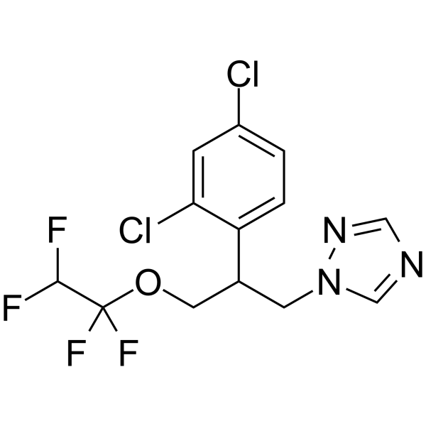
Tetraconazole is a selective irreversible inhibitor of 14-α-sterol demethylase (CYP51) with antifungal activity. Tetraconazole competitively binds to the enzyme to block fungal ergosterol synthesis, resulting in cell membrane damage. The EC50 of tetraconazole against wheat pathogens is 0.382-0.802 mg/L, and the EC50 against onion root tip meristem cell growth is 6.7 mg/L, and (R)-(+)-Tetraconazole is 1.49-1.98 times more active than (S)-(-)-Tetraconazole. Tetraconazole can also induce oxidative stress and chromosomal aberrations in plant cells.
연구목적의 판매만을 진행합니다. 환자를 대상으로 한 판매는 하지 않습니다.
- Purity : 96.98%
- CAS No.: 112281-77-3
- 화학식: C13H11Cl2F4N3O
- 분자량:372.15
-
보관:Pure form -20°C, 3 years , 4°C, 2 years ; In solvent -80°C, 6 months , -20°C, 1 month

To place orders, for customer services and technical support, please contact: MedChemExpress USA
Tel: 609-228-6898 E-mail: [email protected] [email protected]


-
 Biological Activity
-
Chemical Information
-
용액&용해도
- Protocol
- 순도&문서
- References
-
 Help & FAQs





Biological Activity
제품 설명
In Vitro
Yeast metabolism effect experiment:
Tetraconazole (0.5-1.0 mg/L; 10 d) significantly reduces the content of methyl mercaptan in wine yeast fermentation, while increasing the production of fatty acids and ethyl esters[2].
Plant cell toxicity experiment:
Tetraconazole (1.0-10.0 mg/L; 3 d) induces oxidative stress in onion root tip cells, resulting in a 3-fold increase in MDA content, a 1-2-fold increase in SOD and CAT enzyme activities, and inhibits germination rate (reduced to 50%) and root length (reduced by 81%)[3].
MedChemExpress (MCE) has not independently confirmed the accuracy of these methods. They are for reference only.
- Protocol for Cell Counting and Cell Density Analysis
- ROS/oxidative-stress fluorescent staining
- Somatic Cell Culture
- CCK-8/WST-8 Cell Proliferation Assay
- Cell Counting-Based Growth Curve Assay
- MTT Cell Proliferation Assay
- Directly Induced Neuron Culture
- Cell Viability Determination by MTT Colorimetric Assay


Chemical Information
-
CAS No. 112281-77-3
-
Appearance Viscous Liquid
-
분자량 372.15
-
화학식 C13H11Cl2F4N3O
-
Color Colorless to light yellow
-
SMILES
FC(F)C(F)(F)OCC(C1=CC=C(Cl)C=C1Cl)CN2N=CN=C2
-
선적
Room temperature in continental US; may vary elsewhere.
-
보관
Pure form -20°C 3 years 4°C 2 years In solvent -80°C 6 months -20°C 1 month


용액&용해도
In Vitro:
DMSO : 125 mg/mL (335.89 mM; Need ultrasonic; Hygroscopic DMSO has a significant impact on the solubility of product, please use newly opened DMSO)
Please refer to the solubility information to select the appropriate solvent. Once prepared, please aliquot and store the solution to prevent product inactivation from repeated freeze-thaw cycles.
Storage method and period of stock solution: -80°C, 6 months; -20°C, 1 month. When stored at -80°C, please use it within 6 months. When stored at -20°C, please use it within 1 month.
Please refer to the solubility information to select the appropriate solvent. Once prepared, please aliquot and store the solution to prevent product inactivation from repeated freeze-thaw cycles.
Storage method and period of stock solution: -80°C, 6 months; -20°C, 1 month. When stored at -80°C, please use it within 6 months. When stored at -20°C, please use it within 1 month.
Concentration (start) × Volume (start) = Concentration (final) × Volume (final)
In Vivo:
Select the appropriate dissolution method based on your experimental animal and administration route.
- For the following dissolution methods, please ensure to first prepare a clear stock solution using an In Vitro approach and then sequentially add co-solvents:
- To ensure reliable experimental results, the clarified stock solution can be appropriately stored based on storage conditions. As for the working solution for In Vivo experiments, it is recommended to prepare freshly and use it on the same day.
- The percentages shown for the solvents indicate their volumetric ratio in the final prepared solution. If precipitation or phase separation occurs during preparation, heat and/or sonication can be used to aid dissolution.
Add each solvent one by one: 10% DMSO 40% PEG300 5% Tween-80 45% Saline
Solubility: ≥ 2.08 mg/mL (5.59 mM); Clear solution
This protocol yields a clear solution of ≥ 2.08 mg/mL (saturation unknown).
Taking 1 mL working solution as an example, add 100 μL DMSO stock solution (20.8 mg/mL) to 400 μL PEG300, and mix evenly; then add 50 μL Tween-80 and mix evenly; then add 450 μL Saline to adjust the volume to 1 mL.
Preparation of Saline: Dissolve 0.9 g sodium chloride in ddH₂O and dilute to 100 mL to obtain a clear Saline solution.
Add each solvent one by one: 10% DMSO 90% (20% SBE-β-CD in Saline)
Solubility: ≥ 2.08 mg/mL (5.59 mM); Clear solution
This protocol yields a clear solution of ≥ 2.08 mg/mL (saturation unknown).
Taking 1 mL working solution as an example, add 100 μL DMSO stock solution (20.8 mg/mL) to 900 μL 20% SBE-β-CD in Saline, and mix evenly.
Preparation of 20% SBE-β-CD in Saline (4°C, storage for one week): 2 g SBE-β-CD powder is dissolved in 10 mL Saline, completely dissolve until clear.
In Vivo Dissolution Calculator
Please enter the basic information of animal experiments:
-
-
-
-
Recommended: Prepare an additional quantity of animals to account for potential losses during experiments.
Please enter your animal formula composition:
-
%DMSO +
Recommended: Keep the proportion of DMSO in working solution below 2% if your animal is weak.
-
%+
-
+%Tween-80 + +
-
%Saline +
The co-solvents required include: DMSO, . All of co-solvents are available by MedChemExpress (MCE). , Tween 80. All of co-solvents are available by MedChemExpress (MCE).
Working solution concentration: 0.22 mg/mL
Method for preparing stock solution: mg drug dissolved in μL DMSO. Stock solution concentration: mg/mL.
1. Take μL DMSO stock solution;
2. Add μL .
μL , mix evenly;
3. Then add μL Tween 80, mix evenly;
4. Then add μL
Please ensure that the stock solution in the first step is dissolved to a clear state, and add co-solvents in sequence. You can use ultrasonic heating (ultrasonic cleaner, recommended frequency 20-40 kHz), vortexing, etc. to assist dissolution.


순도&문서
-
Data Sheet (274 KB)
-
SDS (458 KB)
- English - EN (458 KB)
- Français - FR (458 KB)
- Deutsch - DE (458 KB)
- Norwegian - NO (458 KB)
- Español - ES (458 KB)
- Swedish - SV (458 KB)
- Italian - IT (458 KB)
- Korean - KR (458 KB)
- Portuguese - PT (458 KB)
-
Handling Instructions (2659 KB)
References
[1]. Tong Z, et al. Enantioselective effects of the chiral fungicide tetraconazole in wheat: Fungicidal activity and degradation behavior. Environ Pollut. 2019;247:1-8. [Content Brief]
[2]. Sieiro-Sampedro T, et al. Tetraconazole alters the methionine and ergosterol biosynthesis pathways in Saccharomyces yeasts promoting changes on volatile derived compounds. Food Res Int. 2020;130:108930. [Content Brief]
[3]. Macar O. Multiple toxic effects of tetraconazole in Allium cepa L. meristematic cells. Environ Sci Pollut Res Int. 2021 Feb;28(8):10092-10099. [Content Brief]


Complete Stock Solution Preparation Table
Please refer to the solubility information to select the appropriate solvent. Once prepared, please aliquot and store the solution to prevent product inactivation from repeated freeze-thaw cycles.
Storage method and period of stock solution: -80°C, 6 months; -20°C, 1 month. When stored at -80°C, please use it within 6 months. When stored at -20°C, please use it within 1 month.
| Optional Solvent | Concentration Solvent Mass | 1 mg | 5 mg | 10 mg | 25 mg |
|---|---|---|---|---|---|
| DMSO | 1 mM | 2.6871 mL | 13.4354 mL | 26.8709 mL | 67.1772 mL |
| 5 mM | 0.5374 mL | 2.6871 mL | 5.3742 mL | 13.4354 mL | |
| 10 mM | 0.2687 mL | 1.3435 mL | 2.6871 mL | 6.7177 mL | |
| 15 mM | 0.1791 mL | 0.8957 mL | 1.7914 mL | 4.4785 mL | |
| 20 mM | 0.1344 mL | 0.6718 mL | 1.3435 mL | 3.3589 mL | |
| 25 mM | 0.1075 mL | 0.5374 mL | 1.0748 mL | 2.6871 mL | |
| 30 mM | 0.0896 mL | 0.4478 mL | 0.8957 mL | 2.2392 mL | |
| 40 mM | 0.0672 mL | 0.3359 mL | 0.6718 mL | 1.6794 mL | |
| 50 mM | 0.0537 mL | 0.2687 mL | 0.5374 mL | 1.3435 mL | |
| 60 mM | 0.0448 mL | 0.2239 mL | 0.4478 mL | 1.1196 mL | |
| 80 mM | 0.0336 mL | 0.1679 mL | 0.3359 mL | 0.8397 mL | |
| 100 mM | 0.0269 mL | 0.1344 mL | 0.2687 mL | 0.6718 mL |


Tetraconazole Related Classifications
HY-117089 Related Classifications
 Powered by Bioz