



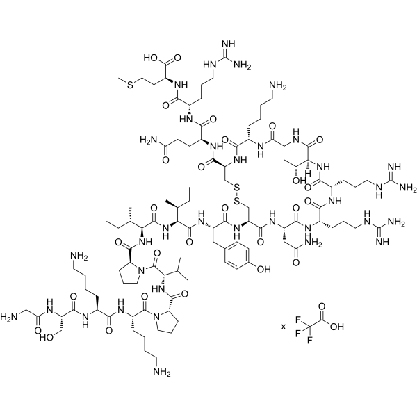
Thanatin TFA

Based on 1 Customer Validation
Thanatin TFA is an inducible cationic antimicrobial peptide. Thanatin TFA s a pathogen-inducible single-disulfide-bond-containing β-hairpin AMP. Thanatin TFA displays broad-spectrum activity against both Gram-negative and Gram-positive bacteria as well as against various species of fungi with MICs of 0.3-40 µM, 0.6-40 µM and 0.6-20 µM, respectively. Thanatin TFA has the property of competitive replacement of divalent cations from bacterial outer membrane (OM), leading to OM disruption.
For research use only. We do not sell to patients.
- Purity : 99.68%
- Formula: C103H177N35O27S3.xC2HF3O2
- Molecular Weight:2433.92 (free base)
-
Storage:
Sealed storage, away from moisture and light.
Powder -80°C, 2 years , -20°C, 1 year* In solvent : -80°C, 6 months; -20°C, 1 month (sealed storage, away from moisture and light)

To place orders, for customer services and technical support, please contact: MedChemExpress USA
Tel: 609-228-6898 E-mail: [email protected] [email protected]


-
 Biological Activity
-
Chemical Information
-
Solvent & Solubility
- Protocols
- Purity & Documentation
- References
-
 Help & FAQs




Biological Activity
Description
In Vitro
Thanatin TFA is strongly cationic (pI of 10.48) and contains a distinct short eight-residue basic loop created through a disulfide bond formation between residues Cys11 and Cys18 at the C-terminus. [1].
Thanatin TFA exhibits potent inhibitory effect on the growth of all New Delhi metallo-β-lactamase-1 (NDM-1)-producing E. coli and K. pneumoniae strains at 0.4-3.2 μM of the minimum inhibitory concentration (MIC) values[2].
MedChemExpress (MCE) has not independently confirmed the accuracy of these methods. They are for reference only.
In Vivo
MedChemExpress (MCE) has not independently confirmed the accuracy of these methods. They are for reference only.
-
Animal Model:Male BALB/c mice aged 8-10 weeks and weighing 18-22 g with CFU E. coli XJ141026
-
Dosage:1, 3, 6 mg/kg
-
Administration:Intraperitoneally injected; at 1 and 6 h
-
Result:1, 3, and 6 mg/kg markedly increased the survival rate from 0 to 30%, 70%, and 100%, respectively.
Showed that the bacterial titers decreased with increasing drug dose.
Rescued the pathological damages (large amounts of inflammatory cell infiltration, alveolar fusion, congestion in the spleen red pulp area, hepatic sinusoidal dilation and congestion) in a dose-dependent manner.


Chemical Information
-
Appearance Solid
-
Molecular Weight 2433.92 (free base)
-
Formula C103H177N35O27S3.xC2HF3O2
-
Color White to off-white
-
Sequence
Gly-Ser-Lys-Lys-Pro-Val-Pro-Ile-Ile-Tyr-Cys-Asn-Arg-Arg-Thr-Gly-Lys-Cys-Gln-Arg-Met (Disulfide bridge:Cys11-Cys18)
-
Sequence Shortening
GSKKPVPIIYCNRRTGKCQRM (Disulfide bridge:Cys11-Cys18)
-
Shipping
Room temperature in continental US; may vary elsewhere.
-
Storage
Sealed storage, away from moisture and light
Powder -80°C 2 years -20°C 1 year * In solvent : -80°C, 6 months; -20°C, 1 month (sealed storage, away from moisture and light)


Solvent & Solubility
In Vitro:
H2O : 100 mg/mL (Need ultrasonic)
DMSO : 100 mg/mL (Need ultrasonic; Hygroscopic DMSO has a significant impact on the solubility of product, please use newly opened DMSO)


Purity & Documentation
-
Data Sheet (283 KB)
-
SDS (252 KB)
- English - EN (252 KB)
- Français - FR (252 KB)
- Deutsch - DE (252 KB)
- Norwegian - NO (252 KB)
- Español - ES (252 KB)
- Swedish - SV (252 KB)
- Italian - IT (252 KB)
- Korean - KR (252 KB)
- Portuguese - PT (252 KB)
-
Handling Instructions (2659 KB)
References
[1]. Rachita Dash, et al. Thanatin: An Emerging Host Defense Antimicrobial Peptide with Multiple Modes of Action. Int J Mol Sci. 2021 Feb 3;22(4):1522. [Content Brief]
[2]. Bo Ma, et al. The antimicrobial peptide thanatin disrupts the bacterial outer membrane and inactivates the NDM-1 metallo-β-lactamase. Nat Commun. 2019 Aug 6;10(1):3517. [Content Brief]

Calculators
Concentration (start) × Volume (start) = Concentration (final) × Volume (final)


Thanatin TFA Related Classifications
HY-P5601A Related Classifications
 Powered by Bioz