



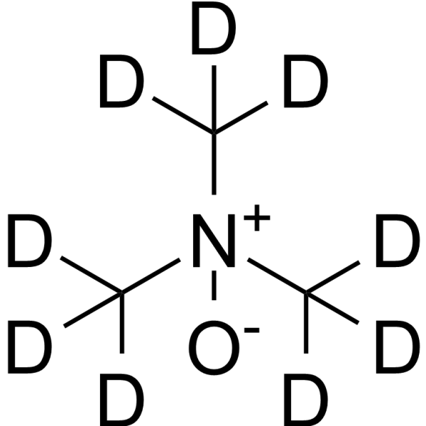
Trimethylamine N-oxide-d9
Based on 1 publication(s) in Google Scholar
Trimethylamine N-oxide-d9 is the deuterium labeled Trimethylamine N-oxide. Trimethylamine N-oxide is a gut microbe-dependent metabolite of dietary choline and other trimethylamine-containing nutrients. Trimethylamine N-oxide induces inflammation by activating the ROS/NLRP3 inflammasome. Trimethylamine N-oxide also accelerates fibroblast-myofibroblast differentiation and induces cardiac fibrosis by activating the TGF-β/smad2 signaling pathway.
For research use only. We do not sell to patients.
- Purity : 99.76%
- CAS No.: 1161070-49-0
- Formula: C3D9NO
- Molecular Weight:84.17
-
Storage:Powder -20°C, 3 years ; In solvent -80°C, 6 months , -20°C, 1 month

To place orders, for customer services and technical support, please contact: MedChemExpress USA
Tel: 609-228-6898 E-mail: [email protected] [email protected]


-
 Biological Activity
-
Chemical Information
-
Solvent & Solubility
- Protocols
- Purity & Documentation
- References
-
 Help & FAQs


-
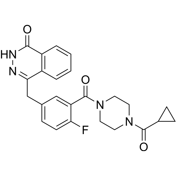
HY-10162
OlaparibPARP Inhibitor -
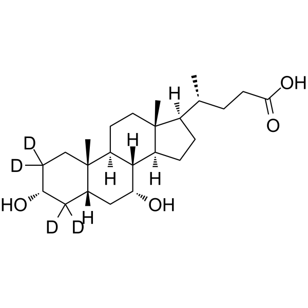
HY-76847S
Chenodeoxycholic Acid-d4Stable Isotope -
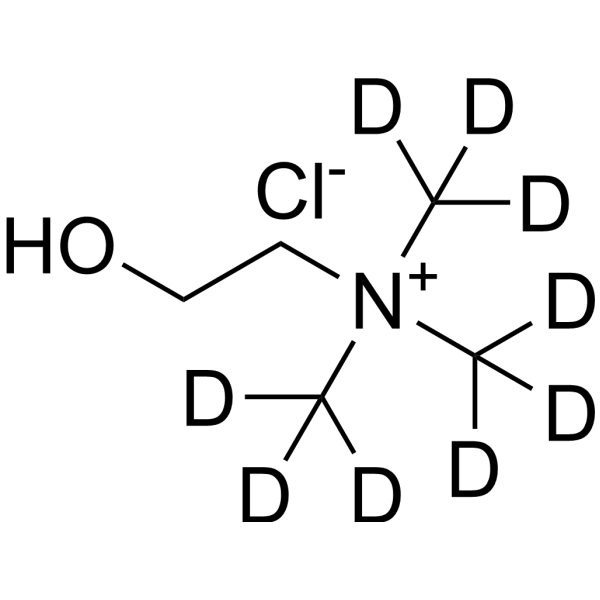
HY-B1337S1
Choline-d9 chlorideStable Isotope -
HY-B0762S
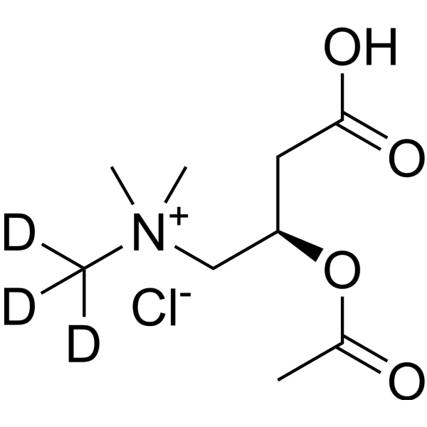
Acetyl-L-carnitine-d3 hydrochlorideStable Isotope -

HY-116084
Trimethylamine N-oxideROS/NLRP3 Inflammasome Activator -
HY-I1124
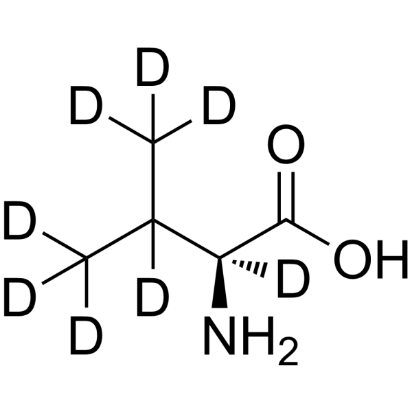
L-Valine-d8Stable Isotope -
HY-W010347S
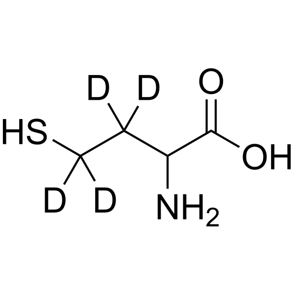
DL-Homocysteine-d4Stable Isotope -
HY-B0351S
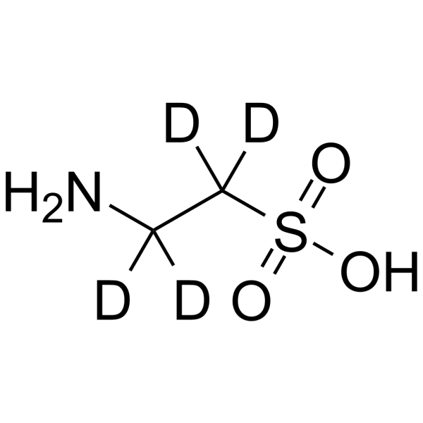
Taurine-d4Stable Isotope -
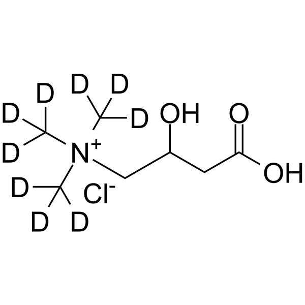
HY-B1453S1
(±)-Carnitine-d9 chlorideStable Isotope -
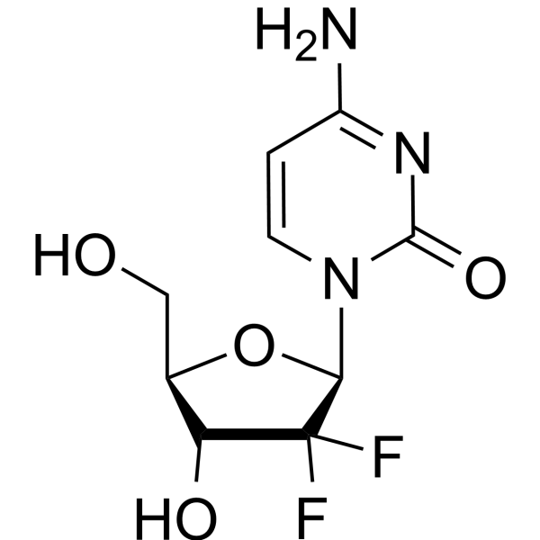
HY-17026
GemcitabineDNA Synthesis Inhibitor -
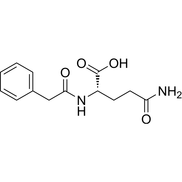
HY-W050026
Phenylacetylglutamine -
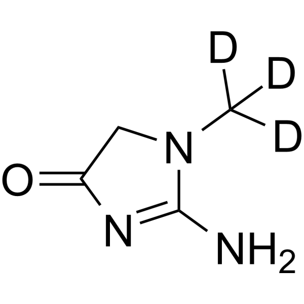
HY-B0504S
Creatinine-d3Stable Isotope
Publications Citing Use of MedChemExpress (MCE) Trimethylamine N-oxide-d9
MoreAll Endogenous Metabolite Isoforms
More

Biological Activity
Description
In Vitro
Stable heavy isotopes of hydrogen, carbon, and other elements have been incorporated into drug molecules, largely as tracers for quantitation during the drug development process. Deuteration has gained attention because of its potential to affect the pharmacokinetic and metabolic profiles of drugs[1].
MedChemExpress (MCE) has not independently confirmed the accuracy of these methods. They are for reference only. Further protocols information, click here.
Application
1. This compound can be used as a tracer
2. This compound can be used as an internal standard for quantitative analysis by NMR, GC-MS, or LC-MS.


Chemical Information
-
CAS No. 1161070-49-0
-
Unlabeled CAS 1184-78-7
-
Appearance Solid
-
Molecular Weight 84.17
-
Formula C3D9NO
-
Color White to off-white
-
SMILES
[2H]C([2H])([N+](C([2H])([2H])[2H])([O-])C([2H])([2H])[2H])[2H]
-
Shipping
Room temperature in continental US; may vary elsewhere.
-
Storage
Powder -20°C 3 years In solvent -80°C 6 months -20°C 1 month


Publications (1)
-
Journal Impact Factor
-
Most Recent
-
J Chromatogr B Analyt Technol Biomed Life Sci
Simultaneous determination of choline, L-carnitine, betaine, trimethylamine, trimethylamine N-oxide, and creatinine in plasma, liver, and feces of hyperlipidemic rats by UHPLC-MS/MS. [Abstract]2024 Aug 1:1243:124210. PMID: 38936270


Solvent & Solubility
In Vitro:
H2O : 100 mg/mL (1188.07 mM; Need ultrasonic)
DMSO : 100 mg/mL (1188.07 mM; Need ultrasonic; Hygroscopic DMSO has a significant impact on the solubility of product, please use newly opened DMSO)
Please refer to the solubility information to select the appropriate solvent. Once prepared, please aliquot and store the solution to prevent product inactivation from repeated freeze-thaw cycles.
Storage method and period of stock solution: -80°C, 6 months; -20°C, 1 month. When stored at -80°C, please use it within 6 months. When stored at -20°C, please use it within 1 month.
* Note: If you choose water as the stock solution, please dilute it to the working solution, then filter and sterilize it with a 0.22 μm filter before use.
Please refer to the solubility information to select the appropriate solvent. Once prepared, please aliquot and store the solution to prevent product inactivation from repeated freeze-thaw cycles.
Storage method and period of stock solution: -80°C, 6 months; -20°C, 1 month. When stored at -80°C, please use it within 6 months. When stored at -20°C, please use it within 1 month.
* Note: If you choose water as the stock solution, please dilute it to the working solution, then filter and sterilize it with a 0.22 μm filter before use.
Concentration (start) × Volume (start) = Concentration (final) × Volume (final)

Protocols
-
Fibrosis/Collagen Morphometry
Fibrosis and collagen morphometry is based on the quantitative visualization of fibrillar collagen deposition in tissue sections using histochemical stains such as Sirius Red (Picrosirius Red) or Masson's trichrome, followed by image-based or polarization-enhanced analysis to estimate collagen proportional area as a surrogate of extracellular matrix accumulation during fibrotic remodeling. Sirius Red combined with polarized light microscopy enhances detection of collagen fibers due to birefringence properties, enabling more specific visualization of collagen type I and III fibrils compared to conventional bright-field histology, while whole-section or region-restricted digital morphometry reduces field-selection bias in fibrosis assessment. Alternative quantitative approaches include second harmonic generation (SHG) and two-photon excited fluorescence microscopy, which enable label-free detection of fibrillar collagen and have been validated against histological staining and biochemica
-
Collagen: Sirius Red Staining
Sirius Red or picrosirius red staining is a histochemical method for visualizing collagen-rich extracellular matrix in tissue sections, and collagen fibers are detected as red-stained structures under bright-field microscopy with enhanced birefringence under polarized light. Picrosirius red is useful for assessing total collagen organization, distribution, and fibrosis burden, but polarized color should not be interpreted as a definitive collagen type I versus type III readout because color is affected by fiber orientation, thickness, and packing.
-
ROS/oxidative-stress fluorescent staining
ROS/oxidative-stress fluorescent staining uses cell-permeant fluorogenic probes that become fluorescent after oxidation inside cells or tissues; commonly used examples include DCFH-DA/DCFDA for broad cellular oxidant detection, DHE for superoxide-related signal detection, MitoSOX for mitochondrial superoxide-related signal detection, and CellROX probes for oxidative-stress-associated fluorescence readouts. The assay detects probe oxidation rather than a single ROS species unless the probe and analysis method have been chemically validated for that species. DCFH-DA enters cells, is deacetylated by intracellular esterases to DCFH, and produces fluorescent DCF after oxidation, so the readout is used as an operational measure of total cellular oxidative stress rather than a species-specific ROS measurement. DHE and MitoSOX can report superoxide-related oxidation, but red fluorescence alone can include non-specific ethidium-like oxidation products; HPLC or optimized spectral approaches are
-
Connective Tissue: Masson's Trichrome/Collagen Trichrome Staining
Masson’s Trichrome (collagen/trichrome staining) is a histological technique that differentially stains tissue compartments using sequential acidic dyes to distinguish collagen from muscle and cytoplasmic components based on dye affinity and tissue permeability differences, enabling visualization of fibrosis and connective tissue architecture in histological sections. The classical formulation typically uses Weigert's iron hematoxylin for nuclear staining, Biebrich scarlet-acid fuchsin for cytoplasm and muscle, and aniline blue (or light green variants) for collagen, producing a characteristic blue/green collagen signal contrasted against red cytoplasm and dark nuclei. The staining principle relies on selective displacement of smaller dye molecules by larger anionic dyes in collagen-rich regions under controlled acidified conditions, which enhances collagen-specific dye retention. This property makes the method widely used for fibrosis assessment in organs such as heart, liver, lung, a
-
Research Protocol for Inflammation-related Diseases
The NLRP3 inflammasome is a cytosolic innate immune signaling platform that integrates priming signals and danger-signal activation to promote caspase-1 activation, maturation of IL-1β and IL-18, and gasdermin D-mediated pyroptotic cell death. The core experimental logic is to determine whether inflammatory disease phenotypes are driven by increased NLRP3 expression, ASC-containing inflammasome assembly, caspase-1 cleavage, GSDMD cleavage, and extracellular release of IL-1β/IL-18 rather than by nonspecific cell injury alone. The pathway is strongly linked to inflammation-related disease phenotypes because monosodium urate crystals activate NALP3/NLRP3 inflammasome signaling in gout-like crystal inflammation, cholesterol crystals activate NLRP3 inflammasomes in atherogenesis models, and DSS-induced intestinal inflammation has been reported to involve NLRP3 inflammasome activity. However, experimental colitis studies also show context-dependent protective effects of NLRP3 inflammasome co
-
Pyroptosis Solutions
Pyroptosis is a lytic inflammatory cell-death pathway executed by gasdermin pores, most classically through inflammasome-mediated activation of caspase-1, cleavage of gasdermin D, membrane pore formation, LDH release, and secretion of IL-1β and IL-18. The canonical pathway is commonly modeled by priming cells with an inflammatory signal such as LPS to induce pro-IL-1β and inflammasome components, followed by an activation signal such as ATP or nigericin to activate NLRP3, ASC speck formation, caspase-1 cleavage, GSDMD cleavage, cytokine release, and pyroptotic membrane rupture. The non-canonical pathway is triggered when cytosolic LPS activates mouse caspase-11 or human caspase-4/5, leading to GSDMD cleavage and pyroptosis, and this can secondarily activate NLRP3-dependent IL-1β release. Pyroptosis is linked to inflammatory injury, infection, cancer, liver disease, ocular disease, placental inflammation, and other disease phenotypes, but unresolved questions include which gasdermin fam


Purity & Documentation
-
Data Sheet (281 KB)
-
SDS (394 KB)
- English - EN (394 KB)
- Français - FR (394 KB)
- Deutsch - DE (394 KB)
- Norwegian - NO (394 KB)
- Español - ES (394 KB)
- Swedish - SV (394 KB)
- Italian - IT (394 KB)
- Korean - KR (394 KB)
- Portuguese - PT (394 KB)
-
Handling Instructions (2659 KB)
References
[1]. Russak EM, et al. Impact of Deuterium Substitution on the Pharmacokinetics of Pharmaceuticals. Ann Pharmacother. 2019;53(2):211-216. [Content Brief]
[2]. Wei Shuai, et al. High-choline Diet Exacerbates Cardiac Dysfunction, Fibrosis, and Inflammation in a Mouse Model of Heart Failure With Preserved Ejection Fraction. J Card Fail. 2020 May 14;S1071-9164(19)31802-0. [Content Brief]
[3]. Wenlong Yang, et al. Gut Microbe-Derived Metabolite Trimethylamine N-oxide Accelerates Fibroblast-Myofibroblast Differentiation and Induces Cardiac Fibrosis. J Mol Cell Cardiol. 2019 Sep;134:119-130. [Content Brief]
[4]. Manuel T Velasquez, et al. Trimethylamine N-Oxide: The Good, the Bad and the Unknown. Toxins (Basel). 2016 Nov 8;8(11):326. [Content Brief]


Complete Stock Solution Preparation Table
Please refer to the solubility information to select the appropriate solvent. Once prepared, please aliquot and store the solution to prevent product inactivation from repeated freeze-thaw cycles.
Storage method and period of stock solution: -80°C, 6 months; -20°C, 1 month. When stored at -80°C, please use it within 6 months. When stored at -20°C, please use it within 1 month.
| Optional Solvent | Concentration Solvent Mass | 1 mg | 5 mg | 10 mg | 25 mg |
|---|---|---|---|---|---|
| H2O / DMSO | 1 mM | 11.8807 mL | 59.4036 mL | 118.8072 mL | 297.0179 mL |
| 5 mM | 2.3761 mL | 11.8807 mL | 23.7614 mL | 59.4036 mL | |
| 10 mM | 1.1881 mL | 5.9404 mL | 11.8807 mL | 29.7018 mL | |
| 15 mM | 0.7920 mL | 3.9602 mL | 7.9205 mL | 19.8012 mL | |
| 20 mM | 0.5940 mL | 2.9702 mL | 5.9404 mL | 14.8509 mL | |
| 25 mM | 0.4752 mL | 2.3761 mL | 4.7523 mL | 11.8807 mL | |
| 30 mM | 0.3960 mL | 1.9801 mL | 3.9602 mL | 9.9006 mL | |
| 40 mM | 0.2970 mL | 1.4851 mL | 2.9702 mL | 7.4254 mL | |
| 50 mM | 0.2376 mL | 1.1881 mL | 2.3761 mL | 5.9404 mL | |
| 60 mM | 0.1980 mL | 0.9901 mL | 1.9801 mL | 4.9503 mL | |
| 80 mM | 0.1485 mL | 0.7425 mL | 1.4851 mL | 3.7127 mL | |
| 100 mM | 0.1188 mL | 0.5940 mL | 1.1881 mL | 2.9702 mL |
* Note: If you choose water as the stock solution, please dilute it to the working solution, then filter and sterilize it with a 0.22 μm filter before use.


Trimethylamine N-oxide-d9 Related Classifications
HY-116084S Related Classifications
 Powered by Bioz