



Tuparstobart

Based on 1 Customer Validation
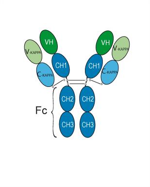
Tuparstobart (Incagn-02385) is an IgG1κ antibody targeting LAG-3. LAG-3 is an immune checkpoint receptor protein mainly expressed on activated T cells, NK cells, B cells and plasmacytoid dendritic cells.
Para uso exclusivo en investigación. No vendemos a pacientes.
- Pureza : 97.65%
- No. CAS: 2649466-03-3
- Peso molecular:145.699 kDa
-
Almacenamiento:
Please store the product under the recommended conditions in the Certificate of Analysis.


To place orders, for customer services and technical support, please contact: MedChemExpress USA
Tel: 609-228-6898 E-mail: [email protected] [email protected]


-
 Actividad biológica
-
Chemical Information
- Protocolo
- Pureza y Documentación
-
 Help & FAQs




Actividad biológica
Descripciòn
Isotype
Human IgG1 kappa
Recommend Isotype Controls
Species Reactivity
Human
IC50 & Target
LAG3/CD223
Ensayo clínico
| NCT Number | Sponsor | Condition | Start Date |
Phase

|
|---|---|---|---|---|
| NCT01329991 | Plexxikon| | 2011-05 | PHASE1 |
Gene ID
Accession
Conjugated
Unconjugated
Reconsititution
The product can be reconstituted/diluted with sterile PBS or saline.
Format
-
Human IgG1 kappa
Aplicación
ELISA, FACS, Functional assay
Verified Bioactivity
-
 Immobilized LAG-3 Protein, Human (HEK293, His, HY-P70722) can bind Tuparstobart. The EC50 for this effect is 67.31 ng/mL.
 -
 Flow cytometric analysis of 1X10⁶ Jurkat cells with Tuparstobart (HY-P990046, red). Cells were fixed with 4% paraformaldehyde. Then stained with the primary antibody at 1/200 dilution for an hour at 4℃. Alexa Fluor 488-conjugated AffiniPure Goat Anti-Human IgG H&L (HY-P83776) was used as the secondary antibody at 1/1,000 dilution for 30 minutes at 4℃. Human IgG1 kappa (HY-P99001, blue) was used as the isotype control, cells without incubation with primary antibody were used as the unlabeled control (black).


Chemical Information
-
No. CAS 2649466-03-3
-
Appearance Liquid
-
Peso molecular 145.699 kDa
-
Color Colorless to light yellow
-
SMILES
[Tuparstobart]
-
Synonyms
Incagn-02385
-
Envío
Shipping with dry ice.
-
Formulation
Please refer to the lot-specific COA for specific buffer information.
-
Almacenamiento
Please store the product under the recommended conditions in the Certificate of Analysis.


Pureza y Documentación
-
Ficha de datos (229 KB)
-
SDS (251 KB)
- English - EN (251 KB)
- Français - FR (251 KB)
- Deutsch - DE (251 KB)
- Norwegian - NO (251 KB)
- Español - ES (251 KB)
- Swedish - SV (251 KB)
- Italian - IT (251 KB)
- Korean - KR (251 KB)
- Portuguese - PT (251 KB)
-
Inhibitory Antibodies User Guide (603 KB)

Calculators
Concentration (start) × Volume (start) = Concentration (final) × Volume (final)


Tuparstobart Related Classifications
HY-P990046 Related Classifications
 Powered by Bioz