



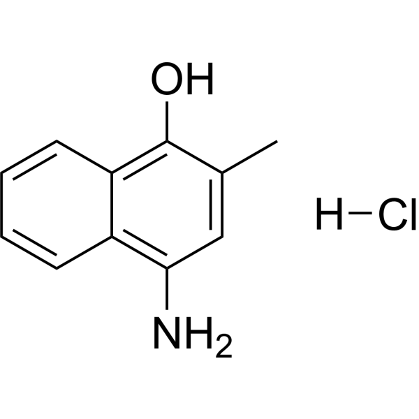
Vitamin K5 hydrochloride

Based on 1 Customer Validation
Vitamin K5 hydrochloride is a photosensitizer and a antimicrobial agent. Vitamin K5 hydrochloride is a specific PKM2 inhibitor with IC50 values of 28, 191 and 120 μM for PKM2, PKM1 and PKL. Vitamin K5 hydrochloride induces apoptosis of colon 26 cells. Vitamin K5 hydrochloride can be used for the research of infection and cancer, and it also can be used as a preservative for pharmaceuticals, foods, and beverages.
For research use only. We do not sell to patients.
- Purity : 99.25%
- CAS No.: 130-24-5
- Formula: C11H12ClNO
- Molecular Weight:209.68
-
Storage:
-20°C, protect from light, stored under nitrogen
* In solvent : -80°C, 6 months; -20°C, 1 month (protect from light, stored under nitrogen)

To place orders, for customer services and technical support, please contact: MedChemExpress USA
Tel: 609-228-6898 E-mail: [email protected] [email protected]


-
 Biological Activity
-
Chemical Information
-
Solvent & Solubility
- Protocols
- Purity & Documentation
- References
-
 Help & FAQs




Biological Activity
Description
In Vitro
Vitamin K5 (0-1600 μM) hydrochloride increases the antibacterial effects of UVA irradiation to Escherichia coli, Bacillus cereus, Pseudomonas aeruginosa, Staphylococcus aureus, Staphylococcus epidermidis and Klebsiella pneumoniae[1]. Vitamin K5 (250-1000 μM) hydrochloride generates superoxide anion radical and hydroxyl radical with a fixed UVA dose of 2.9 J cm-2[1]. Vitamin K5 (0-100 μM; 1 h) hydrochloride dose-dependently inhibits the cellular glucose consumption and lactate production[2]. Vitamin K5 hydrochloride dose-dependently suppresses the proliferation of colon 26 cells[3]. Vitamin K5 hydrochloride induces cell apoptosis and increases activity of caspase-3 in colon 26 cells[3].
MedChemExpress (MCE) has not independently confirmed the accuracy of these methods. They are for reference only. Further protocols information, click here.


Chemical Information
-
CAS No. 130-24-5
-
Appearance Solid
-
Molecular Weight 209.68
-
Formula C11H12ClNO
-
Color Light yellow to light brown
-
SMILES
OC1=C2C=CC=CC2=C(N)C=C1C.[H]Cl
-
Shipping
Room temperature in continental US; may vary elsewhere.
-
Storage
-20°C, protect from light, stored under nitrogen
* In solvent : -80°C, 6 months; -20°C, 1 month (protect from light, stored under nitrogen)


Solvent & Solubility
In Vitro:
DMSO : 125 mg/mL (596.16 mM; Need ultrasonic; Hygroscopic DMSO has a significant impact on the solubility of product, please use newly opened DMSO)
Please refer to the solubility information to select the appropriate solvent. Once prepared, please aliquot and store the solution to prevent product inactivation from repeated freeze-thaw cycles.
Storage method and period of stock solution: -80°C, 6 months; -20°C, 1 month (protect from light, stored under nitrogen). When stored at -80°C, please use it within 6 months. When stored at -20°C, please use it within 1 month.
Please refer to the solubility information to select the appropriate solvent. Once prepared, please aliquot and store the solution to prevent product inactivation from repeated freeze-thaw cycles.
Storage method and period of stock solution: -80°C, 6 months; -20°C, 1 month (protect from light, stored under nitrogen). When stored at -80°C, please use it within 6 months. When stored at -20°C, please use it within 1 month.
Concentration (start) × Volume (start) = Concentration (final) × Volume (final)

Protocols
-
Research Protocol for Infectious Diseases
Infectious-disease experiments test how pathogens interact with host barriers, innate immune receptors, inflammatory signaling, pathogen replication, and tissue injury; pattern-recognition receptors such as TLRs, RIG-I-like receptors, NOD-like receptors, and inflammasomes detect microbial molecules and activate NF-κB, interferon, and cytokine responses. The central hypothesis is that infection severity reflects the balance between pathogen burden and host response: protective inflammation restricts pathogen growth, whereas excessive or mislocalized inflammation contributes to tissue damage and disease phenotype. Unresolved questions include which host pathways are protective versus pathogenic, why some infection models fail to translate to human disease, and which combined readouts best predict clinically relevant infection outcomes.
-
Apoptosis
Apoptosis, also called programmed cell death, is generally characterized by distinct morphological characteristics.
-
TUNEL staining for apoptotic DNA fragmentation
TUNEL staining detects DNA strand breaks by using terminal deoxynucleotidyl transferase to add labeled nucleotides to exposed 3′-OH DNA termini, generating either microscopic staining in fixed cells or tissue sections, or fluorescence/cytometric signal in cell suspensions. TUNEL positivity reflects DNA fragmentation but should not be interpreted alone as definitive apoptosis, because TUNEL can also label necrotic, autolytic, mechanically damaged, or DNA-repair-associated DNA breaks.
-
Cell Cytotoxicity Assay
Cytotoxicity assays are usually based on the assessment of cell membrane damage, which can also be indirectly detected by measuring cell viability. Detection methods include MTT assay, CKK-8 assay, LDH assay and ATP assay, etc.
-
Annexin V plus membrane-impermeant dye apoptosis staining
Annexin V-based apoptosis assays rely on the detection of phosphatidylserine (PS) externalization from the inner leaflet of the plasma membrane to the outer leaflet, an early biochemical hallmark of apoptosis. Fluorescently labeled Annexin V binds PS in a calcium-dependent manner, enabling identification of early apoptotic cells by flow cytometry or fluorescence microscopy. When combined with a membrane-impermeant DNA-binding dye (e. g. , propidium iodide), this approach allows discrimination between viable (Annexin V−/dye−), early apoptotic (Annexin V+/dye−), and late apoptotic or necrotic (Annexin V+/dye+) cell populations by assessing membrane integrity and PS exposure.
-
Apoptosis Solutions
Apoptosis is a regulated, generally non-lytic cell-death pathway that removes unwanted, damaged, infected, or abnormal cells through coordinated morphological changes, caspase activation, DNA fragmentation, and membrane remodeling. The intrinsic apoptosis pathway is controlled mainly by mitochondrial outer membrane permeabilization, BCL-2 family proteins, cytochrome c release, apoptosome formation, caspase-9 activation, and downstream executioner caspase-3/7 activation. The extrinsic apoptosis pathway is initiated by death receptors such as Fas, TNFR, and TRAIL receptors, which recruit adaptor proteins and activate caspase-8 before engaging executioner caspases or mitochondrial amplification through BID cleavage. Apoptosis is linked to many phenotypes, including cancer cell killing, tissue homeostasis, immune regulation, neurodegeneration, infection response, and treatment-induced cytotoxicity; unresolved questions include how apoptosis interacts with necroptosis, pyroptosis, ferroptos


Purity & Documentation
-
Data Sheet (273 KB)
-
SDS (394 KB)
- English - EN (394 KB)
- Français - FR (394 KB)
- Deutsch - DE (394 KB)
- Norwegian - NO (394 KB)
- Español - ES (394 KB)
- Swedish - SV (394 KB)
- Italian - IT (394 KB)
- Korean - KR (394 KB)
- Portuguese - PT (394 KB)
-
Handling Instructions (2659 KB)
References
[1]. Xu F, et al. Vitamin K5 is an efficient photosensitizer for ultraviolet A light inactivation of bacteria. FEMS Microbiol Lett. 2018 Feb 1;365(4). [Content Brief]
[2]. Chen J, et al. Vitamin K(3) and K(5) are inhibitors of tumor pyruvate kinase M2. Cancer Lett. 2012 Mar 28;316(2):204-10. [Content Brief]
[3]. Ogawa M, et al. Vitamins K2, K3 and K5 exert antitumor effects on established colorectal cancer in mice by inducing apoptotic death of tumor cells. Int J Oncol. 2007 Aug;31(2):323-31. [Content Brief]


Complete Stock Solution Preparation Table
Please refer to the solubility information to select the appropriate solvent. Once prepared, please aliquot and store the solution to prevent product inactivation from repeated freeze-thaw cycles.
Storage method and period of stock solution: -80°C, 6 months; -20°C, 1 month (protect from light, stored under nitrogen). When stored at -80°C, please use it within 6 months. When stored at -20°C, please use it within 1 month.
| Optional Solvent | Concentration Solvent Mass | 1 mg | 5 mg | 10 mg | 25 mg |
|---|---|---|---|---|---|
| DMSO | 1 mM | 4.7693 mL | 23.8463 mL | 47.6926 mL | 119.2316 mL |
| 5 mM | 0.9539 mL | 4.7693 mL | 9.5385 mL | 23.8463 mL | |
| 10 mM | 0.4769 mL | 2.3846 mL | 4.7693 mL | 11.9232 mL | |
| 15 mM | 0.3180 mL | 1.5898 mL | 3.1795 mL | 7.9488 mL | |
| 20 mM | 0.2385 mL | 1.1923 mL | 2.3846 mL | 5.9616 mL | |
| 25 mM | 0.1908 mL | 0.9539 mL | 1.9077 mL | 4.7693 mL | |
| 30 mM | 0.1590 mL | 0.7949 mL | 1.5898 mL | 3.9744 mL | |
| 40 mM | 0.1192 mL | 0.5962 mL | 1.1923 mL | 2.9808 mL | |
| 50 mM | 0.0954 mL | 0.4769 mL | 0.9539 mL | 2.3846 mL | |
| 60 mM | 0.0795 mL | 0.3974 mL | 0.7949 mL | 1.9872 mL | |
| 80 mM | 0.0596 mL | 0.2981 mL | 0.5962 mL | 1.4904 mL | |
| 100 mM | 0.0477 mL | 0.2385 mL | 0.4769 mL | 1.1923 mL |


Vitamin K5 hydrochloride Related Classifications
HY-N2552 Related Classifications
 Powered by Bioz