



Xenin-8
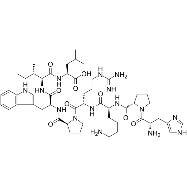
Xenin-8 is the C-terminal octapeptide fragment of xenin, which belongs to the xenopsin family of peptides. Xenin-8 stimulates basal insulin secretion, dose-dependently enhances glucose (EC50 = 0.16 nM) and arginine-induced insulin release, and enhances arginine- and Carbamoylcholine chloride (HY-B1208)-induced glucagon secretion. Xenin-8 antagonizes the inhibition of glucagon release by elevated glucose. Xenin-8 can be used in research on type 2 diabetes and obesity-related glucose metabolic disorders.
Para uso exclusivo en investigación. No vendemos a pacientes.
- No. CAS: 117442-28-1
- Fòrmula: C51H79N15O9
- Peso molecular:1046.27
-
Almacenamiento:
Please store the product under the recommended conditions in the Certificate of Analysis.

To place orders, for customer services and technical support, please contact: MedChemExpress USA
Tel: 609-228-6898 E-mail: [email protected] [email protected]


-
 Actividad biológica
-
Chemical Information
-
Solvente y solubilidad
- Protocolo
- Pureza y Documentación
- Referencias
-
 Help & FAQs


-
HY-P0234
NeurotensinNeurotensin Receptor Agonist -
HY-P1255
KinetensinEndogenous Metabolite -
HY-107664
SR 142948NT Antagonist -
HY-P1256
JMV 449Neurotensin Receptor Agonist -
HY-P1777
IGF-I (24-41)IGF-I Fragment -

HY-107664R
SR 142948 (Standard)Reference Standard -
HY-P1773
IGF-I (30-41)Anti-Inflammatory Agent -

HY-P1901
Insulin alpha-chain (1-13)(HLA)-DR4-Restricted Epitope -

HY-P2080
GIP (1-30) amide,humanGIP Fragment


Actividad biológica
Descripciòn
In Vitro
Xenin-8 is able to antagonize the inhibition of glucagon release caused by elevated glucose concentrations[1].
MedChemExpress (MCE) has not independently confirmed the accuracy of these methods. They are for reference only.


Chemical Information
-
No. CAS 117442-28-1
-
Peso molecular 1046.27
-
Fòrmula C51H79N15O9
-
Sequence
His-Pro-Lys-Arg-Pro-Trp-Ile-Leu
-
Sequence Shortening
HPKRPWIL
-
Envío
Room temperature in continental US; may vary elsewhere.
-
Almacenamiento
Please store the product under the recommended conditions in the Certificate of Analysis.


Solvente y solubilidad
In Vitro
H2O
Peptide Solubility and Storage Guidelines:
1. Calculate the length of the peptide.
2. Calculate the overall charge of the entire peptide according to the following table:
| Contents | Assign value | |
|---|---|---|
| Acidic amino acid | Asp (D), Glu (E), and the C-terminal -COOH. | -1 |
| Basic amino acid | Arg (R), Lys (K), His (H), and the N-terminal -NH2 | +1 |
| Neutral amino acid | Gly (G), Ala (A), Leu (L), Ile (I), Val (V), Cys (C), Met (M), Thr (T), Ser (S), Phe (F), Tyr (Y), Trp (W), Pro (P), Asn (N), Gln (Q) | 0 |
3. Recommended solution:
| Overall charge of peptide | Details |
|---|---|
| Negative (<0) |
1. Try to dissolve the peptide in water first. 2. If water fails, add NH4OH (<50 μL). 3. If the peptide still does not dissolve, add DMSO (50-100 μL) to solubilize the peptide. |
| Positive (>0) |
1. Try to dissolve the peptide in water first. 2. If water fails, try dissolving the peptide in a 10%-30% acetic acid solution. 3. If the peptide still does not dissolve, try dissolving the peptide in a small amount of DMSO. |
| Zero (=0) |
1. Try to dissolve the peptide in organic solvent (acetonitrile, methanol, etc.) first. 2. For very hydrophobic peptides, try dissolving the peptide in a small amount of DMSO, and then dilute the solution with water to the desired concentration. |


Pureza y Documentación
Referencias

Calculators
Concentration (start) × Volume (start) = Concentration (final) × Volume (final)


Xenin-8 Related Classifications
HY-P1257 Related Classifications
 Powered by Bioz