



α-Secretase Substrate II, Fluorogenic
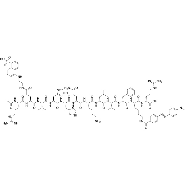
α-Secretase Substrate II, Fluorogenic is an internally quenched fluorogenic peptide substrate for α-Secretase that contains the α-secretase cleavage site of β-Amyloid precursor protein (APP).Ex/Em = 340/490 nm
For research use only. We do not sell to patients.
- Formula: C100H144N30O20S
- Molecular Weight:2118.47
-
Storage:
Please store the product under the recommended conditions in the Certificate of Analysis.

To place orders, for customer services and technical support, please contact: MedChemExpress USA
Tel: 609-228-6898 E-mail: [email protected] [email protected]


-
 Biological Activity
-
Chemical Information
- Protocols
- Purity & Documentation
- References
-
 Help & FAQs




Biological Activity
Description


Chemical Information
-
Molecular Weight 2118.47
-
Formula C100H144N30O20S
-
Sequence
Ac-Arg-{Glu(EDANS)}-Val-His-His-Gln-Lys-Leu-Val-Phe-{Lys(DABCYL)}-Arg
-
Sequence Shortening
Ac-R-{Glu(EDANS)}-VHHQKLVF-{Lys(DABCYL)}-R
-
Shipping
Room temperature in continental US; may vary elsewhere.
-
Storage
Please store the product under the recommended conditions in the Certificate of Analysis.


Purity & Documentation
References

Calculators
Concentration (start) × Volume (start) = Concentration (final) × Volume (final)


α-Secretase Substrate II, Fluorogenic Related Classifications
HY-P10163 Related Classifications
 Powered by Bioz