



18-Hydroxy-11-deoxy corticosterone

Based on 1 Customer Validation
18-Hydroxy-11-deoxy corticosterone (18-OH-DOC) is a mineralocorticoid whose synthesis is regulated by adrenocorticotropic hormone (ACTH) and angiotensin II. 18-Hydroxy-11-deoxy corticosterone is an intermediate in the metabolism of progesterone and plays an important role in regulating blood pressure and water-salt balance. Continuous infusion of 18-Hydroxy-11-deoxy corticosterone can increase systolic blood pressure in rats, and plasma levels of 18-Hydroxy-11-deoxy corticosterone are significantly elevated in the db/db mouse model of type 2 diabetes, suggesting its potential involvement in metabolic dysregulation and diabetes-related regulation. 18-Hydroxy-11-deoxy corticosterone holds promise for research in areas such as hypertension, diabetes, and other related fields.
For research use only. We do not sell to patients.
- CAS No.: 379-68-0
- Formula: C21H30O4
- Molecular Weight:346.46
-
Storage:
Please store the product under the recommended conditions in the Certificate of Analysis.


To place orders, for customer services and technical support, please contact: MedChemExpress USA
Tel: 609-228-6898 E-mail: [email protected] [email protected]


-
 Biological Activity
-
Chemical Information
- Protocols
- Purity & Documentation
- References
-
 Help & FAQs


-
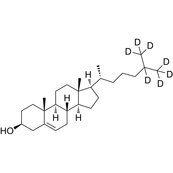
HY-N0322S
Cholesterol-d7Stable Isotope -
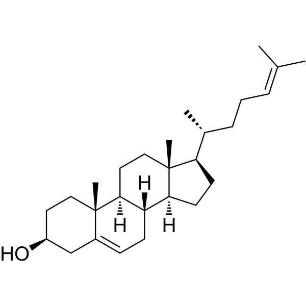
HY-113224
DesmosterolEndogenous Metabolite -
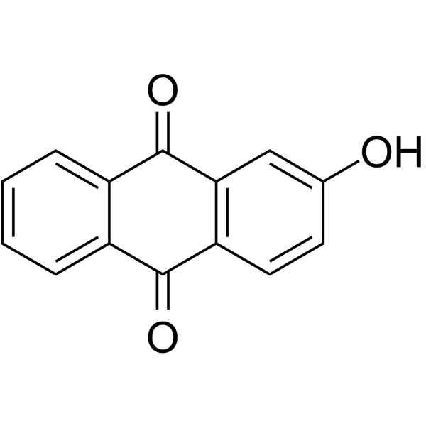
HY-W012352
2-HydroxyanthraquinoneWnt/β-catenin Inhibitor -
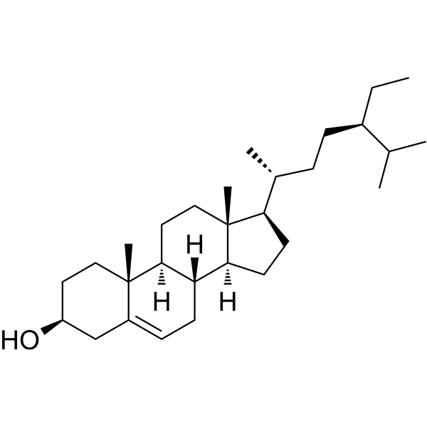
HY-N0171A
Beta-Sitosterol (purity>98%)Dietary Plant Sterols -
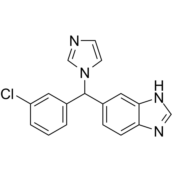
HY-106019
LiarozoleRAMBA -

HY-Z7721
(Rac)-Beraprost sodiumRacemic Isomer of Beraprost sodium -

HY-113224R
Desmosterol (Standard)Endogenous Metabolite -

HY-139414
LysophosphatidylcholinesHemolytic Agent -
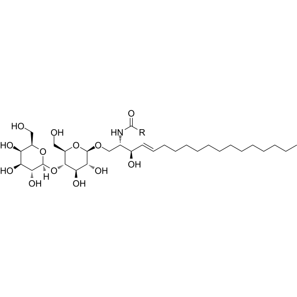
HY-154921
Lactosylceramide (bovine buttermilk)Glycosphingolipid -
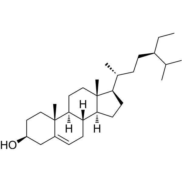
HY-N0171
Beta-Sitosterol (purity>80%)Anticancer Nutraceutical -

HY-P0014
LiraglutideGLP-1R Agonist -
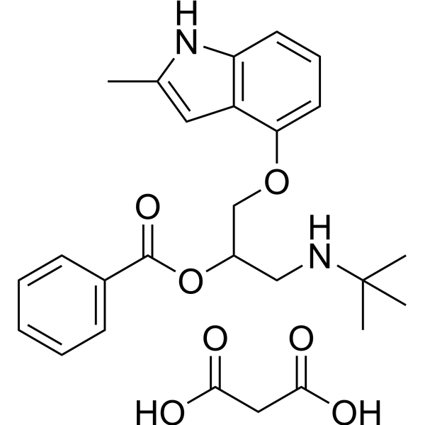
HY-B1562B
Bopindolol malonateβ-Adrenoceptors Antagonist
All Angiotensin Receptor Isoforms
More

Biological Activity
Description


Chemical Information
-
CAS No. 379-68-0
-
Molecular Weight 346.46
-
Formula C21H30O4
-
SMILES
OC[C@@]12[C@](CC[C@@H]2C(CO)=O)([H])[C@@]3([H])[C@]([C@@]4(C(CC3)=CC(CC4)=O)C)([H])CC1
-
Synonyms
18-OH-DOC; 11-Deoxy-18-hydroxycorticosterone
-
Shipping
Room temperature in continental US; may vary elsewhere.
-
Storage
Please store the product under the recommended conditions in the Certificate of Analysis.

Protocols
-
Research Protocol for Endocrine Diseases
Endocrine diseases often arise from disrupted hormone production, hormone signaling, or target-tissue responsiveness; for diabetes-focused endocrine disease models, insulin signaling regulates glucose uptake, hepatic glucose output, lipid metabolism, and β-cell compensation. Type 2 diabetes develops through interacting defects in insulin resistance, β-cell dysfunction, adipose inflammation, hepatic glucose overproduction, altered incretin signaling, and ectopic lipid metabolism. A major unresolved question is whether endocrine dysfunction is driven primarily by target-tissue insulin resistance, intrinsic β-cell failure, immune/inflammatory stress, or combined multi-organ failure that differs by disease stage.
-
Research Protocol for Cardiovascular Diseases
Cardiovascular disease can be modeled as maladaptive cardiac remodeling, where ischemic injury or pressure overload activates inflammatory signaling, fibroblast activation, extracellular-matrix deposition, cardiomyocyte hypertrophy, vascular remodeling, and progressive ventricular dysfunction. The TGF-β/SMAD axis is a central profibrotic pathway after myocardial injury and pressure overload, while innate immune and cytokine pathways regulate leukocyte recruitment, scar formation, and adverse remodeling. Key unresolved questions include which inflammatory signals are reparative versus harmful, when fibrosis is protective versus maladaptive, and whether pathway inhibition improves function without weakening necessary infarct healing or compensatory remodeling.
-
Research Protocol for Metabolic Diseases
AMP-activated protein kinase, AMPK, is a conserved cellular energy sensor that responds to reduced cellular energy status and coordinates metabolism by increasing ATP-generating catabolic pathways while suppressing ATP-consuming anabolic processes. In metabolic disease research, the AMPK pathway is experimentally relevant because it regulates hepatic lipid synthesis, fatty acid oxidation, glucose production, skeletal-muscle glucose disposal, mTORC1-linked biosynthesis, autophagy, mitochondrial homeostasis, and whole-body energy balance. The central pathway logic is that energy stress, metformin, exercise-like stimulation, or direct AMPK activators increase AMPKα Thr172 phosphorylation and downstream substrate phosphorylation, including ACC and RAPTOR. Phosphorylation of ACC suppresses lipogenesis and supports fatty acid oxidation, whereas phosphorylation of RAPTOR suppresses mTORC1 signaling and links cellular energy status to growth and protein synthesis control. The pathway is linked
-
How to Choose the Right Model Animal
Choosing the right model animal is a validity-driven decision in which the species, strain, sex, age, genetic background, disease-induction method, outcome measures, and welfare burden must match the scientific question rather than laboratory tradition or convenience. A model should be selected by judging face validity, construct validity, and predictive validity: whether it resembles the human phenotype, whether it reproduces relevant mechanisms, and whether results are likely to predict human biology or treatment response. Animal studies often fail to translate because of species differences, weak disease resemblance, poor experimental design, inadequate reporting, publication bias, and underuse of randomization, blinding, and sample-size justification. Unresolved questions include how to rank competing models objectively, how much human-disease complexity must be reproduced for a given objective, and when non-animal systems such as organoids, ex vivo tissue, or computational models


Purity & Documentation
References
[1]. Braley LM, et al. The effect of angiotensin II and saralasin on 18-hydroxy-11-deoxycorticosterone production by isolated human adrenal glomerulosa cells. J Clin Endocrinol Metab. 1979 Oct;49(4):600-3. [Content Brief]
[2]. Carroll J, et al. The relationship between plasma 18-hydroxy-11-deoxycorticosterone levels and production of hypertension in the rat. J Steroid Biochem. 1981 Oct;14(10):989-95. [Content Brief]
[3]. Müller J. The conversion of 18-hydroxycorticosterone and 18-hydroxy-11-deoxycorticosterone to aldosterone by rat adrenal tissue: evidence for an alternative biosynthetic pathway. J Steroid Biochem. 1980 Mar;13(3):245-51. [Content Brief]
[4]. Slominski A, et al. Metabolism of progesterone to DOC, corticosterone and 18OHDOC in cultured human melanoma cells. FEBS Lett. 1999 Jul 23;455(3):364-6. [Content Brief]
[5]. Giesbertz P, et al. Metabolite profiling in plasma and tissues of ob/ob and db/db mice identifies novel markers of obesity and type 2 diabetes. Diabetologia. 2015 Sep;58(9):2133-43. [Content Brief]

Calculators
Concentration (start) × Volume (start) = Concentration (final) × Volume (final)


18-Hydroxy-11-deoxy corticosterone Related Classifications
HY-W738639 Related Classifications
 Powered by Bioz