



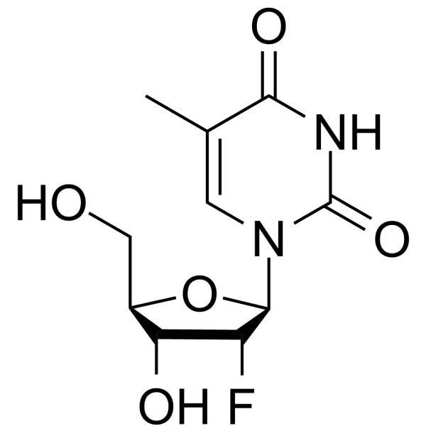
2'-Fluorothymidine

Based on 1 Customer Validation
2'-Fluorothymidine (2'-Fluoro-2'-deoxythymidine) is a nucleoside analog and hCNT inhibitor, with an IC50 of 1.1 μM against hCNT1 and an IC50 of 9.7 μM against hCNT3. 2'-Fluorothymidine undergoes phosphorylation by cytosolic thymidine kinase (TK1) and mitochondrial thymidine kinase (TK2). 2'-Fluorothymidine acts as a backbone stabilizer in oligonucleotide synthesis and can also form radiolabeled candidates. 2'-Fluorothymidine is applicable in tumor-related research.
For research use only. We do not sell to patients.
- Purity : 99.07%
- CAS No.: 122799-38-6
- Formula: C10H13FN2O5
- Molecular Weight:260.22
-
Storage:Powder -20°C, 3 years , 4°C, 2 years ; In solvent -80°C, 6 months , -20°C, 1 month

To place orders, for customer services and technical support, please contact: MedChemExpress USA
Tel: 609-228-6898 E-mail: [email protected] [email protected]


-
 Biological Activity
-
Chemical Information
-
Solvent & Solubility
- Protocols
- Purity & Documentation
- References
-
 Help & FAQs




Biological Activity
Description
In Vitro
2'-Fluorothymidine (0.0001-1 mM; 3 days) exhibits minimal cytotoxicity toward murine EMT-6, KBALB, KBALB-STK cells, and human 143B cells, with an IC50 above 1 mM, while human 143B-LTK cells show 50% survival at 1 mM[1].
2'-Fluorothymidine (Graded concentrations; 10 min) inhibits [3H]uridine uptake via recombinant human nucleoside transporters expressed in Saccharomyces cerevisiae with IC50 values of 107 µM (hENT1), 342 µM (hENT2), 1.1 µM (hCNT1), 326 µM (hCNT2), and 9.7 µM (hCNT3), showing higher affinity than FLT for all tested transporters[1].
2'-Fluorothymidine (10-100 µM (TK1); 12-50 µM (TK2); 20 min) is phosphorylated by human TK1 at 58% of the activity of thymidine (327 nmol/mg/min) and by human TK2 at 39% of the activity of thymidine (377 nmol/mg/min), with TK2 activity toward FT being ~6-fold higher than toward FLT[1].
2'-Fluorothymidine (0.2 mCi/well; 1 h, 6 h, 12 h, 24 h) uptake increases ~10-fold over 24 h in murine KBALB, KBALB-STK cells, and human 143B, 143B-LTK cells, with significantly higher uptake in HSV-1 TK-expressing KBALB-STK and 143B-LTK cells compared to their non-transduced counterparts[1].
MedChemExpress (MCE) has not independently confirmed the accuracy of these methods. They are for reference only.
-
Cell Line:murine EMT-6, KBALB, KBALB-STK cells, human 143B, 143B-LTK cells
-
Concentration:0.0001-1 mM
-
Incubation Time:3 days
-
Result:Exhibits minimal cytotoxicity at concentrations below 0.1 mM across all five cell lines.
Induces 50% survival in 143B-LTK cells at 1 mM.
Requires a concentration above 1 mM to achieve 50% survival in all other cell lines.
In Vivo
MedChemExpress (MCE) has not independently confirmed the accuracy of these methods. They are for reference only.


Chemical Information
-
CAS No. 122799-38-6
-
Appearance Solid
-
Molecular Weight 260.22
-
Formula C10H13FN2O5
-
Color White to off-white
-
SMILES
OC[C@@H]1[C@@H](O)[C@@H](F)[C@H](N2C(NC(C(C)=C2)=O)=O)O1
-
Synonyms
2'-Deoxy-2'-fluoro-5-Methyluridine
-
Shipping
Room temperature in continental US; may vary elsewhere.
-
Storage
Powder -20°C 3 years 4°C 2 years In solvent -80°C 6 months -20°C 1 month


Solvent & Solubility
In Vitro:
DMSO : 125 mg/mL (480.36 mM; Need ultrasonic; Hygroscopic DMSO has a significant impact on the solubility of product, please use newly opened DMSO)
Please refer to the solubility information to select the appropriate solvent. Once prepared, please aliquot and store the solution to prevent product inactivation from repeated freeze-thaw cycles.
Storage method and period of stock solution: -80°C, 6 months; -20°C, 1 month. When stored at -80°C, please use it within 6 months. When stored at -20°C, please use it within 1 month.
Please refer to the solubility information to select the appropriate solvent. Once prepared, please aliquot and store the solution to prevent product inactivation from repeated freeze-thaw cycles.
Storage method and period of stock solution: -80°C, 6 months; -20°C, 1 month. When stored at -80°C, please use it within 6 months. When stored at -20°C, please use it within 1 month.
Concentration (start) × Volume (start) = Concentration (final) × Volume (final)


Purity & Documentation
-
Data Sheet (277 KB)
-
SDS (252 KB)
- English - EN (252 KB)
- Français - FR (252 KB)
- Deutsch - DE (252 KB)
- Norwegian - NO (252 KB)
- Español - ES (252 KB)
- Swedish - SV (252 KB)
- Italian - IT (252 KB)
- Korean - KR (252 KB)
- Portuguese - PT (252 KB)
-
Handling Instructions (2659 KB)
References


Complete Stock Solution Preparation Table
Please refer to the solubility information to select the appropriate solvent. Once prepared, please aliquot and store the solution to prevent product inactivation from repeated freeze-thaw cycles.
Storage method and period of stock solution: -80°C, 6 months; -20°C, 1 month. When stored at -80°C, please use it within 6 months. When stored at -20°C, please use it within 1 month.
| Optional Solvent | Concentration Solvent Mass | 1 mg | 5 mg | 10 mg | 25 mg |
|---|---|---|---|---|---|
| DMSO | 1 mM | 3.8429 mL | 19.2145 mL | 38.4290 mL | 96.0726 mL |
| 5 mM | 0.7686 mL | 3.8429 mL | 7.6858 mL | 19.2145 mL | |
| 10 mM | 0.3843 mL | 1.9215 mL | 3.8429 mL | 9.6073 mL | |
| 15 mM | 0.2562 mL | 1.2810 mL | 2.5619 mL | 6.4048 mL | |
| 20 mM | 0.1921 mL | 0.9607 mL | 1.9215 mL | 4.8036 mL | |
| 25 mM | 0.1537 mL | 0.7686 mL | 1.5372 mL | 3.8429 mL | |
| 30 mM | 0.1281 mL | 0.6405 mL | 1.2810 mL | 3.2024 mL | |
| 40 mM | 0.0961 mL | 0.4804 mL | 0.9607 mL | 2.4018 mL | |
| 50 mM | 0.0769 mL | 0.3843 mL | 0.7686 mL | 1.9215 mL | |
| 60 mM | 0.0640 mL | 0.3202 mL | 0.6405 mL | 1.6012 mL | |
| 80 mM | 0.0480 mL | 0.2402 mL | 0.4804 mL | 1.2009 mL | |
| 100 mM | 0.0384 mL | 0.1921 mL | 0.3843 mL | 0.9607 mL |


2'-Fluorothymidine Related Classifications
HY-128710 Related Classifications
 Powered by Bioz