



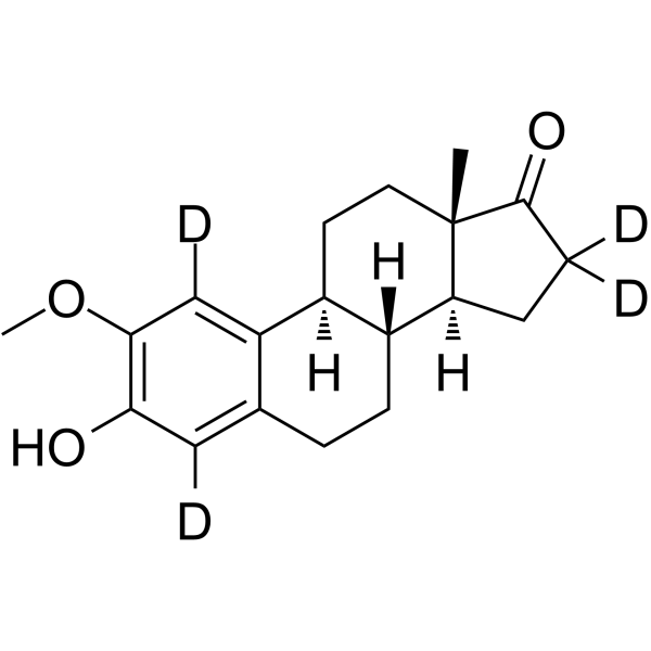
2-Methoxyestrone-d4

Based on 1 Customer Validation
2-Methoxyestrone-d4 is the deuterated-labeled 2-Methoxyestrone (HY-113252). 2-Methoxyestrone is an estrogen metabolite with antimitotic activity. 2-Methoxyestrone acts as a liver-specific prohormone that undergoes demethylation to form active 2-hydroxyestrogens, and it can also interconvert with 2-Methoxyestradiol (HY-12033) via the 17β-hydroxysteroid dehydrogenase pathway. In the presence of all-trans retinoic acid, 2-Methoxyestrone exerts a strong, concentration-dependent inhibitory effect on cell growth. 2-Methoxyestrone delays the progression of pulmonary hypertension in rats, alleviates right ventricular and pulmonary vascular remodeling, and relieves proliferative pulmonary hypertension in rats. 2-Methoxyestrone can be used in studies related to proliferative pulmonary hypertension.
For research use only. We do not sell to patients.
- Purity : 98.0%
- CAS No.: 949885-90-9
- Formula: C19H20D4O3
- Molecular Weight:304.42
-
Storage:Powder -20°C, 3 years , 4°C, 2 years ; In solvent -80°C, 6 months , -20°C, 1 month

To place orders, for customer services and technical support, please contact: MedChemExpress USA
Tel: 609-228-6898 E-mail: [email protected] [email protected]


-
 Biological Activity
-
Chemical Information
-
Solvent & Solubility
- Protocols
- Purity & Documentation
- References
-
 Help & FAQs


All Endogenous Metabolite Isoforms
More

Biological Activity
Description
In Vitro
Stable heavy isotopes of hydrogen, carbon, and other elements have been incorporated into drug molecules, largely as tracers for quantitation during the drug development process. Deuteration has gained attention because of its potential to affect the pharmacokinetic and metabolic profiles of drugs.
MedChemExpress (MCE) has not independently confirmed the accuracy of these methods. They are for reference only.
- Protocol for Cell Counting and Cell Density Analysis
- Research Protocol for Endocrine Diseases
- Somatic Cell Culture
- CCK-8/WST-8 Cell Proliferation Assay
- Research Protocol for Cardiovascular Diseases
- Cell Counting-Based Growth Curve Assay
- MTT Cell Proliferation Assay
- Cell Viability Determination by MTT Colorimetric Assay
Application
1. This compound can be used as a tracer
2. This compound can be used as an internal standard for quantitative analysis by NMR, GC-MS, or LC-MS.


Chemical Information
-
CAS No. 949885-90-9
-
Unlabeled CAS 362-08-3
-
Appearance Solid
-
Molecular Weight 304.42
-
Formula C19H20D4O3
-
Color White to yellow
-
SMILES
C[C@@]12[C@](CC([2H])([2H])C2=O)([H])[C@@]3([H])[C@@](CC1)([H])C4=C([2H])C(OC)=C(O)C([2H])=C4CC3
-
Shipping
Room temperature in continental US; may vary elsewhere.
-
Storage
Powder -20°C 3 years 4°C 2 years In solvent -80°C 6 months -20°C 1 month


Solvent & Solubility
In Vitro:
DMSO : 50 mg/mL (164.25 mM; Need ultrasonic; Hygroscopic DMSO has a significant impact on the solubility of product, please use newly opened DMSO)
Please refer to the solubility information to select the appropriate solvent. Once prepared, please aliquot and store the solution to prevent product inactivation from repeated freeze-thaw cycles.
Storage method and period of stock solution: -80°C, 6 months; -20°C, 1 month. When stored at -80°C, please use it within 6 months. When stored at -20°C, please use it within 1 month.
Please refer to the solubility information to select the appropriate solvent. Once prepared, please aliquot and store the solution to prevent product inactivation from repeated freeze-thaw cycles.
Storage method and period of stock solution: -80°C, 6 months; -20°C, 1 month. When stored at -80°C, please use it within 6 months. When stored at -20°C, please use it within 1 month.
Concentration (start) × Volume (start) = Concentration (final) × Volume (final)


Purity & Documentation
-
Data Sheet (287 KB)
-
SDS (251 KB)
- English - EN (251 KB)
- Français - FR (251 KB)
- Deutsch - DE (251 KB)
- Norwegian - NO (251 KB)
- Español - ES (251 KB)
- Swedish - SV (251 KB)
- Italian - IT (251 KB)
- Korean - KR (251 KB)
- Portuguese - PT (251 KB)
-
Handling Instructions (2659 KB)
References


Complete Stock Solution Preparation Table
Please refer to the solubility information to select the appropriate solvent. Once prepared, please aliquot and store the solution to prevent product inactivation from repeated freeze-thaw cycles.
Storage method and period of stock solution: -80°C, 6 months; -20°C, 1 month. When stored at -80°C, please use it within 6 months. When stored at -20°C, please use it within 1 month.
| Optional Solvent | Concentration Solvent Mass | 1 mg | 5 mg | 10 mg | 25 mg |
|---|---|---|---|---|---|
| DMSO | 1 mM | 3.2849 mL | 16.4247 mL | 32.8494 mL | 82.1234 mL |
| 5 mM | 0.6570 mL | 3.2849 mL | 6.5699 mL | 16.4247 mL | |
| 10 mM | 0.3285 mL | 1.6425 mL | 3.2849 mL | 8.2123 mL | |
| 15 mM | 0.2190 mL | 1.0950 mL | 2.1900 mL | 5.4749 mL | |
| 20 mM | 0.1642 mL | 0.8212 mL | 1.6425 mL | 4.1062 mL | |
| 25 mM | 0.1314 mL | 0.6570 mL | 1.3140 mL | 3.2849 mL | |
| 30 mM | 0.1095 mL | 0.5475 mL | 1.0950 mL | 2.7374 mL | |
| 40 mM | 0.0821 mL | 0.4106 mL | 0.8212 mL | 2.0531 mL | |
| 50 mM | 0.0657 mL | 0.3285 mL | 0.6570 mL | 1.6425 mL | |
| 60 mM | 0.0547 mL | 0.2737 mL | 0.5475 mL | 1.3687 mL | |
| 80 mM | 0.0411 mL | 0.2053 mL | 0.4106 mL | 1.0265 mL | |
| 100 mM | 0.0328 mL | 0.1642 mL | 0.3285 mL | 0.8212 mL |


2-Methoxyestrone-d4 Related Classifications
HY-113252S2 Related Classifications
 Powered by Bioz
Keywords
- 2-Methoxyestrone-d4
- 949885-90-9
- Isotope-Labeled Compounds
- Endogenous Metabolite
- Drug Metabolite
- hLF
- rats
- pulmonary arterial hypertension
- angioproliferative pulmonary arterial hypertension
- all-trans retinoic acid
- α-fetoprotein
- HeLa cells
- right ventricle
- hPASMC
- pulmonary vascular remodeling
- Inhibitor
- inhibitor
- inhibit