



4-Methoxychalcone-1

Based on 1 Customer Validation
4-Methoxychalcone is a naturally occurring chalcone compound. 4-Methoxychalcone has antioxidant activity, anti-inflammatory activity, antitumor activity and antibacterial activity. 4-Methoxychalcone can be used to study inflammation and tumor models.
For research use only. We do not sell to patients.
- Purity : 99.20%
- CAS No.: 959-33-1
- Formula: C16H14O2
- Molecular Weight:238.28
-
Storage:
Store at room temperature 3 years.
In solvent -80°C, 2 years , -20°C, 1 year


To place orders, for customer services and technical support, please contact: MedChemExpress USA
Tel: 609-228-6898 E-mail: [email protected] [email protected]


-
 Biological Activity
-
Chemical Information
-
Solvent & Solubility
- Protocols
- Purity & Documentation
- References
-
 Help & FAQs


-
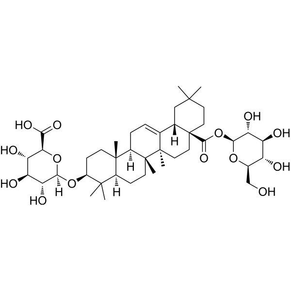
HY-N0818
Chikusetsusaponin IvaAnti-inflammatory Agent -
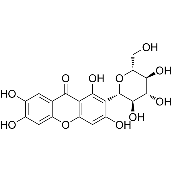
HY-N0290
MangiferinNrf2 Activator -

HY-N6653
LentinanNrf2 Activator -
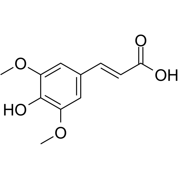
HY-W009732
Sinapinic acid -
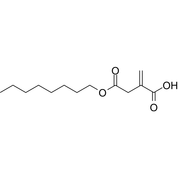
HY-112675
4-Octyl itaconateNrf2 Activator -
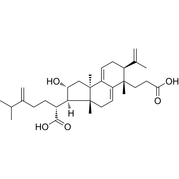
HY-N2995
Poricoic acid ANatural Product -

HY-N0290R
Mangiferin (Standard)Nrf2 Activator -
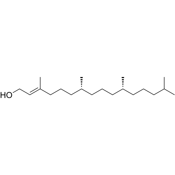
HY-N3075
PhytolDiterpene Alcohol -
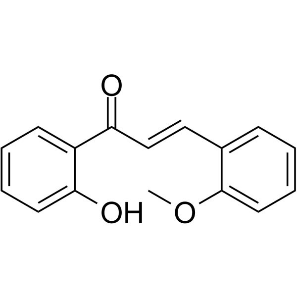
HY-128452
2'-Hydroxy-2-methoxychalconeBacterial Inhibitor -
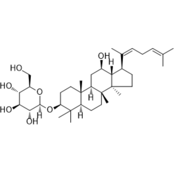
HY-N0606
Ginsenoside Rh3Nrf2 Activator -

HY-W009732R
Sinapinic acid (Standard) -

HY-N3075R
Phytol (Standard)Diterpene Alcohol


Biological Activity
Description
Cellular Effect
|
Cell Line

|
Type | Value | Description | References |
|---|---|---|---|---|
| A2780 | GI50 |
14.4 μM
Compound: 9
|
Antiproliferative activity against human A2780 cells assessed as cell growth inhibition measured after 72 hrs by trypan blue assay
Antiproliferative activity against human A2780 cells assessed as cell growth inhibition measured after 72 hrs by trypan blue assay
|
[PMID: 34262643] |
| A549 | IC50 |
5.2 μM
Compound: 5
|
Antiproliferative activity against human A549 cells assessed as cell growth inhibition measured after 48 hrs by CellTiter Aqueous One Solution MTS assay
Antiproliferative activity against human A549 cells assessed as cell growth inhibition measured after 48 hrs by CellTiter Aqueous One Solution MTS assay
|
[PMID: 37666364] |
| CAL-51 | GI50 |
2.69 μM
Compound: SSE14106
|
Antiproliferative activity against human CAL51 cells assessed as growth inhibition after 3 days by SRB assay
Antiproliferative activity against human CAL51 cells assessed as growth inhibition after 3 days by SRB assay
|
[PMID: 28743509] |
| CCRF-CEM | IC50 |
33.5 μM
Compound: 4c
|
Inhibitory concentration against CEM T-lymphocytes
Inhibitory concentration against CEM T-lymphocytes
|
[PMID: 12086496] |
| DLD-1 | EC50 |
52 μM
Compound: 9
|
Cytotoxicity against human DLD-1 cells assessed as reduction in cell viability incubated for 48 hrs by AlamarBlue-based assay
Cytotoxicity against human DLD-1 cells assessed as reduction in cell viability incubated for 48 hrs by AlamarBlue-based assay
|
[PMID: 36356534] |
| FHC | CC50 |
99 μM
Compound: 9
|
Cytotoxicity against human FHC cells assessed as reduction in cell viability incubated for 48 hrs by AlamarBlue-based assay
Cytotoxicity against human FHC cells assessed as reduction in cell viability incubated for 48 hrs by AlamarBlue-based assay
|
[PMID: 36356534] |
| HCT-116 | EC50 |
22 μM
Compound: 9
|
Cytotoxicity against human HCT-116 cells assessed as reduction in cell viability incubated for 48 hrs by AlamarBlue-based assay
Cytotoxicity against human HCT-116 cells assessed as reduction in cell viability incubated for 48 hrs by AlamarBlue-based assay
|
[PMID: 36356534] |
| HCT-116 | EC50 |
30 μM
Compound: 9
|
Cytotoxicity against p53-/- human HCT-116 cells assessed as reduction in cell viability incubated for 48 hrs by AlamarBlue-based assay
Cytotoxicity against p53-/- human HCT-116 cells assessed as reduction in cell viability incubated for 48 hrs by AlamarBlue-based assay
|
[PMID: 36356534] |
| HCT-116 | GI50 |
3.86 μM
Compound: SSE14106
|
Antiproliferative activity against human HCT116 cells assessed as growth inhibition after 3 days by SRB assay
Antiproliferative activity against human HCT116 cells assessed as growth inhibition after 3 days by SRB assay
|
[PMID: 28743509] |
| HepG2 | IC50 |
>100 μM
Compound: 4
|
Cytotoxicity against human HepG2 cells after 48 hrs by MTT assay
Cytotoxicity against human HepG2 cells after 48 hrs by MTT assay
|
[PMID: 22658085] |
| HK-2 | IC50 |
23.2 μM
Compound: 5
|
Growth inhibition of HK2 cells by sulforhodamine assay
Growth inhibition of HK2 cells by sulforhodamine assay
|
[PMID: 17383189] |
| HT-29 | EC50 |
32 μM
Compound: 9
|
Cytotoxicity against human HT-29 cells assessed as reduction in cell viability incubated for 48 hrs by AlamarBlue-based assay
Cytotoxicity against human HT-29 cells assessed as reduction in cell viability incubated for 48 hrs by AlamarBlue-based assay
|
[PMID: 36356534] |
| HT-29 | GI50 |
16.7 μM
Compound: 9
|
Antiproliferative activity against human HT-29 cells assessed as cell growth inhibition measured after 72 hrs by trypan blue assay
Antiproliferative activity against human HT-29 cells assessed as cell growth inhibition measured after 72 hrs by trypan blue assay
|
[PMID: 34262643] |
| HT-29 | IC50 |
54 μM
Compound: 5
|
Growth inhibition of HT29 cells by sulforhodamine assay
Growth inhibition of HT29 cells by sulforhodamine assay
|
[PMID: 17383189] |
| K562 | IC50 |
29 μM
Compound: 4-Methoxychalcone
|
Inhibition of NF-kappaB transactivation in TNF-alpha-stimulated human K562 cells preincubated for 2 hrs followed by TNF-alpha challenge measured after 6 hrs by dual luciferase reporter gene assay
Inhibition of NF-kappaB transactivation in TNF-alpha-stimulated human K562 cells preincubated for 2 hrs followed by TNF-alpha challenge measured after 6 hrs by dual luciferase reporter gene assay
|
[PMID: 24775915] |
| L1210 | IC50 |
50.4 μM
Compound: 4c
|
Inhibitory activity against L1210 leukemia cells
Inhibitory activity against L1210 leukemia cells
|
[PMID: 12086496] |
| MCF7 | IC50 |
90 μM
Compound: 5
|
Growth inhibition of MCF7 cells by sulforhodamine assay
Growth inhibition of MCF7 cells by sulforhodamine assay
|
[PMID: 17383189] |
| MSTO-211H | GI50 |
10.4 μM
Compound: 9
|
Antiproliferative activity against human MSTO-211H cells assessed as cell growth inhibition measured after 72 hrs by trypan blue assay
Antiproliferative activity against human MSTO-211H cells assessed as cell growth inhibition measured after 72 hrs by trypan blue assay
|
[PMID: 34262643] |
| P388 | IC50 |
10.63 μM
Compound: 4c
|
Inhibitory activity against Murine P388 cells
Inhibitory activity against Murine P388 cells
|
[PMID: 12086496] |
| RBL-1 | IC50 |
20 μM
Compound: 27
|
Inhibition of 5-lipoxygenase in rat RBL1 cells
Inhibition of 5-lipoxygenase in rat RBL1 cells
|
10.1007/s00044-013-0745-7 |
| RBL-1 | IC50 |
20 μM
Compound: 27
|
Inhibition of cyclooxygenase in rat RBL1 cells
Inhibition of cyclooxygenase in rat RBL1 cells
|
10.1007/s00044-013-0745-7 |
| TK-10 | IC50 |
53 μM
Compound: 5
|
Growth inhibition of TK10 cells by sulforhodamine assay
Growth inhibition of TK10 cells by sulforhodamine assay
|
[PMID: 17383189] |


Chemical Information
-
CAS No. 959-33-1
-
Appearance Solid
-
Molecular Weight 238.28
-
Formula C16H14O2
-
Color Off-white to light yellow
-
SMILES
O=C(C1=CC=CC=C1)/C=C/C2=CC=C(OC)C=C2
-
Structure Classification
-
Initial Source
-
Shipping
Room temperature in continental US; may vary elsewhere.
-
Storage
Store at room temperature 3 years
In solvent -80°C 2 years -20°C 1 year


Solvent & Solubility
In Vitro:
DMSO : 100 mg/mL (419.67 mM; Need ultrasonic; Hygroscopic DMSO has a significant impact on the solubility of product, please use newly opened DMSO)
Please refer to the solubility information to select the appropriate solvent. Once prepared, please aliquot and store the solution to prevent product inactivation from repeated freeze-thaw cycles.
Storage method and period of stock solution: -80°C, 2 years; -20°C, 1 year. When stored at -80°C, please use it within 2 years. When stored at -20°C, please use it within 1 year.
Please refer to the solubility information to select the appropriate solvent. Once prepared, please aliquot and store the solution to prevent product inactivation from repeated freeze-thaw cycles.
Storage method and period of stock solution: -80°C, 2 years; -20°C, 1 year. When stored at -80°C, please use it within 2 years. When stored at -20°C, please use it within 1 year.
Concentration (start) × Volume (start) = Concentration (final) × Volume (final)
In Vivo:
Select the appropriate dissolution method based on your experimental animal and administration route.
- For the following dissolution methods, please ensure to first prepare a clear stock solution using an In Vitro approach and then sequentially add co-solvents:
- To ensure reliable experimental results, the clarified stock solution can be appropriately stored based on storage conditions. As for the working solution for In Vivo experiments, it is recommended to prepare freshly and use it on the same day.
- The percentages shown for the solvents indicate their volumetric ratio in the final prepared solution. If precipitation or phase separation occurs during preparation, heat and/or sonication can be used to aid dissolution.
In Vivo Dissolution Calculator
Please enter the basic information of animal experiments:
-
-
-
-
Recommended: Prepare an additional quantity of animals to account for potential losses during experiments.
Please enter your animal formula composition:
-
%DMSO +
Recommended: Keep the proportion of DMSO in working solution below 2% if your animal is weak.
-
%+
-
+%Tween-80 + +
-
%Saline +
The co-solvents required include: DMSO, . All of co-solvents are available by MedChemExpress (MCE). , Tween 80. All of co-solvents are available by MedChemExpress (MCE).
Working solution concentration: 0.22 mg/mL
Method for preparing stock solution: mg drug dissolved in μL DMSO. Stock solution concentration: mg/mL.
1. Take μL DMSO stock solution;
2. Add μL .
μL , mix evenly;
3. Then add μL Tween 80, mix evenly;
4. Then add μL
Please ensure that the stock solution in the first step is dissolved to a clear state, and add co-solvents in sequence. You can use ultrasonic heating (ultrasonic cleaner, recommended frequency 20-40 kHz), vortexing, etc. to assist dissolution.

Protocols
-
Research Protocol for Infectious Diseases
Infectious-disease experiments test how pathogens interact with host barriers, innate immune receptors, inflammatory signaling, pathogen replication, and tissue injury; pattern-recognition receptors such as TLRs, RIG-I-like receptors, NOD-like receptors, and inflammasomes detect microbial molecules and activate NF-κB, interferon, and cytokine responses. The central hypothesis is that infection severity reflects the balance between pathogen burden and host response: protective inflammation restricts pathogen growth, whereas excessive or mislocalized inflammation contributes to tissue damage and disease phenotype. Unresolved questions include which host pathways are protective versus pathogenic, why some infection models fail to translate to human disease, and which combined readouts best predict clinically relevant infection outcomes.
-
Cotton Pellet Granuloma
Cotton pellet granuloma is a classical in vivo chronic inflammation model used to evaluate the anti-inflammatory potential of test substances by measuring their ability to inhibit granuloma tissue formation around an implanted foreign body (cotton pellet) in rodents. The method is based on the biological response to a sterile implanted material, which induces proliferative phase inflammation characterized by fibroblast proliferation and collagen-rich granuloma formation, and the final readout reflects the extent of chronic inflammatory tissue growth surrounding the pellet. In multiple preclinical pharmacological evaluations, inhibition of cotton pellet-induced granuloma formation has been used as an indicator of anti-inflammatory activity in both synthetic and natural product screening contexts.
-
Carrageenan-Induced Paw Edema
Carrageenan-induced paw edema is an acute inflammation model in which intraplantar injection of carrageenan induces localized inflammatory swelling characterized by vascular permeability, leukocyte infiltration, and production of inflammatory mediators such as prostaglandins and cytokines, making it widely used to evaluate anti-inflammatory agents in vivo. The resulting paw volume or thickness increase is quantified over time as a direct readout of inflammatory intensity and drug efficacy, typically reflecting cyclooxygenase-mediated prostaglandin-driven edema formation and immune cell recruitment in peripheral tissue[20].
-
Research Protocol for Inflammation-related Diseases
The NLRP3 inflammasome is a cytosolic innate immune signaling platform that integrates priming signals and danger-signal activation to promote caspase-1 activation, maturation of IL-1β and IL-18, and gasdermin D-mediated pyroptotic cell death. The core experimental logic is to determine whether inflammatory disease phenotypes are driven by increased NLRP3 expression, ASC-containing inflammasome assembly, caspase-1 cleavage, GSDMD cleavage, and extracellular release of IL-1β/IL-18 rather than by nonspecific cell injury alone. The pathway is strongly linked to inflammation-related disease phenotypes because monosodium urate crystals activate NALP3/NLRP3 inflammasome signaling in gout-like crystal inflammation, cholesterol crystals activate NLRP3 inflammasomes in atherogenesis models, and DSS-induced intestinal inflammation has been reported to involve NLRP3 inflammasome activity. However, experimental colitis studies also show context-dependent protective effects of NLRP3 inflammasome co
-
Subcutaneous Cell-Line-Derived Xenograft
Subcutaneous cell-line-derived xenograft (CDX) models are established by implanting cultured human cancer cell lines into immunodeficient mice, where the injected cells form localized tumors that can be monitored in vivo as a measure of tumorigenic potential, growth kinetics, and treatment response. These models are widely used in oncology research because they allow reproducible tumor formation and enable comparative assessment of tumor growth between different cell lines or genetic manipulations in a controlled in vivo microenvironment. Subcutaneous implantation of cancer cells in immunodeficient mice is a standard approach for evaluating tumor growth behavior and therapeutic response across multiple cancer types, including prostate, esophageal, pancreatic, and colon cancer models.
-
Bacterial live/dead nucleic-acid viability staining
The LIVE/DEAD bacterial viability staining method is based on differential permeability of nucleic-acid-binding fluorescent dyes, most commonly SYTO 9 and propidium iodide (PI), which enables discrimination of bacterial populations with intact versus compromised cytoplasmic membranes. SYTO 9 penetrates both intact and damaged bacterial membranes and binds nucleic acids to produce green fluorescence, whereas propidium iodide penetrates only cells with compromised membranes and fluoresces red while also reducing SYTO 9 signal through competitive binding and fluorescence interactions. The resulting fluorescence pattern is interpreted as a proxy for membrane integrity, which is widely used as an indicator of bacterial viability in microscopy, flow cytometry, and spectroscopic platforms. However, mechanistic studies show that SYTO 9 and PI interactions involve displacement and fluorescence resonance energy transfer effects, which can influence signal interpretation depending on dye ratios a


Purity & Documentation
-
Data Sheet (269 KB)
-
SDS (392 KB)
- English - EN (392 KB)
- Français - FR (392 KB)
- Deutsch - DE (392 KB)
- Norwegian - NO (392 KB)
- Español - ES (392 KB)
- Swedish - SV (392 KB)
- Italian - IT (392 KB)
- Korean - KR (392 KB)
- Portuguese - PT (392 KB)
-
Handling Instructions (2659 KB)
References


Complete Stock Solution Preparation Table
Please refer to the solubility information to select the appropriate solvent. Once prepared, please aliquot and store the solution to prevent product inactivation from repeated freeze-thaw cycles.
Storage method and period of stock solution: -80°C, 2 years; -20°C, 1 year. When stored at -80°C, please use it within 2 years. When stored at -20°C, please use it within 1 year.
| Optional Solvent | Concentration Solvent Mass | 1 mg | 5 mg | 10 mg | 25 mg |
|---|---|---|---|---|---|
| DMSO | 1 mM | 4.1967 mL | 20.9837 mL | 41.9674 mL | 104.9186 mL |
| 5 mM | 0.8393 mL | 4.1967 mL | 8.3935 mL | 20.9837 mL | |
| 10 mM | 0.4197 mL | 2.0984 mL | 4.1967 mL | 10.4919 mL | |
| 15 mM | 0.2798 mL | 1.3989 mL | 2.7978 mL | 6.9946 mL | |
| 20 mM | 0.2098 mL | 1.0492 mL | 2.0984 mL | 5.2459 mL | |
| 25 mM | 0.1679 mL | 0.8393 mL | 1.6787 mL | 4.1967 mL | |
| 30 mM | 0.1399 mL | 0.6995 mL | 1.3989 mL | 3.4973 mL | |
| 40 mM | 0.1049 mL | 0.5246 mL | 1.0492 mL | 2.6230 mL | |
| 50 mM | 0.0839 mL | 0.4197 mL | 0.8393 mL | 2.0984 mL | |
| 60 mM | 0.0699 mL | 0.3497 mL | 0.6995 mL | 1.7486 mL | |
| 80 mM | 0.0525 mL | 0.2623 mL | 0.5246 mL | 1.3115 mL | |
| 100 mM | 0.0420 mL | 0.2098 mL | 0.4197 mL | 1.0492 mL |


4-Methoxychalcone-1 Related Classifications
HY-W083376A Related Classifications
 Powered by Bioz