



Acacetin
Based on 16 publication(s) in Google Scholar
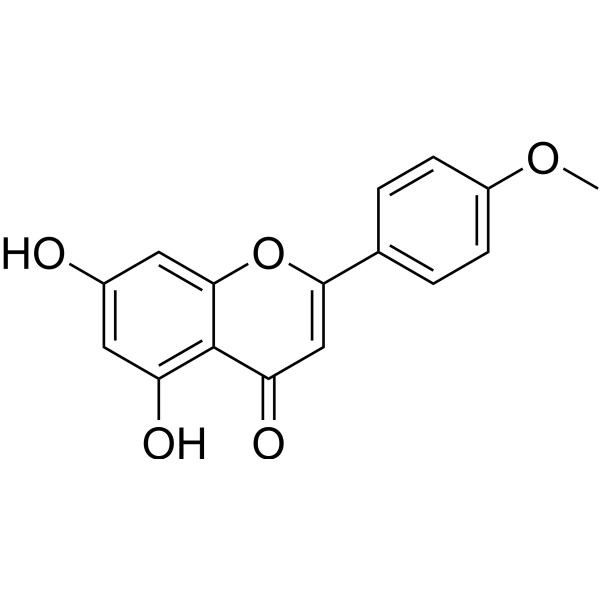
Acacetin (5,7-Dihydroxy-4'-methoxyflavone) is an orally active flavonoid derived from Dendranthema morifolium. Acacetin docks in the ATP binding pocket of PI3Kγ. Acacetin causes cell cycle arrest and induces apoptosis and autophagy in cancer cells. Acacetin has potent anti-cancer and anti-inflammatory activity and has the potential for pain-related diseases research.
For research use only. We do not sell to patients.
- Purity : 99.71%
- CAS No.: 480-44-4
- Formula: C16H12O5
- Molecular Weight:284.26
-
Storage:Powder -20°C, 3 years , 4°C, 2 years ; In solvent -80°C, 2 years , -20°C, 1 year

To place orders, for customer services and technical support, please contact: MedChemExpress USA
Tel: 609-228-6898 E-mail: [email protected] [email protected]


-
 Biological Activity
-
Chemical Information
-
Solvent & Solubility
- Protocols
- Purity & Documentation
- References
-
 Help & FAQs


-
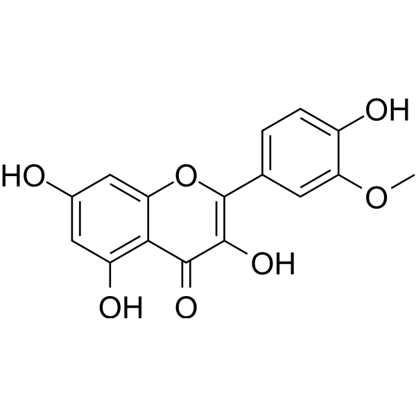
HY-N0776
IsorhamnetinEndogenous Metabolite -
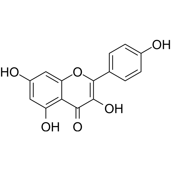
HY-14590
KaempferolEstrogen Receptor α Inhibitor -
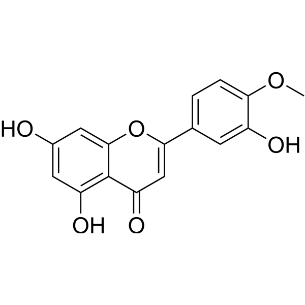
HY-N0125
DiosmetinCytochrome P450 Inhibitor -
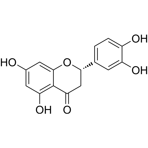
HY-N0637
EriodictyolEndogenous Metabolite -
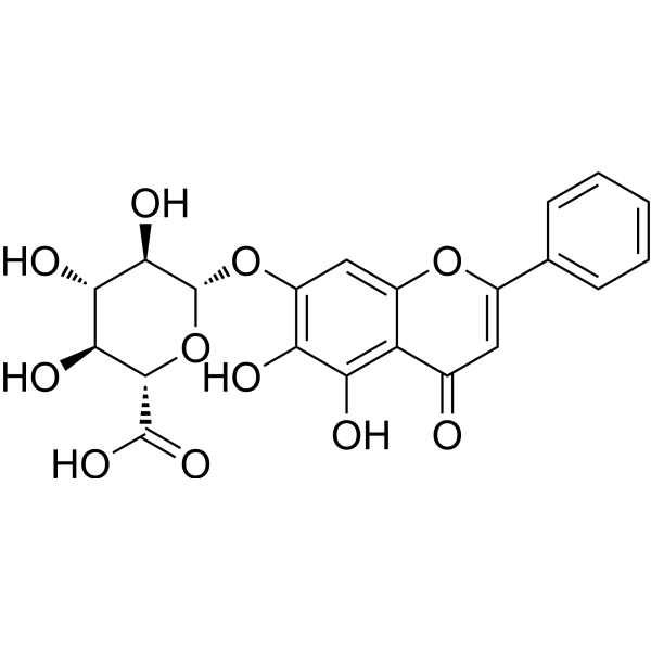
HY-N0197
BaicalinAutophagy Inducer -
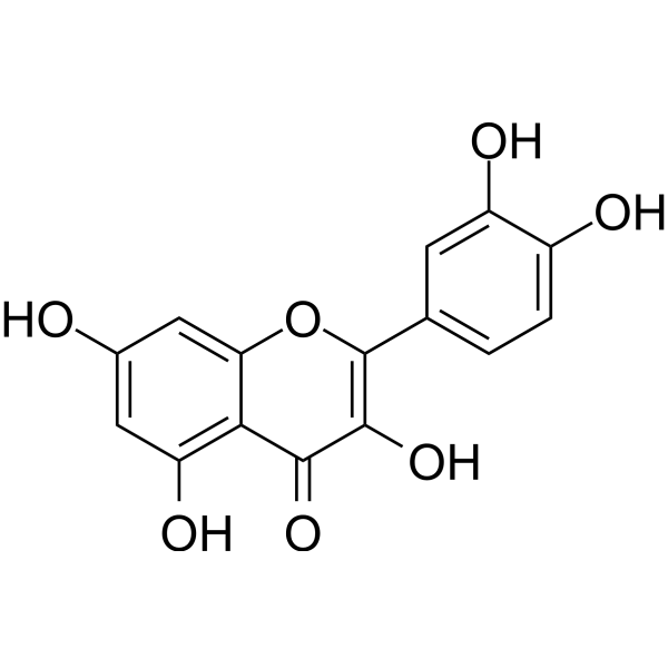
HY-18085
QuercetinFlavonoid Antioxidant -

HY-13259
MG-132Proteasome Inhibitor -
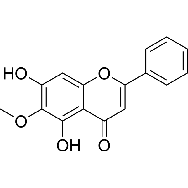
HY-N0560
Oroxylin AAutophagy Inducer -
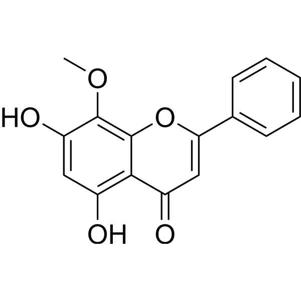
HY-N0400
WogoninCDK Inhibitor -
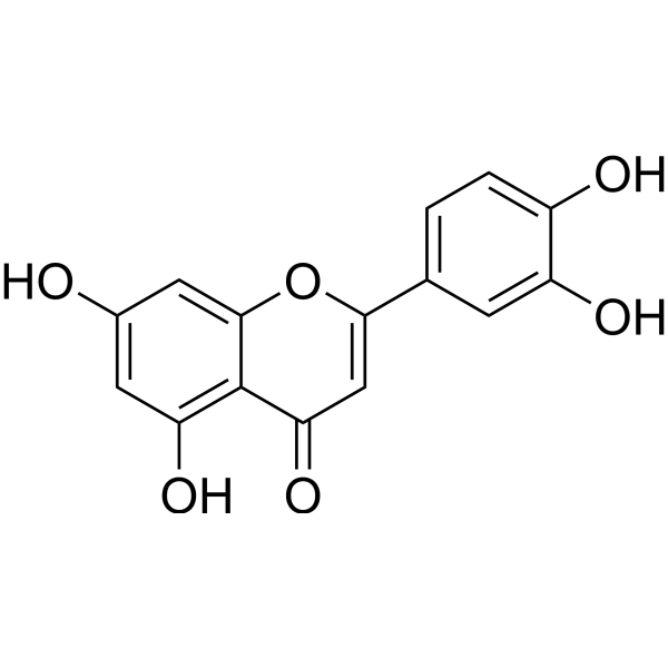
HY-N0162
LuteolinNrf2 Inhibitor -
HY-N0016
GlyciteinApoptosis Inducer -
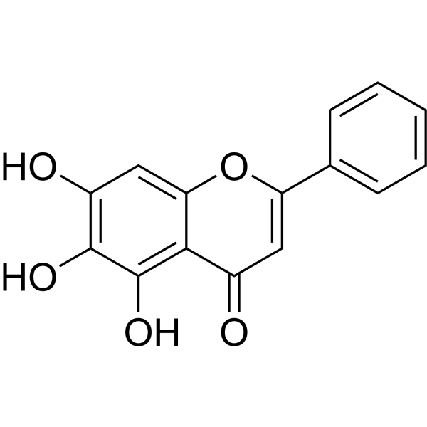
HY-N0196
BaicaleinXanthine Oxidase Inhibitor
Publications Citing Use of MedChemExpress (MCE) Acacetin
More- Metabolism. 2026 Jun 7:182:156670. [Abstract]
- Acta Pharm Sin B. 2021 Jan;11(1):143-155. [Abstract]
- Pharmacol Res. 2020 May:155:104751. [Abstract]
- Phytomedicine. 2025 Nov 25:148:157405. [Abstract]
- Food Chem. 2025 Dec 30:497:146992. [Abstract]
- Food Chem. 2025 Oct 15:489:144992. [Abstract]
- EMBO Rep. 2022 Jun 7;23(6):e53932. [Abstract]
- Int J Mol Sci. 2025 Nov 21;26(23):11262. [Abstract]
- Sci Rep. 2024 Jan 29;14(1):2348. [Abstract]
- Chem Biol Drug Des. 2026 May;107(5):e70308. [Abstract]
- Neurogastroenterol Motil. 2026 Apr;38(4):e70298. [Abstract]
- World J Gastrointest Oncol. 2024 Aug 15;16(8):3624-3634. [Abstract]
- Future Microbiol. 2020 May:15:485-496. [Abstract]
- Biol Pharm Bull. 2022;45(8):1116-1123. [Abstract]
- SSRN. 2025 May 7.
- Heliyon. 2024 Mar 25;10(7):e28693. [Abstract]
 Customer Validation & Images

-
Cell Imaging/Staining
-
Flow Cytometry
-
Cell Imaging/Staining
-
WB
-
Cell Imaging/Staining


Biological Activity
Description
Cellular Effect
|
Cell Line

|
Type | Value | Description | References |
|---|---|---|---|---|
| A-431 | IC50 |
4 x 10⁻¹μg/mL
Compound: 39
|
Inhibition of EGFR in human A431 cells
Inhibition of EGFR in human A431 cells
|
[PMID: 1479375] |
| A549 | IC50 |
0.4 μg/mL
Compound: Acacetin
|
Cytotoxicity against human A549 cells by SRB assay
Cytotoxicity against human A549 cells by SRB assay
|
[PMID: 31784199] |
| HCT-15 | IC50 |
0.4 μg/mL
Compound: Acacetin
|
Cytotoxicity against human HCT15 cells by SRB assay
Cytotoxicity against human HCT15 cells by SRB assay
|
[PMID: 31784199] |
| KB | ED50 |
>100 μg/mL
Compound: NSC-76061
|
Cytotoxicity against human KB cells
Cytotoxicity against human KB cells
|
[PMID: 469554] |
| MOLM-13 | IC50 |
9.1 μM
Compound: 28
|
Cytotoxicity against human MOLM-13 cells assessed as reduction in cell viability measured after 72 hrs by MTT assay
Cytotoxicity against human MOLM-13 cells assessed as reduction in cell viability measured after 72 hrs by MTT assay
|
[PMID: 33393294] |
| Monocyte | IC50 |
22 μM
Compound: Acacetin
|
Inhibition of TNFalpha expression in LPS-stimulated human monocytes treated 30 mins before LPS challenge measured after 14 hrs by ELISA
Inhibition of TNFalpha expression in LPS-stimulated human monocytes treated 30 mins before LPS challenge measured after 14 hrs by ELISA
|
[PMID: 10096854] |
| MV4-11 | IC50 |
6.8 μM
Compound: 28
|
Cytotoxicity against human MV4-11 cells assessed as reduction in cell viability measured after 72 hrs by MTT assay
Cytotoxicity against human MV4-11 cells assessed as reduction in cell viability measured after 72 hrs by MTT assay
|
[PMID: 33393294] |
| Neutrophil | IC50 |
7 μM
Compound: 4d
|
Inhibition of oxidative burst in PMA-stimulated human neutrophils assessed as inhibition of superoxide anion radical-induced lucigenin oxidation incubated for 5 mins prior to PMA challenge by chemiluminescence assay
Inhibition of oxidative burst in PMA-stimulated human neutrophils assessed as inhibition of superoxide anion radical-induced lucigenin oxidation incubated for 5 mins prior to PMA challenge by chemiluminescence assay
|
[PMID: 23871908] |
| RAW264.7 | CC50 |
60.9 μM
Compound: 17
|
Cytotoxicity against mouse RAW264.7 cells assessed as reduction in cell viability measured after 24 hrs by MTT assay
Cytotoxicity against mouse RAW264.7 cells assessed as reduction in cell viability measured after 24 hrs by MTT assay
|
[PMID: 33667099] |
| RAW264.7 | IC50 |
27.7 μM
Compound: 27
|
Inhibition of LPS-stimulated nitric oxide production in mouse RAW264.7 cells by Greiss method
Inhibition of LPS-stimulated nitric oxide production in mouse RAW264.7 cells by Greiss method
|
[PMID: 27955927] |
| RAW264.7 | IC50 |
5.9 μM
Compound: 17
|
Inhibition of LPS induced NO production in mouse RAW264.7 cells measured after 24 hrs by Griess reagent based assay
Inhibition of LPS induced NO production in mouse RAW264.7 cells measured after 24 hrs by Griess reagent based assay
|
[PMID: 33667099] |
| RAW264.7 | IC50 |
53.7 μM
Compound: 27
|
Inhibition of LPS/INF-gamma-stimulated nitric oxide production in mouse RAW264.7 cells by Greiss method
Inhibition of LPS/INF-gamma-stimulated nitric oxide production in mouse RAW264.7 cells by Greiss method
|
[PMID: 27955927] |
| RAW264.7 | IC50 |
6.2 μM
Compound: 11
|
Antiinflammatory activity against LPS-stimulated mouse RAW264.7 cells assessed as decrease in PGE2 production preincubated for 1 hr followed by LPS stimulation and measured after 24 hrs by ELISA
Antiinflammatory activity against LPS-stimulated mouse RAW264.7 cells assessed as decrease in PGE2 production preincubated for 1 hr followed by LPS stimulation and measured after 24 hrs by ELISA
|
[PMID: 31747281] |
| SK-MEL-2 | IC50 |
0.4 μg/mL
Compound: Acacetin
|
Cytotoxicity against human SK-MEL-2 cells by SRB assay
Cytotoxicity against human SK-MEL-2 cells by SRB assay
|
[PMID: 31784199] |
| SK-OV-3 | IC50 |
0.4 μg/mL
Compound: Acacetin
|
Cytotoxicity against human SKOV3 cells by SRB assay
Cytotoxicity against human SKOV3 cells by SRB assay
|
[PMID: 31784199] |
| XF498 | IC50 |
0.4 μg/mL
Compound: Acacetin
|
Cytotoxicity against human XF498 cells by SRB assay
Cytotoxicity against human XF498 cells by SRB assay
|
[PMID: 31784199] |
In Vitro
Acacetin (5,7-Dihydroxy-4'-methoxyflavone; 10-200 μM; 24 hours) decreases cell viabilities in a dose-dependent manner. Acacetin has little effect on human normal glial cell line HEB and non-tumorigenic epithelial cell line MCF-10A[1].
Acacetin (50-150 μM; 24 hours) causes G2/M cell cycle arrest and induces apoptosis and autophagy[1].
Acacetin (50-150 μM; 24 hours) leads to decreases in levels of PI3Kγ-p110, p-AKT, p-mTOR, p-p70S6K, and p-ULK in a dose-dependent manner[1].
MedChemExpress (MCE) has not independently confirmed the accuracy of these methods. They are for reference only. Further protocols information, click here.
-
Cell Line:Breast cancer MCF-7 cells, hepatocellular carcinoma SMMC-7721 cells, lung adenocarcinoma A549 cells, esophageal carcinoma Eca109 cells
-
Concentration:10, 20, 40, 60, 80, 100, 150, 200 μM
-
Incubation Time:24 hours
-
Result:Decreased cancer cell viabilities in a dose-dependent manner.
Had IC50 values of 82.75 μM, 103.9 μM, 157.4 μM, 54.7 μM in MDA-MB-231, MCF-7, A549, Eca109 cells, respectively.
-
Cell Line:MDA-MB-231 cells
-
Concentration:50, 100, 150 μM
-
Incubation Time:24 hours
-
Result:Resulted in increase in percentage of cells at G2/M phase and decrease in percentage of cells at G1 and S phase in a dose-dependent manner.
-
Cell Line:MDA-MB-231 cells
-
Concentration:50, 100, 150 μM
-
Incubation Time:24 hours
-
Result:Induced apoptosis.
-
Cell Line:MDA-MB-231 cells
-
Concentration:50, 100, 150 μM
-
Incubation Time:24 hours
-
Result:Induced autophagy.
Resulted in marked increases in EGFP-LC3 puncta formation and a dose-dependent accumulation of LC3-II.
-
Cell Line:MDA-MB-231 cells
-
Concentration:50, 100, 150 μM
-
Incubation Time:24 hours
-
Result:Resulted in decrease in levels of Bcl-2 and Bcl-xL and increase in levels of p53.
Led to decreases in levels of PI3Kγ-p110, p-AKT, p-mTOR, p-p70S6K, and p-ULK in a dose-dependent manner.
Had little or no effect on expression of PI3Kα, PI3Kβ, PI3Kδ, p-ERK, p-p38, and p-JNK.
In Vivo
Acacetin (25 mg/kg/day; orally; for 3 days) reduces neuronal cell death in an animal model of ischemia[2].
Acacetin (1.8-56.2 mg/kg/day; ip; single dose) decreases visceral and inflammatory nociception and prevented the formalin-induced oedema[3].
MedChemExpress (MCE) has not independently confirmed the accuracy of these methods. They are for reference only.
-
Animal Model:Male C57BL/6 mice, 7 weeks of age[2]
-
Dosage:5, 20 mg/kg
-
Administration:Orally; once a day for 3 days
-
Result:Significantly suppressed microglial activation in an LPS-induced (ip; 5mg/kg) neuroinflammation mouse model.


Chemical Information
-
CAS No. 480-44-4
-
Appearance Solid
-
Molecular Weight 284.26
-
Formula C16H12O5
-
Color Light yellow to yellow
-
SMILES
O=C1C=C(C2=CC=C(OC)C=C2)OC3=CC(O)=CC(O)=C13
-
Synonyms
5,7-Dihydroxy-4'-methoxyflavone
-
Structure Classification
-
Shipping
Room temperature in continental US; may vary elsewhere.
-
Storage
Powder -20°C 3 years 4°C 2 years In solvent -80°C 2 years -20°C 1 year


Publications (16)
-
Journal Impact Factor
-
Most Recent
-
Metabolism
Acacetin ameliorates MASLD by inhibiting the Notch1 pathway in hepatocytes and reprogramming macrophage polarization via Keap1-Nrf2-mediated restraint of ferroptosis. [Abstract]2026 Jun 7:182:156670. PMID: 42259496 -
Acta Pharm Sin B
Chrysin serves as a novel inhibitor of DGK α/FAK interaction to suppress the malignancy of esophageal squamous cell carcinoma (ESCC). [Abstract]2021 Jan;11(1):143-155. PMID: 33532186 -
Pharmacol Res
Cardamonin retards progression of autosomal dominant polycystic kidney disease via inhibiting renal cyst growth and interstitial fibrosis. [Abstract]2020 May:155:104751. PMID: 32151678 -
Phytomedicine
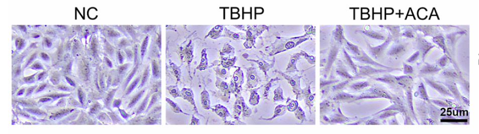
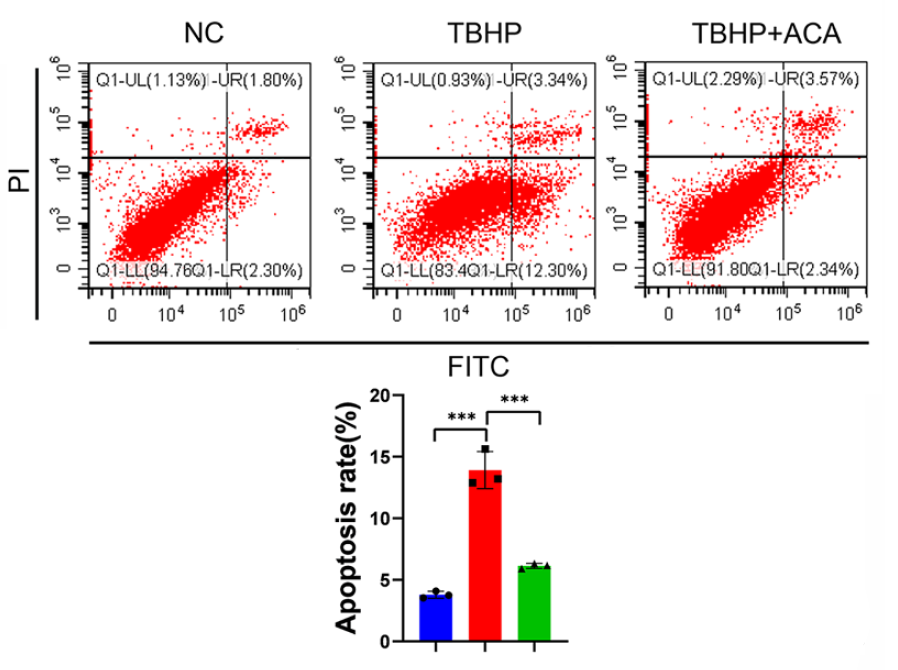
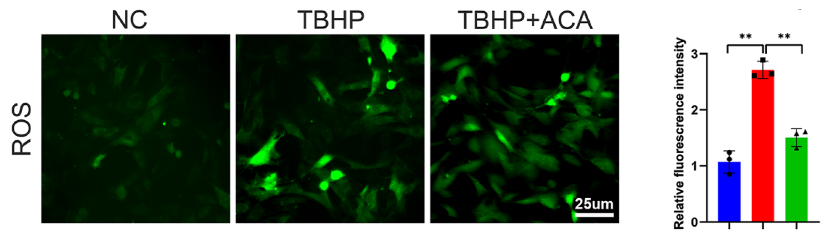
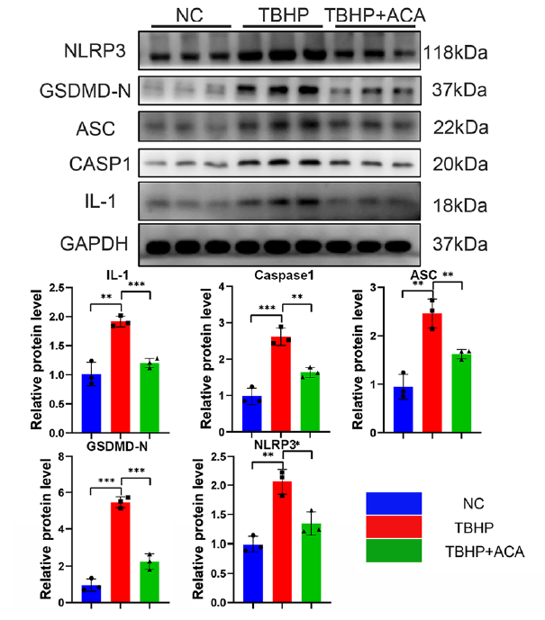
Duhuo Jisheng decoction alleviates intervertebral disc degeneration via the acacetin-mediated MAPK1/HMOX1 axis by inhibiting nucleus pulposus cell pyroptosis. [Abstract]2025 Nov 25:148:157405. PMID: 41135274
Acacetin purchased from MedChemExpress. Usage Cited in: Phytomedicine. 2025 Nov 25:148:157405. [Abstract]
Acacetin (ACA) (10 μM; 24 h) significantly alleviated TBHP-induced pyroptotic alterations in NPCs.
Acacetin purchased from MedChemExpress. Usage Cited in: Phytomedicine. 2025 Nov 25:148:157405. [Abstract]
Acacetin (ACA) (10 μM; 24 h) significantly inhibited TBHP-induced NPC programmed cell death.
Acacetin purchased from MedChemExpress. Usage Cited in: Phytomedicine. 2025 Nov 25:148:157405. [Abstract]
Acacetin (ACA) (10 μM; 24 h) significantly reduced ROS production in NPCs exposed to TBHP.
Acacetin purchased from MedChemExpress. Usage Cited in: Phytomedicine. 2025 Nov 25:148:157405. [Abstract]
Acacetin (ACA) (10 μM; 24 h) downregulated the expression of pyroptosis-related proteins in NPCs exposed to TBHP.
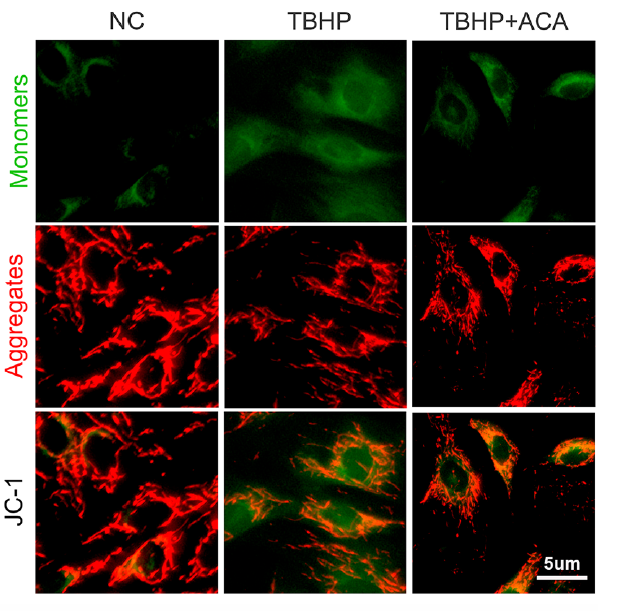
Acacetin purchased from MedChemExpress. Usage Cited in: Phytomedicine. 2025 Nov 25:148:157405. [Abstract]
Acacetin (ACA) (10 μM; 24 h) effectively restored the reduced mitochondrial membrane potential in NPCs induced by TBHP.
-
Food Chem
Effects of sun drying combined with baking processes on the flavor quality of Chongqing Tuocha raw tea. [Abstract]2025 Dec 30:497:146992. PMID: 41285060 -
Food Chem
Flavonoid-mediated metabolic underpinning quality variation in red bud-sport pear mutants. [Abstract]2025 Oct 15:489:144992. PMID: 40466530 -
EMBO Rep
2022 Jun 7;23(6):e53932. PMID: 35403787 -
Int J Mol Sci
Elucidating the Mechanisms of Chrysanthemum Action on Atopic Dermatitis via Network Pharmacology and Machine Learning. [Abstract]2025 Nov 21;26(23):11262. PMID: 41373428 -
Sci Rep
Acacetin inhibited non-small-cell lung cancer (NSCLC) cell growth via upregulating miR-34a in vitro and in vivo. [Abstract]2024 Jan 29;14(1):2348. PMID: 38287075 -
Chem Biol Drug Des
Acacetin Attenuates Heatstroke-Induced Acute Liver Injury by Targeting the c-Jun/PTGS2 Pathway. [Abstract]2026 May;107(5):e70308. PMID: 42068089 -
Neurogastroenterol Motil
Acacetin Alleviates Loperamide-Induced Functional Constipation by Inhibiting P53-Mediated Apoptosis in Colonic Epithelial Cells. [Abstract]2026 Apr;38(4):e70298. PMID: 41913080 -
World J Gastrointest Oncol
Effect of acacetin on inhibition of apoptosis in Helicobacter pylori-infected gastric epithelial cell line. [Abstract]2024 Aug 15;16(8):3624-3634. PMID: 39171164 -
Future Microbiol
Characterization of the sortase A from Lactobacillus acidophilus ATCC 4356 involved in adherence to intestinal cells. [Abstract]2020 May:15:485-496. PMID: 32476478 -
Biol Pharm Bull
Regioselective Glucuronidation of Flavones at C5, C7, and C4' Positions in Human Liver and Intestinal Microsomes: Comparison among Apigenin, Acacetin, and Genkwanin. [Abstract]2022;45(8):1116-1123. PMID: 35908893 -
-
Heliyon
Mechanism of acacetin regulating hepatic stellate cell apoptosis based on network pharmacology and experimental verification. [Abstract]2024 Mar 25;10(7):e28693. PMID: 38571642


Solvent & Solubility
In Vitro:
DMSO : 125 mg/mL (439.74 mM; Need ultrasonic; Hygroscopic DMSO has a significant impact on the solubility of product, please use newly opened DMSO)
Please refer to the solubility information to select the appropriate solvent. Once prepared, please aliquot and store the solution to prevent product inactivation from repeated freeze-thaw cycles.
Storage method and period of stock solution: -80°C, 2 years; -20°C, 1 year. When stored at -80°C, please use it within 2 years. When stored at -20°C, please use it within 1 year.
Please refer to the solubility information to select the appropriate solvent. Once prepared, please aliquot and store the solution to prevent product inactivation from repeated freeze-thaw cycles.
Storage method and period of stock solution: -80°C, 2 years; -20°C, 1 year. When stored at -80°C, please use it within 2 years. When stored at -20°C, please use it within 1 year.
Concentration (start) × Volume (start) = Concentration (final) × Volume (final)
In Vivo:
Select the appropriate dissolution method based on your experimental animal and administration route.
- For the following dissolution methods, please ensure to first prepare a clear stock solution using an In Vitro approach and then sequentially add co-solvents:
- To ensure reliable experimental results, the clarified stock solution can be appropriately stored based on storage conditions. As for the working solution for In Vivo experiments, it is recommended to prepare freshly and use it on the same day.
- The percentages shown for the solvents indicate their volumetric ratio in the final prepared solution. If precipitation or phase separation occurs during preparation, heat and/or sonication can be used to aid dissolution.
For the following dissolution methods, please prepare the working solution directly:
It is recommended to prepare fresh solutions and use them promptly within a short period of time.
The percentages shown for the solvents indicate their volumetric ratio in the final prepared solution. If precipitation or phase separation occurs during preparation, heat and/or sonication can be used to aid dissolution.
In Vivo Dissolution Calculator
Please enter the basic information of animal experiments:
-
-
-
-
Recommended: Prepare an additional quantity of animals to account for potential losses during experiments.
Please enter your animal formula composition:
-
%DMSO +
Recommended: Keep the proportion of DMSO in working solution below 2% if your animal is weak.
-
%+
-
+%Tween-80 + +
-
%Saline +
The co-solvents required include: DMSO, . All of co-solvents are available by MedChemExpress (MCE). , Tween 80. All of co-solvents are available by MedChemExpress (MCE).
Working solution concentration: 0.22 mg/mL
Method for preparing stock solution: mg drug dissolved in μL DMSO. Stock solution concentration: mg/mL.
1. Take μL DMSO stock solution;
2. Add μL .
μL , mix evenly;
3. Then add μL Tween 80, mix evenly;
4. Then add μL
Please ensure that the stock solution in the first step is dissolved to a clear state, and add co-solvents in sequence. You can use ultrasonic heating (ultrasonic cleaner, recommended frequency 20-40 kHz), vortexing, etc. to assist dissolution.

Protocols
-
Annexin V plus membrane-impermeant dye apoptosis staining
Annexin V-based apoptosis assays rely on the detection of phosphatidylserine (PS) externalization from the inner leaflet of the plasma membrane to the outer leaflet, an early biochemical hallmark of apoptosis. Fluorescently labeled Annexin V binds PS in a calcium-dependent manner, enabling identification of early apoptotic cells by flow cytometry or fluorescence microscopy. When combined with a membrane-impermeant DNA-binding dye (e. g. , propidium iodide), this approach allows discrimination between viable (Annexin V−/dye−), early apoptotic (Annexin V+/dye−), and late apoptotic or necrotic (Annexin V+/dye+) cell populations by assessing membrane integrity and PS exposure.
-
BrdU Incorporation Assay
Bromodeoxyuridine (BrdU) incorporation assay is based on the principle that BrdU, a thymidine analog, is incorporated into newly synthesized DNA during the S phase of the cell cycle, thereby serving as a marker of DNA replication and cellular proliferation. Incorporated BrdU can be detected using anti-BrdU antibodies following DNA denaturation, enabling visualization or quantification of proliferating cells through immunochemical detection methods such as immunofluorescence or immunohistochemistry.
-
Protocol for Cell Cycle
Cell-cycle analysis by flow cytometry measures DNA content in single cells to estimate the fraction of cells in G0/G1, S, and G2/M phases. Propidium iodide intercalates into DNA, and after RNA removal with RNase, fluorescence intensity reflects cellular DNA content: 2N cells are assigned to G0/G1, cells between 2N and 4N to S phase, and 4N cells to G2/M. DNA-content analysis alone cannot reliably separate G0 from G1 or G2 from M. Ki-67 can distinguish quiescent G0 cells from cycling cells, EdU or BrdU incorporation marks active DNA synthesis in S phase, and phospho-histone H3 staining identifies mitotic cells within the 4N population.
-
Research Protocol for Inflammation-related Diseases
The NLRP3 inflammasome is a cytosolic innate immune signaling platform that integrates priming signals and danger-signal activation to promote caspase-1 activation, maturation of IL-1β and IL-18, and gasdermin D-mediated pyroptotic cell death. The core experimental logic is to determine whether inflammatory disease phenotypes are driven by increased NLRP3 expression, ASC-containing inflammasome assembly, caspase-1 cleavage, GSDMD cleavage, and extracellular release of IL-1β/IL-18 rather than by nonspecific cell injury alone. The pathway is strongly linked to inflammation-related disease phenotypes because monosodium urate crystals activate NALP3/NLRP3 inflammasome signaling in gout-like crystal inflammation, cholesterol crystals activate NLRP3 inflammasomes in atherogenesis models, and DSS-induced intestinal inflammation has been reported to involve NLRP3 inflammasome activity. However, experimental colitis studies also show context-dependent protective effects of NLRP3 inflammasome co
-
Apoptosis
Apoptosis, also called programmed cell death, is generally characterized by distinct morphological characteristics.
-
TUNEL staining for apoptotic DNA fragmentation
TUNEL staining detects DNA strand breaks by using terminal deoxynucleotidyl transferase to add labeled nucleotides to exposed 3′-OH DNA termini, generating either microscopic staining in fixed cells or tissue sections, or fluorescence/cytometric signal in cell suspensions. TUNEL positivity reflects DNA fragmentation but should not be interpreted alone as definitive apoptosis, because TUNEL can also label necrotic, autolytic, mechanically damaged, or DNA-repair-associated DNA breaks.
-
Flow cytometric DNA-content cell-cycle staining
Flow cytometric DNA-content cell-cycle staining measures the fluorescence intensity of DNA-bound fluorochromes in single cells or nuclei to estimate DNA content distributions, allowing assignment of populations to G0/G1, S, and G2/M phases by DNA histogram deconvolution. Propidium iodide (PI) intercalates into DNA, and PI fluorescence is proportional to cellular DNA content when staining is performed under conditions that make DNA accessible and minimize non-DNA signal. Cells with G2/M DNA content are expected to show approximately twice the fluorescence intensity of G0/G1 cells, while S-phase cells occupy intermediate fluorescence values. PI-based DNA-content analysis can also detect cells with fractional DNA content, often reported as sub-G1, when DNA fragmentation and extraction during staining reduce retained DNA signal in apoptotic cells. DAPI is an alternative DNA fluorochrome for univariate DNA-content analysis, while bivariate approaches combining DNA content with proliferation
-
Cotton Pellet Granuloma
Cotton pellet granuloma is a classical in vivo chronic inflammation model used to evaluate the anti-inflammatory potential of test substances by measuring their ability to inhibit granuloma tissue formation around an implanted foreign body (cotton pellet) in rodents. The method is based on the biological response to a sterile implanted material, which induces proliferative phase inflammation characterized by fibroblast proliferation and collagen-rich granuloma formation, and the final readout reflects the extent of chronic inflammatory tissue growth surrounding the pellet. In multiple preclinical pharmacological evaluations, inhibition of cotton pellet-induced granuloma formation has been used as an indicator of anti-inflammatory activity in both synthetic and natural product screening contexts.
-
Carrageenan-Induced Paw Edema
Carrageenan-induced paw edema is an acute inflammation model in which intraplantar injection of carrageenan induces localized inflammatory swelling characterized by vascular permeability, leukocyte infiltration, and production of inflammatory mediators such as prostaglandins and cytokines, making it widely used to evaluate anti-inflammatory agents in vivo. The resulting paw volume or thickness increase is quantified over time as a direct readout of inflammatory intensity and drug efficacy, typically reflecting cyclooxygenase-mediated prostaglandin-driven edema formation and immune cell recruitment in peripheral tissue[20].
-
Autophagy
Autophagy is a process in which eukaryotic cells use lysosomes to degrade their own cytoplasmic proteins and damaged organelles under the regulation of autophagy related gene (Atg). Microtubule-associated proteins light chain 3 (LC3) is recognized as autophagy marker, which transfers from cytoplasmic LC3 (LC3-I) to membrane type (LC3-II). LC3-II/I ratio could be detected by Western Blot and fluorescence microscopy.
-
Apoptosis Solutions
Apoptosis is a regulated, generally non-lytic cell-death pathway that removes unwanted, damaged, infected, or abnormal cells through coordinated morphological changes, caspase activation, DNA fragmentation, and membrane remodeling. The intrinsic apoptosis pathway is controlled mainly by mitochondrial outer membrane permeabilization, BCL-2 family proteins, cytochrome c release, apoptosome formation, caspase-9 activation, and downstream executioner caspase-3/7 activation. The extrinsic apoptosis pathway is initiated by death receptors such as Fas, TNFR, and TRAIL receptors, which recruit adaptor proteins and activate caspase-8 before engaging executioner caspases or mitochondrial amplification through BID cleavage. Apoptosis is linked to many phenotypes, including cancer cell killing, tissue homeostasis, immune regulation, neurodegeneration, infection response, and treatment-induced cytotoxicity; unresolved questions include how apoptosis interacts with necroptosis, pyroptosis, ferroptos
-
Lysosome and acidic-vesicle live-cell staining
Lysosome and acidic-vesicle live-cell staining detects acidic intracellular compartments by using membrane-permeant acidotropic probes that accumulate in low-pH vesicles, including lysosomes, late endosomes, autolysosomes, and acidic phagosomes. LysoTracker staining is commonly used as an intensity-based readout of acidic lysosomal compartment abundance or enlargement, while acridine orange produces green fluorescence in less concentrated compartments and red fluorescence after concentration-dependent accumulation in acidic vesicular organelles. Loss or reduction of acridine-orange red signal can be used as a readout of lysosomal membrane permeabilization or reduced acidic-vesicle integrity. This protocol is designed for live cultured cells and can be adapted for fluorescence microscopy, high-content imaging, plate-reader readout, or flow cytometry when the selected literature supports the readout. Because these dyes report acidotropic accumulation rather than lysosome identity alone,
-
Macroautophagy Solutions
Macroautophagy is a conserved lysosome-dependent degradation pathway in which cytoplasmic material is sequestered into double-membrane autophagosomes and delivered to lysosomes for degradation and recycling. The pathway supports cellular homeostasis during nutrient limitation, organelle stress, protein-aggregate accumulation, infection, differentiation, and tissue remodeling by coupling cargo sequestration, autophagosome maturation, lysosomal fusion, and degradation of cargo-derived macromolecules. The core molecular sequence includes initiation by nutrient- and stress-regulated autophagy machinery, autophagosome nucleation, LC3/ATG8-family conjugation to autophagosomal membranes, cargo selection through receptors such as SQSTM1/p62, autophagosome-lysosome fusion, and lysosomal degradation. LC3 was identified as a mammalian homolog of yeast Atg8 that localizes to autophagosomal membranes after processing, and p62/SQSTM1 was shown to connect ubiquitinated cargo with autophagic degradati


Purity & Documentation
-
Data Sheet (280 KB)
-
SDS (394 KB)
- English - EN (394 KB)
- Français - FR (394 KB)
- Deutsch - DE (394 KB)
- Norwegian - NO (394 KB)
- Español - ES (394 KB)
- Swedish - SV (394 KB)
- Italian - IT (394 KB)
- Korean - KR (394 KB)
- Portuguese - PT (394 KB)
-
Handling Instructions (2659 KB)
References
[1]. Hong-Wei Zhang, et al. Flavonoids inhibit cell proliferation and induce apoptosis and autophagy through downregulation of PI3Kγ mediated PI3K/AKT/mTOR/p70S6K/ULK signaling pathway in human breast cancer cells. Sci Rep. 2018 Jul 26;8(1):11255. [Content Brief]
[2]. Sang Keun Ha, et al. Acacetin attenuates neuroinflammation via regulation the response to LPS stimuli in vitro and in vivo. Neurochem Res. 2012 Jul;37(7):1560-7. [Content Brief]
[3]. A I Carballo-Villalobos, et al. Evidence of mechanism of action of anti-inflammatory/antinociceptive activities of acacetin. Eur J Pain. 2014 Mar;18(3):396-405. [Content Brief]


Complete Stock Solution Preparation Table
Please refer to the solubility information to select the appropriate solvent. Once prepared, please aliquot and store the solution to prevent product inactivation from repeated freeze-thaw cycles.
Storage method and period of stock solution: -80°C, 2 years; -20°C, 1 year. When stored at -80°C, please use it within 2 years. When stored at -20°C, please use it within 1 year.
| Optional Solvent | Concentration Solvent Mass | 1 mg | 5 mg | 10 mg | 25 mg |
|---|---|---|---|---|---|
| DMSO | 1 mM | 3.5179 mL | 17.5895 mL | 35.1791 mL | 87.9477 mL |
| 5 mM | 0.7036 mL | 3.5179 mL | 7.0358 mL | 17.5895 mL | |
| 10 mM | 0.3518 mL | 1.7590 mL | 3.5179 mL | 8.7948 mL | |
| 15 mM | 0.2345 mL | 1.1726 mL | 2.3453 mL | 5.8632 mL | |
| 20 mM | 0.1759 mL | 0.8795 mL | 1.7590 mL | 4.3974 mL | |
| 25 mM | 0.1407 mL | 0.7036 mL | 1.4072 mL | 3.5179 mL | |
| 30 mM | 0.1173 mL | 0.5863 mL | 1.1726 mL | 2.9316 mL | |
| 40 mM | 0.0879 mL | 0.4397 mL | 0.8795 mL | 2.1987 mL | |
| 50 mM | 0.0704 mL | 0.3518 mL | 0.7036 mL | 1.7590 mL | |
| 60 mM | 0.0586 mL | 0.2932 mL | 0.5863 mL | 1.4658 mL | |
| 80 mM | 0.0440 mL | 0.2199 mL | 0.4397 mL | 1.0993 mL | |
| 100 mM | 0.0352 mL | 0.1759 mL | 0.3518 mL | 0.8795 mL |


Acacetin Related Classifications
HY-N0451 Related Classifications
 Powered by Bioz