



Danshensu
Based on 9 publication(s) in Google Scholar
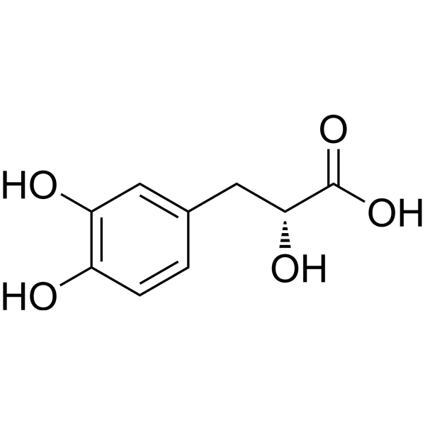
Danshensu (Dan shen suan A), an orally active phenolic compound, can induce Nrf2/HO-1 activation and inhibition of NF-κB pathway. Danshensu reduces reactive oxygen species (ROS) production, upregulates antioxidant defense mechanism and inhibits intrinsic apoptotic pathway. Danshensu displays a potent antiviral activity against SARS-CoV-2 with EC50 of 0.97 μM. Danshensu has anti-oxidation, anti-apoptosis, anti-lung inflammatory and has the potential for COVID-19, cardiovascular and cerebrovascular diseases research.
For research use only. We do not sell to patients.
- Purity : 99.90%
- CAS No.: 76822-21-4
- Formula: C9H10O5
- Molecular Weight:198.17
-
Storage:Powder -20°C, 3 years , 4°C, 2 years ; In solvent -80°C, 6 months , -20°C, 1 month

To place orders, for customer services and technical support, please contact: MedChemExpress USA
Tel: 609-228-6898 E-mail: [email protected] [email protected]


-
 Biological Activity
-
Chemical Information
-
Solvent & Solubility
- Protocols
- Purity & Documentation
- References
-
 Help & FAQs


-
HY-112675
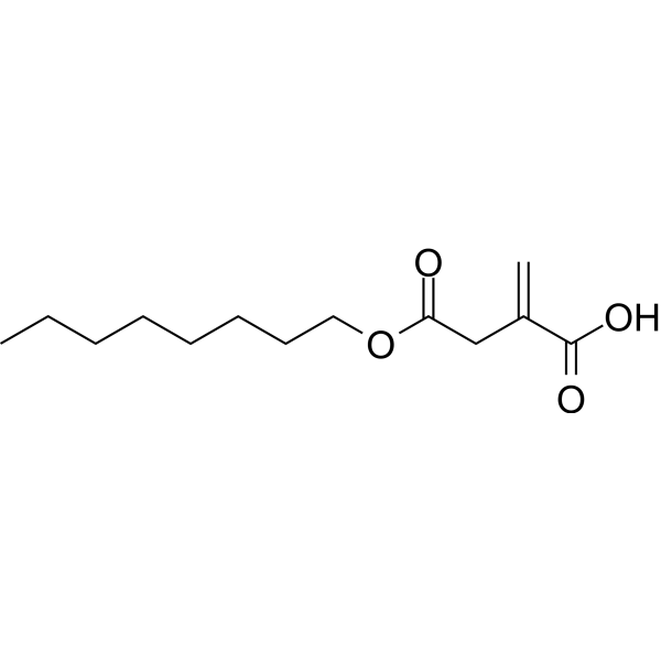
4-Octyl itaconateNrf2 Activator -
HY-N1362
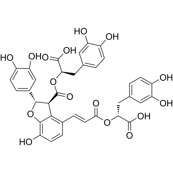
Salvianolic acid BAntioxidant -
HY-N0005
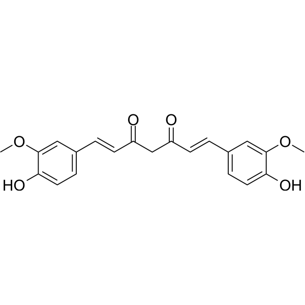
Curcuminp300 Histone Acetylatransferase Inhibitor -
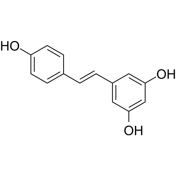
HY-16561
ResveratrolPolyphenolic Phytoalexin -
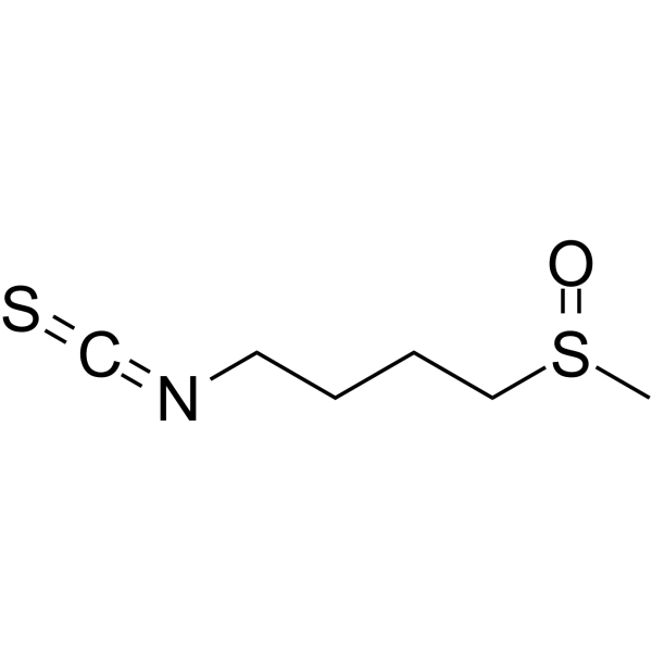
HY-13755
SulforaphaneKeap1/Nrf2/ARE Inducer -
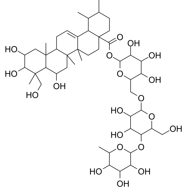
HY-N0568
MadecassosideEndogenous Metabolite -
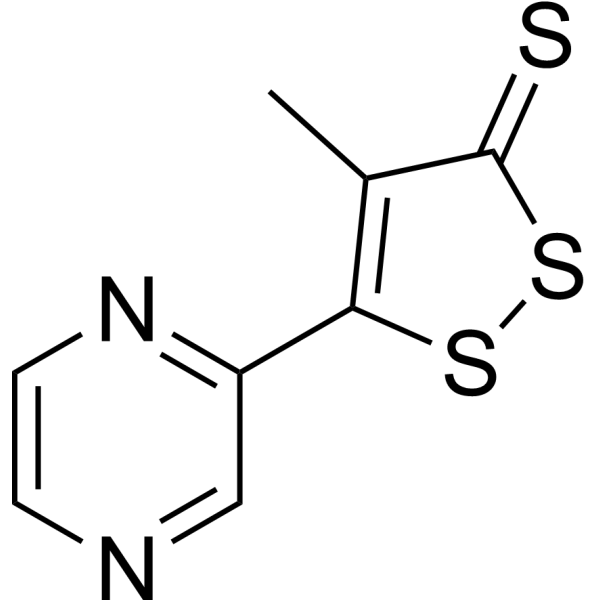
HY-12519
OltiprazHIF-1α Inhibitor -
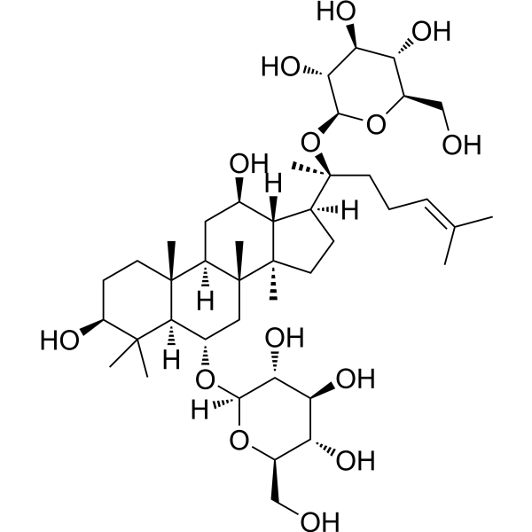
HY-N0045
Ginsenoside Rg1Apoptosis Inducer -
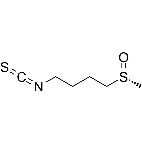
HY-13755A
(R)-SulforaphaneKeap1/Nrf2/ARE Inducer -
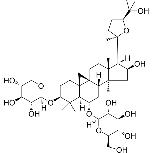
HY-N0431
Astragaloside IVMMP Inhibitor -
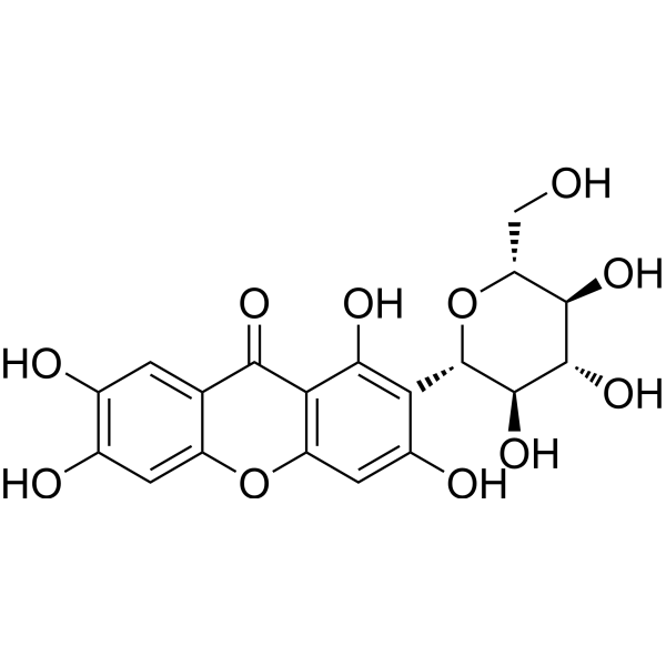
HY-N0290
MangiferinNrf2 Activator -
HY-N0741
LeonurineAutophagy Inhibitor
Publications Citing Use of MedChemExpress (MCE) Danshensu
More- Phytomedicine. 2025 Feb:137:156378. [Abstract]
- Phytomedicine. 2023 May:113:154743. [Abstract]
- Food Sci Hum Wellness. 2025.
- Cancer Immunol Immunother. 2025 Jun 30;74(8):256. [Abstract]
- Eur J Pharmacol. 2025 Apr 5:992:177296. [Abstract]
- J Inflamm Res. 2026 Jun 19:19:596461. [Abstract]
- J Tradit Complement Med. 2025 Feb 7;15(4):434-445. [Abstract]
- Breast Cancer Res Treat. 2024 Dec;208(3):657-671. [Abstract]
- Vet Microbiol. 2026 May:316:110992. [Abstract]
 Customer Validation & Images

-
In Vivo Efficacy Study
-
WB
-
IF
-
Histological Imaging/Staining
-
IF


Biological Activity
Description
In Vitro
Danshensu (Dan shen suan A) potently inhibits the entry of SARS-CoV-2 S protein-pseudo-typed virus (SARS-CoV-2 S) into ACE2-overexpressed HEK-293T cells (IC50=0.31 μM) and Vero-E6 cell (IC50=4.97 μM)[1].
Danshensu (0-100 μM; for 24 h) at higher concentrations (50 and 100 μM) causes significant reduction in migration and invasion of both FaDu and Ca9-22 cells[2].
Danshensu (0-100 μM; for 24 h)dose-dependently reduced the phosphorylation of ERK and p38 phosphorylation in FaDu cell[2].
Danshensu (0-100 μM; for 24, 48, 72 h) does not have any cytotoxic effect on human oral cancer cells[2].
MedChemExpress (MCE) has not independently confirmed the accuracy of these methods. They are for reference only. Further protocols information, click here.
-
Cell Line:FaDu and Ca9-22 cells
-
Concentration:25, 50, and 100 μM
-
Incubation Time:24 h
-
Result:At higher concentrations (50 and 100 μM) caused significant reduction in migration and invasion of both FaDu and Ca9-22 cells.
-
Cell Line:FaDu and Ca9-22 cells
-
Concentration:25, 50, and 100 μM
-
Incubation Time:24 h
-
Result:Phosphorylation of ERK reduced dose-dependently after 24 h in FaDu cell.
Caused significant reduction in p38 phosphorylation.
In Vivo
MedChemExpress (MCE) has not independently confirmed the accuracy of these methods. They are for reference only.
-
Animal Model:Adult BALB/c mice (male, 6-8 weeks, 20±2 g)[1]
-
Dosage:25, 50, 100 mg/kg
-
Administration:Oral administration (daily for 7 continuous days) or i.v. (once)
-
Result:Could prevent SARS-CoV-2 S protein-induced acute lung inflammation.
Ameliorated inflammatory cytokines in serum and lung tissue.


Chemical Information
-
CAS No. 76822-21-4
-
Appearance Solid
-
Molecular Weight 198.17
-
Formula C9H10O5
-
Color White to off-white
-
SMILES
OC1=C(C=CC(C[C@H](C(O)=O)O)=C1)O
-
Synonyms
Dan shen suan A; Salvianic acid A
-
Structure Classification
-
Shipping
Room temperature in continental US; may vary elsewhere.
-
Storage
Powder -20°C 3 years 4°C 2 years In solvent -80°C 6 months -20°C 1 month


Publications (9)
-
Journal Impact Factor
-
Most Recent
-
Phytomedicine
Danshensu enhances autophagy and reduces inflammation by downregulating TNF-α to inhibit the NF-κB signaling pathway in ischemic flaps. [Abstract]2025 Feb:137:156378. PMID: 39818119
Danshensu purchased from MedChemExpress. Usage Cited in: Phytomedicine. 2025 Feb:137:156378. [Abstract]
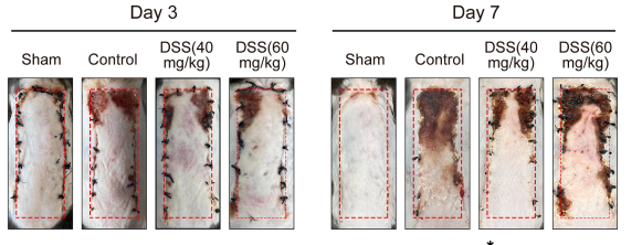
Danshensu (DSS;40, 60 mg/kg; IP; daily for 7 days) significantly increased the flap survival rate in the C57BL/6J mouse.
Danshensu purchased from MedChemExpress. Usage Cited in: Phytomedicine. 2025 Feb:137:156378. [Abstract]
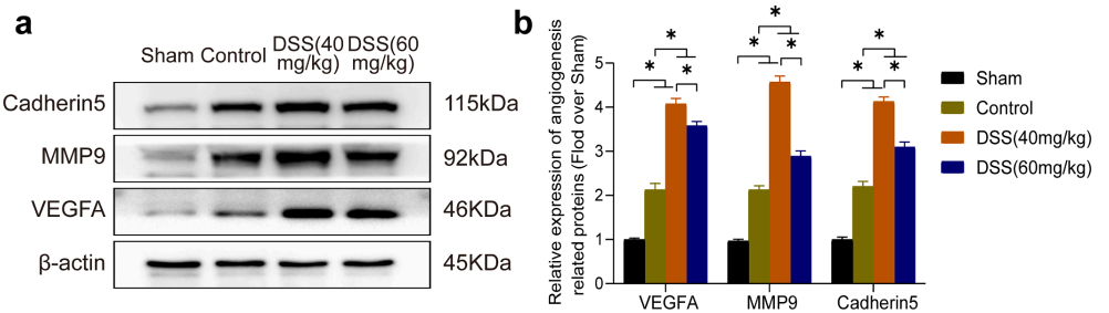
Danshensu (DSS;40, 60 mg/kg; IP; daily for 7 days) increased the protein expression of Cadherin5, MMP9, and VEGFA in the C57BL/6J mouse. Representative western blot images of Cadherin5, MMP9, and VEGFA in mouse skin tissue lysates, with density quantification and statistical analysis of protein levels in each group.
Danshensu purchased from MedChemExpress. Usage Cited in: Phytomedicine. 2025 Feb:137:156378. [Abstract]
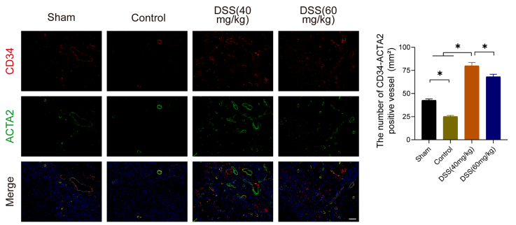
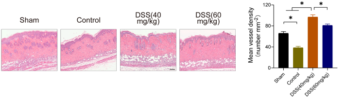
Danshensu (DSS; 40, 60 mg/kg; IP; daily for 7 days) showed the number of blood vessels significantly decreased after ischemic injury of the skin flap. This result was reversed with the use of DSS.
Danshensu purchased from MedChemExpress. Usage Cited in: Phytomedicine. 2025 Feb:137:156378. [Abstract]
Danshensu (DSS; 40, 60 mg/kg; IP; daily for 7 days) increased average vessel density in mouse.
Danshensu purchased from MedChemExpress. Usage Cited in: Phytomedicine. 2025 Feb:137:156378. [Abstract]
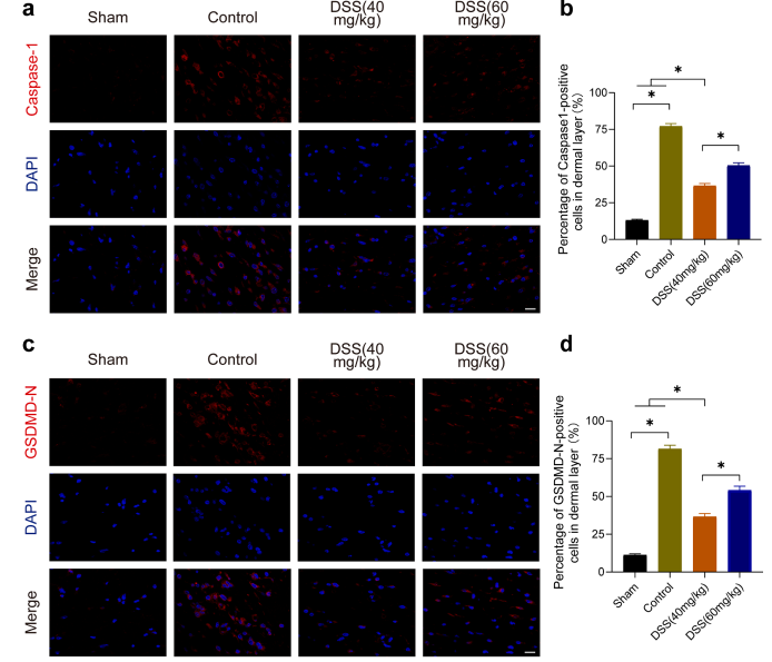
Danshensu (DSS; 40, 60 mg/kg; IP; daily for 7 days) showed that the proportion of GSDMD-N and Caspase-1 positive cells was considerably higher following ischemic injury of the skin flap and notably reduced after DSS treatment. Immunofluorescence staining of Caspase-1 (red) on skin tissue sections from each group, with DAPI staining for cell nuclei (blue).
Danshensu purchased from MedChemExpress. Usage Cited in: Phytomedicine. 2025 Feb:137:156378. [Abstract]
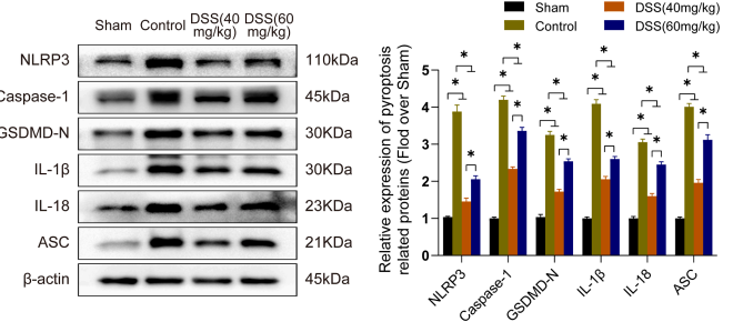
Danshensu (DSS; 40, 60 mg/kg; IP; daily for 7 days) decreased the protein expression of GSDMD-N and Caspase-1 in C57BL/6 J mice.
-
Phytomedicine
Anti-inflammatory effect of Danhong injection through inhibition of GSDMD-mediated pyroptosis. [Abstract]2023 May:113:154743. PMID: 36893672 -
-
Cancer Immunol Immunother
Salvianic acid A enhances anti-PD-1 therapy by promoting HEV-mediated stem-like CD8 T cells infiltration in TNBC. [Abstract]2025 Jun 30;74(8):256. PMID: 40586931 -
Eur J Pharmacol
Sodium Danshensu alleviates bone cancer pain by inhibiting the osteoclast differentiation and CGRP expression. [Abstract]2025 Apr 5:992:177296. PMID: 39900329 -
J Inflamm Res
Salvianic Acid A Regulates Ferroptosis by Activating the Nrf2-GPX4 Pathway Through EGFR to Protect Myocardial Ischemia-Reperfusion Injury. [Abstract]2026 Jun 19:19:596461. PMID: 42345001 -
J Tradit Complement Med
Salvianic acid A promotes osteogenic differentiation of bone marrow mesenchymal stem cells in senile osteoporosis through bromodomain-containing protein 4/Ariadne RBR E3 ubiquitin-protein ligase 1/Rubicon axis. [Abstract]2025 Feb 7;15(4):434-445. PMID: 40677544 -
Breast Cancer Res Treat
Phosphoenolpyruvate carboxykinase-2 (PCK2) is a therapeutic target in triple-negative breast cancer. [Abstract]2024 Dec;208(3):657-671. PMID: 39177932 -
Vet Microbiol
The Chinese medicine monomer Schisandrin C inhibits PRRSV infection by regulating the OGT-PI3K/AKT/mTOR signaling pathway. [Abstract]2026 May:316:110992. PMID: 41865607


Solvent & Solubility
In Vitro:
H2O : 5 mg/mL (25.23 mM; Need ultrasonic)
DMSO : < 1 mg/mL (insoluble or slightly soluble)
Please refer to the solubility information to select the appropriate solvent. Once prepared, please aliquot and store the solution to prevent product inactivation from repeated freeze-thaw cycles.
Storage method and period of stock solution: -80°C, 6 months; -20°C, 1 month. When stored at -80°C, please use it within 6 months. When stored at -20°C, please use it within 1 month.
* Note: If you choose water as the stock solution, please dilute it to the working solution, then filter and sterilize it with a 0.22 μm filter before use.
Please refer to the solubility information to select the appropriate solvent. Once prepared, please aliquot and store the solution to prevent product inactivation from repeated freeze-thaw cycles.
Storage method and period of stock solution: -80°C, 6 months; -20°C, 1 month. When stored at -80°C, please use it within 6 months. When stored at -20°C, please use it within 1 month.
* Note: If you choose water as the stock solution, please dilute it to the working solution, then filter and sterilize it with a 0.22 μm filter before use.
Concentration (start) × Volume (start) = Concentration (final) × Volume (final)
In Vivo:
For the following dissolution methods, please prepare the working solution directly:
It is recommended to prepare fresh solutions and use them promptly within a short period of time.
The percentages shown for the solvents indicate their volumetric ratio in the final prepared solution. If precipitation or phase separation occurs during preparation, heat and/or sonication can be used to aid dissolution.
Add each solvent one by one: PBS
Solubility: 10 mg/mL (50.46 mM); Clear solution; Need ultrasonic and heat to 60°C

Protocols
-
Research Protocol for Infectious Diseases
Infectious-disease experiments test how pathogens interact with host barriers, innate immune receptors, inflammatory signaling, pathogen replication, and tissue injury; pattern-recognition receptors such as TLRs, RIG-I-like receptors, NOD-like receptors, and inflammasomes detect microbial molecules and activate NF-κB, interferon, and cytokine responses. The central hypothesis is that infection severity reflects the balance between pathogen burden and host response: protective inflammation restricts pathogen growth, whereas excessive or mislocalized inflammation contributes to tissue damage and disease phenotype. Unresolved questions include which host pathways are protective versus pathogenic, why some infection models fail to translate to human disease, and which combined readouts best predict clinically relevant infection outcomes.
-
Apoptosis
Apoptosis, also called programmed cell death, is generally characterized by distinct morphological characteristics.
-
TUNEL staining for apoptotic DNA fragmentation
TUNEL staining detects DNA strand breaks by using terminal deoxynucleotidyl transferase to add labeled nucleotides to exposed 3′-OH DNA termini, generating either microscopic staining in fixed cells or tissue sections, or fluorescence/cytometric signal in cell suspensions. TUNEL positivity reflects DNA fragmentation but should not be interpreted alone as definitive apoptosis, because TUNEL can also label necrotic, autolytic, mechanically damaged, or DNA-repair-associated DNA breaks.
-
Annexin V plus membrane-impermeant dye apoptosis staining
Annexin V-based apoptosis assays rely on the detection of phosphatidylserine (PS) externalization from the inner leaflet of the plasma membrane to the outer leaflet, an early biochemical hallmark of apoptosis. Fluorescently labeled Annexin V binds PS in a calcium-dependent manner, enabling identification of early apoptotic cells by flow cytometry or fluorescence microscopy. When combined with a membrane-impermeant DNA-binding dye (e. g. , propidium iodide), this approach allows discrimination between viable (Annexin V−/dye−), early apoptotic (Annexin V+/dye−), and late apoptotic or necrotic (Annexin V+/dye+) cell populations by assessing membrane integrity and PS exposure.
-
ROS/oxidative-stress fluorescent staining
ROS/oxidative-stress fluorescent staining uses cell-permeant fluorogenic probes that become fluorescent after oxidation inside cells or tissues; commonly used examples include DCFH-DA/DCFDA for broad cellular oxidant detection, DHE for superoxide-related signal detection, MitoSOX for mitochondrial superoxide-related signal detection, and CellROX probes for oxidative-stress-associated fluorescence readouts. The assay detects probe oxidation rather than a single ROS species unless the probe and analysis method have been chemically validated for that species. DCFH-DA enters cells, is deacetylated by intracellular esterases to DCFH, and produces fluorescent DCF after oxidation, so the readout is used as an operational measure of total cellular oxidative stress rather than a species-specific ROS measurement. DHE and MitoSOX can report superoxide-related oxidation, but red fluorescence alone can include non-specific ethidium-like oxidation products; HPLC or optimized spectral approaches are
-
Apoptosis Solutions
Apoptosis is a regulated, generally non-lytic cell-death pathway that removes unwanted, damaged, infected, or abnormal cells through coordinated morphological changes, caspase activation, DNA fragmentation, and membrane remodeling. The intrinsic apoptosis pathway is controlled mainly by mitochondrial outer membrane permeabilization, BCL-2 family proteins, cytochrome c release, apoptosome formation, caspase-9 activation, and downstream executioner caspase-3/7 activation. The extrinsic apoptosis pathway is initiated by death receptors such as Fas, TNFR, and TRAIL receptors, which recruit adaptor proteins and activate caspase-8 before engaging executioner caspases or mitochondrial amplification through BID cleavage. Apoptosis is linked to many phenotypes, including cancer cell killing, tissue homeostasis, immune regulation, neurodegeneration, infection response, and treatment-induced cytotoxicity; unresolved questions include how apoptosis interacts with necroptosis, pyroptosis, ferroptos
-
Research Protocol for Inflammation-related Diseases
The NLRP3 inflammasome is a cytosolic innate immune signaling platform that integrates priming signals and danger-signal activation to promote caspase-1 activation, maturation of IL-1β and IL-18, and gasdermin D-mediated pyroptotic cell death. The core experimental logic is to determine whether inflammatory disease phenotypes are driven by increased NLRP3 expression, ASC-containing inflammasome assembly, caspase-1 cleavage, GSDMD cleavage, and extracellular release of IL-1β/IL-18 rather than by nonspecific cell injury alone. The pathway is strongly linked to inflammation-related disease phenotypes because monosodium urate crystals activate NALP3/NLRP3 inflammasome signaling in gout-like crystal inflammation, cholesterol crystals activate NLRP3 inflammasomes in atherogenesis models, and DSS-induced intestinal inflammation has been reported to involve NLRP3 inflammasome activity. However, experimental colitis studies also show context-dependent protective effects of NLRP3 inflammasome co


Purity & Documentation
-
Data Sheet (278 KB)
-
SDS (396 KB)
- English - EN (396 KB)
- Français - FR (396 KB)
- Deutsch - DE (396 KB)
- Norwegian - NO (396 KB)
- Español - ES (396 KB)
- Swedish - SV (396 KB)
- Italian - IT (396 KB)
- Korean - KR (396 KB)
- Portuguese - PT (396 KB)
-
Handling Instructions (2659 KB)
References
[1]. Wei Wang, et al. Danshensu alleviates pseudo-typed SARS-CoV-2 induced mouse acute lung inflammation. Acta Pharmacol Sin. 2022 Apr;43(4):771-780. [Content Brief]
[2]. V Bharath Kumar, et al. Sodium Danshensu Inhibits Oral Cancer Cell Migration and Invasion by Modulating p38 Signaling Pathway. Front Endocrinol (Lausanne). 2020 Sep 30:11:568436. [Content Brief]
[3]. Chen Yu, et al. Danshensu attenuates cisplatin-induced nephrotoxicity through activation of Nrf2 pathway and inhibition of NF-κB. Biomed Pharmacother. 2021 Oct:142:111995. [Content Brief]


Complete Stock Solution Preparation Table
Please refer to the solubility information to select the appropriate solvent. Once prepared, please aliquot and store the solution to prevent product inactivation from repeated freeze-thaw cycles.
Storage method and period of stock solution: -80°C, 6 months; -20°C, 1 month. When stored at -80°C, please use it within 6 months. When stored at -20°C, please use it within 1 month.
| Optional Solvent | Concentration Solvent Mass | 1 mg | 5 mg | 10 mg | 25 mg |
|---|---|---|---|---|---|
| H2O | 1 mM | 5.0462 mL | 25.2309 mL | 50.4617 mL | 126.1543 mL |
| 5 mM | 1.0092 mL | 5.0462 mL | 10.0923 mL | 25.2309 mL | |
| 10 mM | 0.5046 mL | 2.5231 mL | 5.0462 mL | 12.6154 mL | |
| 15 mM | 0.3364 mL | 1.6821 mL | 3.3641 mL | 8.4103 mL | |
| 20 mM | 0.2523 mL | 1.2615 mL | 2.5231 mL | 6.3077 mL | |
| 25 mM | 0.2018 mL | 1.0092 mL | 2.0185 mL | 5.0462 mL |
* Note: If you choose water as the stock solution, please dilute it to the working solution, then filter and sterilize it with a 0.22 μm filter before use.


Danshensu Related Classifications
HY-N1913 Related Classifications
 Powered by Bioz