



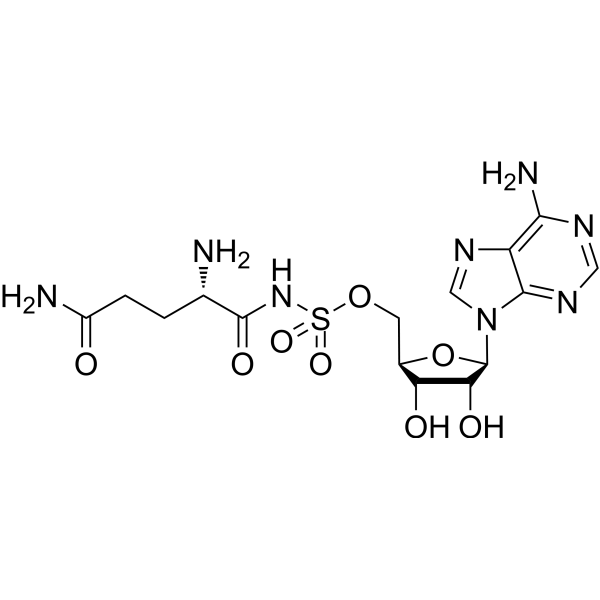
Gln-AMS
Based on 3 publication(s) in Google Scholar
Gln-AMS is a potent inhibitor of Aminoacyl-tRNA Synthetase. Gln-AMS blocks the lactylation modification of downstream targets through competitive binding to AARS1, thereby regulating apoptosis, ferroptosis, and the transcriptional processes of related genes. Gln-AMS can be used in research on breast cancer, diabetic nephropathy, and sepsis-associated encephalopathy.
For research use only. We do not sell to patients.
- Purity : 99.66%
- CAS No.: 209543-57-7
- Formula: C15H22N8O8S
- Molecular Weight:474.45
-
Storage:
-20°C, sealed storage, away from moisture
* In solvent : -80°C, 6 months; -20°C, 1 month (sealed storage, away from moisture)

To place orders, for customer services and technical support, please contact: MedChemExpress USA
Tel: 609-228-6898 E-mail: [email protected] [email protected]


-
 Biological Activity
-
Chemical Information
-
Solvent & Solubility
- Protocols
- Purity & Documentation
- References
-
 Help & FAQs


-
HY-112862
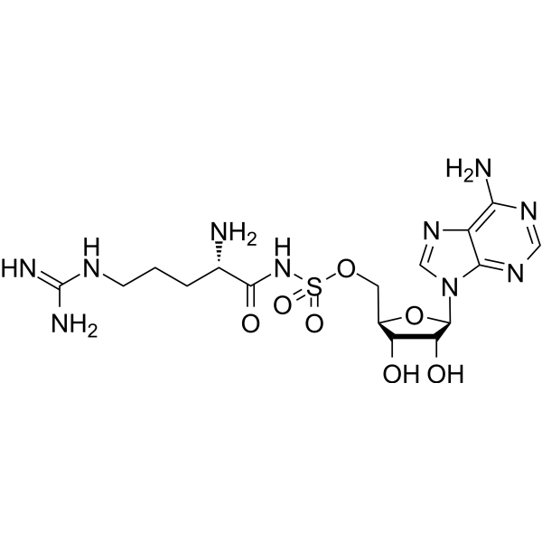
Arg-AMSAminoacyl-tRNA Synthetase Inhibitor -
HY-13823
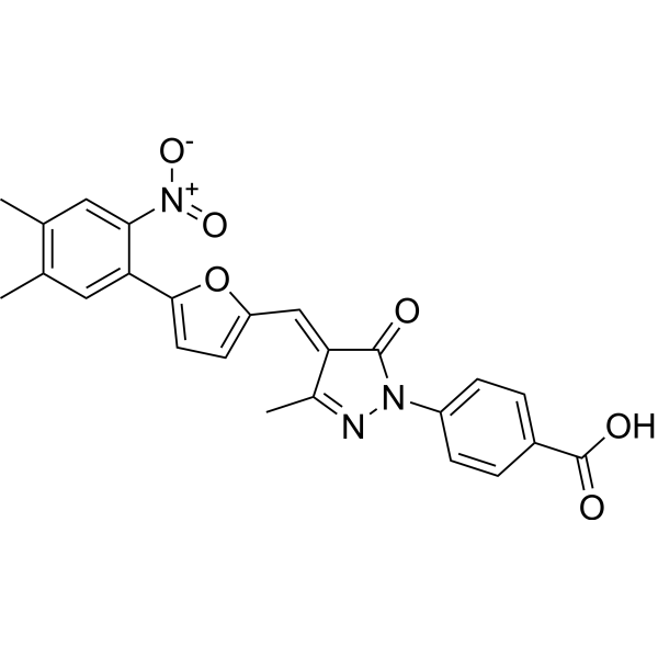
C646p300/CBP HAT Inhibitor -
HY-15887
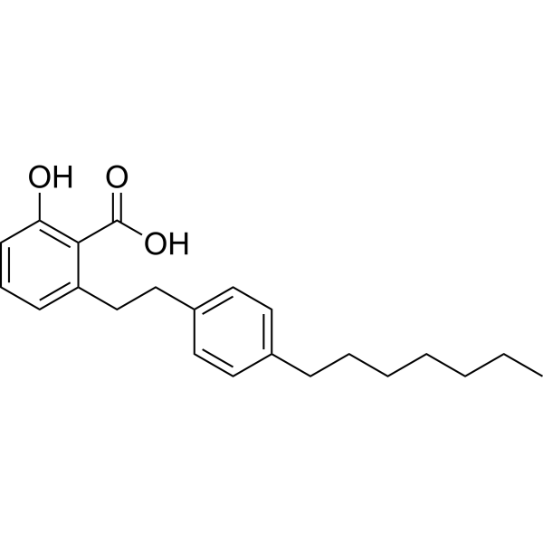
MG 149Tip60 Inhibitor -
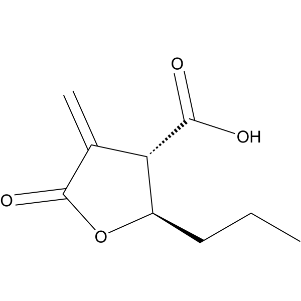
HY-129039
Butyrolactone 3Gcn5 Inhibitor -
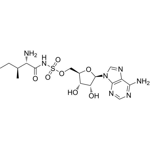
HY-108939
Aminoacyl tRNA synthetase-IN-1aaRS Inhibitor -
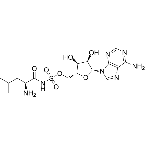
HY-108900
Leu-AMSLeucyl-tRNA Synthetase Inhibitor -
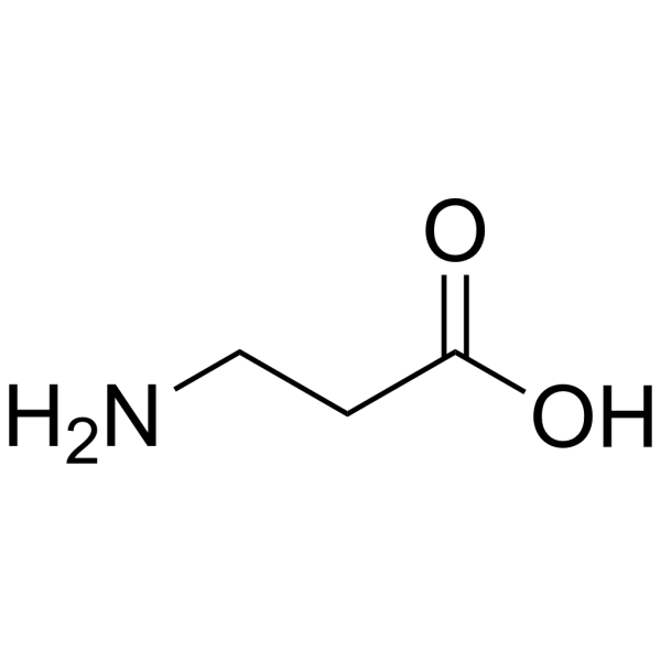
HY-N0230
β-AlanineEndogenous Metabolite -
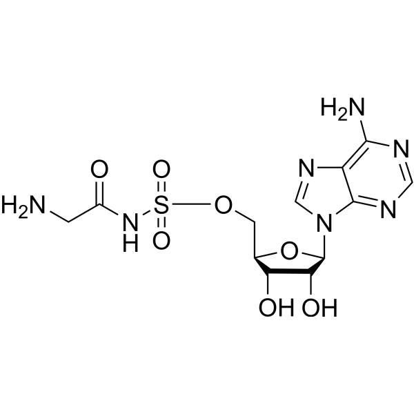
HY-108940
GlyRS-IN-1GlyRS Inhibitor -
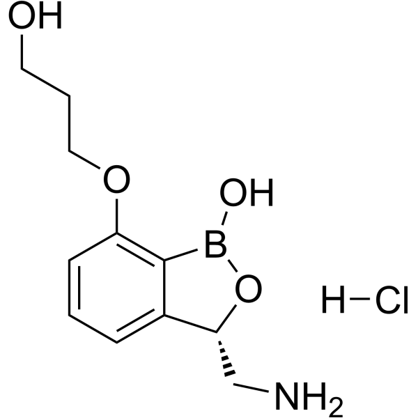
HY-12479A
Epetraborole hydrochlorideLeuRS Inhibitor -
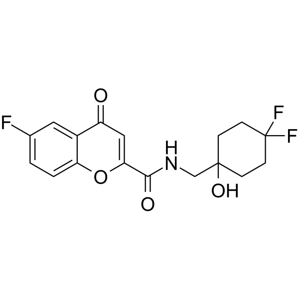
HY-126130
LysRs-IN-2Aminoacyl-tRNA Synthetase Inhibitor -

HY-108939R
Aminoacyl tRNA synthetase-IN-1 (Standard)Reference Standard -

HY-108940R
GlyRS-IN-1 (Standard)Reference Standard
Publications Citing Use of MedChemExpress (MCE) Gln-AMS
More

Biological Activity
Description
IC50 & Target
[1]|
AARS1 |
GlnRS |
Cellular Effect
|
Cell Line

|
Type | Value | Description | References |
|---|---|---|---|---|
| PBMC | IC50 |
0.8 μM
Compound: GlnSA
|
Immunosuppressive activity against human PBMC assessed as inhibition of allogenic mixed lymphocyte reaction
Immunosuppressive activity against human PBMC assessed as inhibition of allogenic mixed lymphocyte reaction
|
[PMID: 18438987] |
In Vitro
Gln-AMS (10 μM; 24 h) reduces global Kla, H3K79la, and H4K91la modification levels in breast cancer cells (MDA-MB-468 and T-47D)[1].
Gln-AMS (1 μM; 72 h) decreased ACSL4 expression, increased GPX4 expression, reduced lipid peroxidation, restored mitochondrial membrane potential, decreased MDA and Fe2+ levels, and reduced mitochondrial superoxide levels in HGECs and HK-2 cells, thereby inhibiting ferroptosis[2].
Gln-AMS (1 μM; 72 h) reversed high glucose-induced elevated expression of AARS1 and H3K18la and reversed cell death in human glomerular endothelial cells (HGECs) and human kidney tubular epithelial cells (HK-2)[2].
Gln-AMS (1 μM; 72 h) decreased ELOVL5 protein and mRNA expression in HGECs and HK-2 cells[2].
Gln-AMS (10 μM; 2 h) inhibits AARS1-mediated global protein lactylation and ATRIP lactylation in primary hippocampal neurons[3].
Gln-AMS (10 μM; 12 h) inhibits Lactic acid (HY-B2227)-induced global protein lactylation and p-ATR, p-CHK1 expression in primary hippocampal neurons[3].
Gln-AMS (10 μM; 12 h) inhibits p-ATR and p-CHK1 expression in HT22 cells[3].
MedChemExpress (MCE) has not independently confirmed the accuracy of these methods. They are for reference only. Further protocols information, click here.
-
Cell Line:MDA-MB-468 and T-47D cells
-
Concentration:10 μM
-
Incubation Time:24 h
-
Result:Decreased global Kla, H4K79la, and H4K91la levels.
-
Cell Line:HT22
-
Concentration:as indicated (Gln-AMS); 1 μg/mL (LPS); varying concentrations (lactate)
-
Incubation Time:12 h
-
Result:Modulated p-ATR (1989T) and p-CHK1 levels in LPS and lactate-treated HT22 cells.
-
Cell Line:Primary hippocampal neurons
-
Concentration:10 μM
-
Incubation Time:2, 12 h
-
Result:Inhibited overall protein lactylation and ATRIP lactylation in the cytoplasm. The product also decreased the expression of p-ATR and p-CHK1 induced by lactate.
-
Cell Line:HT22
-
Concentration:10 μM
-
Incubation Time:12 h
-
Result:Suppressed the expression of p-ATR and p-CHK1.


Chemical Information
-
CAS No. 209543-57-7
-
Appearance Solid
-
Molecular Weight 474.45
-
Formula C15H22N8O8S
-
Color White to off-white
-
SMILES
NC1=C2N=CN([C@@H]3O[C@H](COS(=O)(NC([C@@H](N)CCC(N)=O)=O)=O)[C@@H](O)[C@H]3O)C2=NC=N1
-
Shipping
Room temperature in continental US; may vary elsewhere.
-
Storage
-20°C, sealed storage, away from moisture
* In solvent : -80°C, 6 months; -20°C, 1 month (sealed storage, away from moisture)


Publications (3)
-
Journal Impact Factor
-
Most Recent
-
Adv Sci (Weinh)
Lactylated Apolipoprotein C-II Induces Immunotherapy Resistance by Promoting Extracellular Lipolysis. [Abstract]2024 Oct;11(38):e2406333. PMID: 38981044 -
Cell Death Differ
AARS1-mediated lactylation of H3K18 and STAT1 promotes ferroptosis in diabetic nephropathy. [Abstract]2025 Sep 23. PMID: 40987895 -
Brain Behav Immun
Targeting AARS1-dependent lactylation improves neuronal process plasticity and mitigates cognitive deficits in sepsis-associated encephalopathy. [Abstract]2026 Jul:135:106493. PMID: 41713664


Solvent & Solubility
In Vitro:
DMSO : 100 mg/mL (210.77 mM; Need ultrasonic; Hygroscopic DMSO has a significant impact on the solubility of product, please use newly opened DMSO)
Please refer to the solubility information to select the appropriate solvent. Once prepared, please aliquot and store the solution to prevent product inactivation from repeated freeze-thaw cycles.
Storage method and period of stock solution: -80°C, 6 months; -20°C, 1 month (sealed storage, away from moisture). When stored at -80°C, please use it within 6 months. When stored at -20°C, please use it within 1 month.
Please refer to the solubility information to select the appropriate solvent. Once prepared, please aliquot and store the solution to prevent product inactivation from repeated freeze-thaw cycles.
Storage method and period of stock solution: -80°C, 6 months; -20°C, 1 month (sealed storage, away from moisture). When stored at -80°C, please use it within 6 months. When stored at -20°C, please use it within 1 month.
Concentration (start) × Volume (start) = Concentration (final) × Volume (final)
In Vivo:
Select the appropriate dissolution method based on your experimental animal and administration route.
- For the following dissolution methods, please ensure to first prepare a clear stock solution using an In Vitro approach and then sequentially add co-solvents:
- To ensure reliable experimental results, the clarified stock solution can be appropriately stored based on storage conditions. As for the working solution for In Vivo experiments, it is recommended to prepare freshly and use it on the same day.
- The percentages shown for the solvents indicate their volumetric ratio in the final prepared solution. If precipitation or phase separation occurs during preparation, heat and/or sonication can be used to aid dissolution.
Add each solvent one by one: 10% DMSO 40% PEG300 5% Tween-80 45% Saline
Solubility: ≥ 2.5 mg/mL (5.27 mM); Clear solution
This protocol yields a clear solution of ≥ 2.5 mg/mL (saturation unknown).
Taking 1 mL working solution as an example, add 100 μL DMSO stock solution (25.0 mg/mL) to 400 μL PEG300, and mix evenly; then add 50 μL Tween-80 and mix evenly; then add 450 μL Saline to adjust the volume to 1 mL.
Preparation of Saline: Dissolve 0.9 g sodium chloride in ddH₂O and dilute to 100 mL to obtain a clear Saline solution.
Add each solvent one by one: 10% DMSO 90% (20% SBE-β-CD in Saline)
Solubility: ≥ 2.5 mg/mL (5.27 mM); Clear solution
This protocol yields a clear solution of ≥ 2.5 mg/mL (saturation unknown).
Taking 1 mL working solution as an example, add 100 μL DMSO stock solution (25.0 mg/mL) to 900 μL 20% SBE-β-CD in Saline, and mix evenly.
Preparation of 20% SBE-β-CD in Saline (4°C, storage for one week): 2 g SBE-β-CD powder is dissolved in 10 mL Saline, completely dissolve until clear.
In Vivo Dissolution Calculator
Please enter the basic information of animal experiments:
-
-
-
-
Recommended: Prepare an additional quantity of animals to account for potential losses during experiments.
Please enter your animal formula composition:
-
%DMSO +
Recommended: Keep the proportion of DMSO in working solution below 2% if your animal is weak.
-
%+
-
+%Tween-80 + +
-
%Saline +
The co-solvents required include: DMSO, . All of co-solvents are available by MedChemExpress (MCE). , Tween 80. All of co-solvents are available by MedChemExpress (MCE).
Working solution concentration: 0.22 mg/mL
Method for preparing stock solution: mg drug dissolved in μL DMSO. Stock solution concentration: mg/mL. * In solvent : -80°C, 6 months; -20°C, 1 month (sealed storage, away from moisture)
1. Take μL DMSO stock solution;
2. Add μL .
μL , mix evenly;
3. Then add μL Tween 80, mix evenly;
4. Then add μL
Please ensure that the stock solution in the first step is dissolved to a clear state, and add co-solvents in sequence. You can use ultrasonic heating (ultrasonic cleaner, recommended frequency 20-40 kHz), vortexing, etc. to assist dissolution.

Protocols
-
Apoptosis
Apoptosis, also called programmed cell death, is generally characterized by distinct morphological characteristics.
-
TUNEL staining for apoptotic DNA fragmentation
TUNEL staining detects DNA strand breaks by using terminal deoxynucleotidyl transferase to add labeled nucleotides to exposed 3′-OH DNA termini, generating either microscopic staining in fixed cells or tissue sections, or fluorescence/cytometric signal in cell suspensions. TUNEL positivity reflects DNA fragmentation but should not be interpreted alone as definitive apoptosis, because TUNEL can also label necrotic, autolytic, mechanically damaged, or DNA-repair-associated DNA breaks.
-
LPS-Induced Endotoxemia/Systemic Inflammation
Lipopolysaccharide (LPS)-induced endotoxemia is a widely used in vivo model of acute systemic inflammation in which LPS, a Gram-negative bacterial endotoxin, activates innate immune signaling primarily through TLR4, leading to rapid and transient induction of pro-inflammatory cytokines such as TNF-α, IL-6, and IL-1β in circulation and tissues. This cytokine surge is commonly used as a measurable readout of systemic inflammatory activation and immune dysregulation, and is typically assessed within hours after intraperitoneal LPS administration in mouse models of endotoxemia. The model captures key features of systemic inflammatory response syndrome, including cytokine release, immune cell activation, and downstream tissue responses, and has been used to evaluate anti-inflammatory interventions such as cytokine modulation, lipid mediators, and immune cell-targeting therapies.
-
Annexin V plus membrane-impermeant dye apoptosis staining
Annexin V-based apoptosis assays rely on the detection of phosphatidylserine (PS) externalization from the inner leaflet of the plasma membrane to the outer leaflet, an early biochemical hallmark of apoptosis. Fluorescently labeled Annexin V binds PS in a calcium-dependent manner, enabling identification of early apoptotic cells by flow cytometry or fluorescence microscopy. When combined with a membrane-impermeant DNA-binding dye (e. g. , propidium iodide), this approach allows discrimination between viable (Annexin V−/dye−), early apoptotic (Annexin V+/dye−), and late apoptotic or necrotic (Annexin V+/dye+) cell populations by assessing membrane integrity and PS exposure.
-
Apoptosis Solutions
Apoptosis is a regulated, generally non-lytic cell-death pathway that removes unwanted, damaged, infected, or abnormal cells through coordinated morphological changes, caspase activation, DNA fragmentation, and membrane remodeling. The intrinsic apoptosis pathway is controlled mainly by mitochondrial outer membrane permeabilization, BCL-2 family proteins, cytochrome c release, apoptosome formation, caspase-9 activation, and downstream executioner caspase-3/7 activation. The extrinsic apoptosis pathway is initiated by death receptors such as Fas, TNFR, and TRAIL receptors, which recruit adaptor proteins and activate caspase-8 before engaging executioner caspases or mitochondrial amplification through BID cleavage. Apoptosis is linked to many phenotypes, including cancer cell killing, tissue homeostasis, immune regulation, neurodegeneration, infection response, and treatment-induced cytotoxicity; unresolved questions include how apoptosis interacts with necroptosis, pyroptosis, ferroptos
-
Ferroptosis Solutions
Ferroptosis is an iron-dependent, non-apoptotic form of regulated cell death characterized by lethal lipid peroxidation and sensitivity to suppression by iron chelators or lipophilic radical-trapping antioxidants. The core pathway links cystine uptake through system Xc−, glutathione availability, GPX4-dependent detoxification of phospholipid hydroperoxides, iron-dependent oxidative reactions, and polyunsaturated-phospholipid metabolism into a cell-death program that is biochemically and morphologically distinct from apoptosis, necrosis, and autophagy. The ferroptosis pathway is experimentally linked to phenotype through chemical and genetic perturbation. Erastin induces ferroptosis by inhibiting cystine uptake through system Xc− and weakening antioxidant defenses, while GPX4 inhibition or depletion causes lipid peroxide accumulation and ferroptotic cancer-cell death. ACSL4 and oxidizable arachidonoyl- or adrenoyl-containing phosphatidylethanolamines shape ferroptosis sensitivity by con
-
Breast Cancer Modeling
Breast cancer is a heterogeneous cancer, and it has been distinguished into four subtypes: luminal A, luminal B, HER2-positive and basal-like. Molecular mutations, epigenetic alterations, hormone exposure and immune microenvironment are related to the progression of breast cancer.


Purity & Documentation
-
Data Sheet (281 KB)
-
SDS (252 KB)
- English - EN (252 KB)
- Français - FR (252 KB)
- Deutsch - DE (252 KB)
- Norwegian - NO (252 KB)
- Español - ES (252 KB)
- Swedish - SV (252 KB)
- Italian - IT (252 KB)
- Korean - KR (252 KB)
- Portuguese - PT (252 KB)
-
Handling Instructions (2659 KB)
References
[1]. Liu J, et al. H4K79 and H4K91 histone lactylation, newly identified lactylation sites enriched in breast cancer. Journal of experimental & clinical cancer research : CR. 2025 Aug 23;44(1):252. [Content Brief]
[2]. Hong J, et al. AARS1-mediated lactylation of H3K18 and STAT1 promotes ferroptosis in diabetic nephropathy. Cell death and differentiation. 2026 Mar;33(3):589-604. [Content Brief]
[3]. Luo S, et al. Targeting AARS1-dependent lactylation improves neuronal process plasticity and mitigates cognitive deficits in sepsis-associated encephalopathy. Brain, behavior, and immunity. 2026 Jul;135:106493. [Content Brief]


Complete Stock Solution Preparation Table
Please refer to the solubility information to select the appropriate solvent. Once prepared, please aliquot and store the solution to prevent product inactivation from repeated freeze-thaw cycles.
Storage method and period of stock solution: -80°C, 6 months; -20°C, 1 month (sealed storage, away from moisture). When stored at -80°C, please use it within 6 months. When stored at -20°C, please use it within 1 month.
| Optional Solvent | Concentration Solvent Mass | 1 mg | 5 mg | 10 mg | 25 mg |
|---|---|---|---|---|---|
| DMSO | 1 mM | 2.1077 mL | 10.5385 mL | 21.0770 mL | 52.6926 mL |
| 5 mM | 0.4215 mL | 2.1077 mL | 4.2154 mL | 10.5385 mL | |
| 10 mM | 0.2108 mL | 1.0539 mL | 2.1077 mL | 5.2693 mL | |
| 15 mM | 0.1405 mL | 0.7026 mL | 1.4051 mL | 3.5128 mL | |
| 20 mM | 0.1054 mL | 0.5269 mL | 1.0539 mL | 2.6346 mL | |
| 25 mM | 0.0843 mL | 0.4215 mL | 0.8431 mL | 2.1077 mL | |
| 30 mM | 0.0703 mL | 0.3513 mL | 0.7026 mL | 1.7564 mL | |
| 40 mM | 0.0527 mL | 0.2635 mL | 0.5269 mL | 1.3173 mL | |
| 50 mM | 0.0422 mL | 0.2108 mL | 0.4215 mL | 1.0539 mL | |
| 60 mM | 0.0351 mL | 0.1756 mL | 0.3513 mL | 0.8782 mL | |
| 80 mM | 0.0263 mL | 0.1317 mL | 0.2635 mL | 0.6587 mL | |
| 100 mM | 0.0211 mL | 0.1054 mL | 0.2108 mL | 0.5269 mL |


Gln-AMS Related Classifications
HY-112861 Related Classifications
 Powered by Bioz