



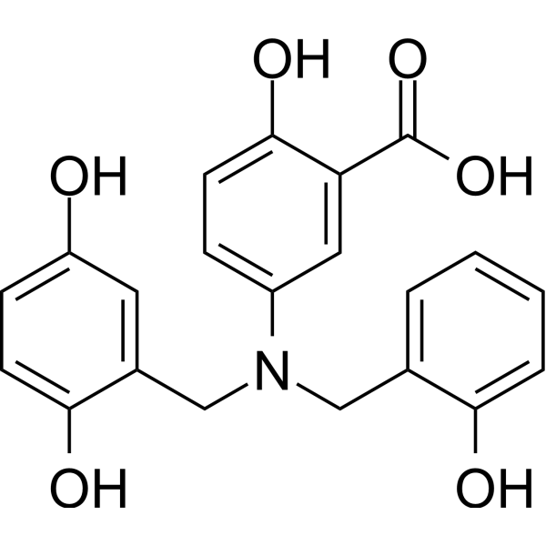
Lavendustin A
Based on 1 publication(s) in Google Scholar
Lavendustin A (RG-14355) is a potent, selective and ATP-competitive inhibitor of epidermal growth factor receptor (EGFR) tyrosine kinase, with an IC50 of 11 nM. Lavendustin A does not inhibit protein kinase A or C. Lavendustin A can suppress VEGF-induced angiogenesis.
Nur für Forschungszwecke. Wir verkaufen nicht an Patienten.
- Reinheit : 99.09%
- CAS. Nr.: 125697-92-9
- Formel: C21H19NO6
- Molecular Weight:381.38
-
Speicherung:
4°C, stored under nitrogen
* In solvent : -80°C, 6 months; -20°C, 1 month (stored under nitrogen)

To place orders, for customer services and technical support, please contact: MedChemExpress USA
Tel: 609-228-6898 E-mail: [email protected] [email protected]


-
 Biologische Aktivität
-
Chemical Information
-
Lösungsmittel & Löslichkeit
- Protokoll
- Reinheit & Dokumentation
- Verweise
-
 Help & FAQs


-
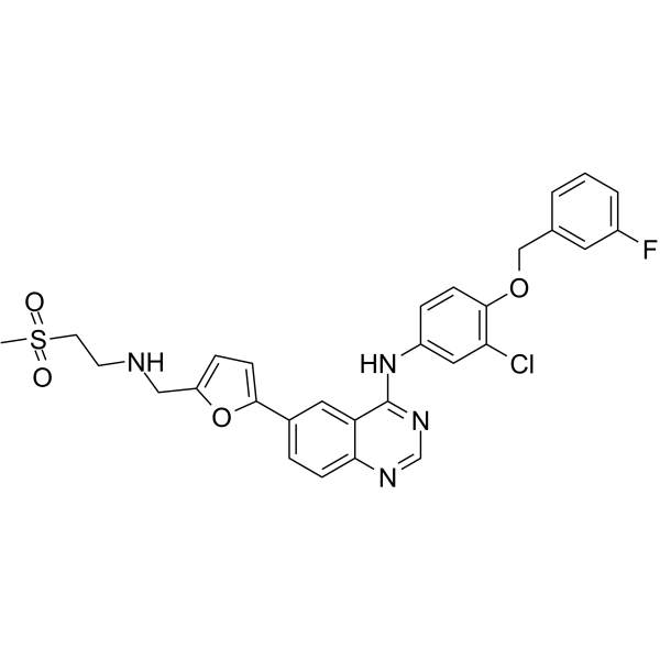
HY-50898
LapatinibEGFR/HER2 Inhibitor -
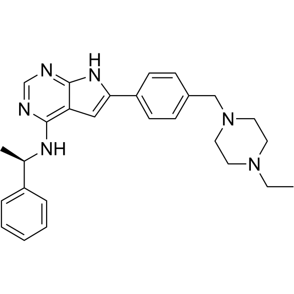
HY-10045
AEE788EGFR/ErbB2 Inhibitor -

HY-18963R
Lavendustin A (Standard)Tyrosine Kinase Inhibitor -
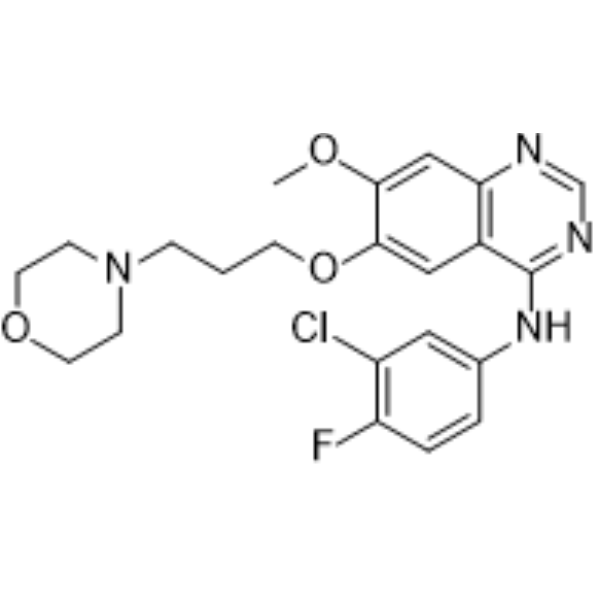
HY-50895
GefitinibEGFR Inhibitor -
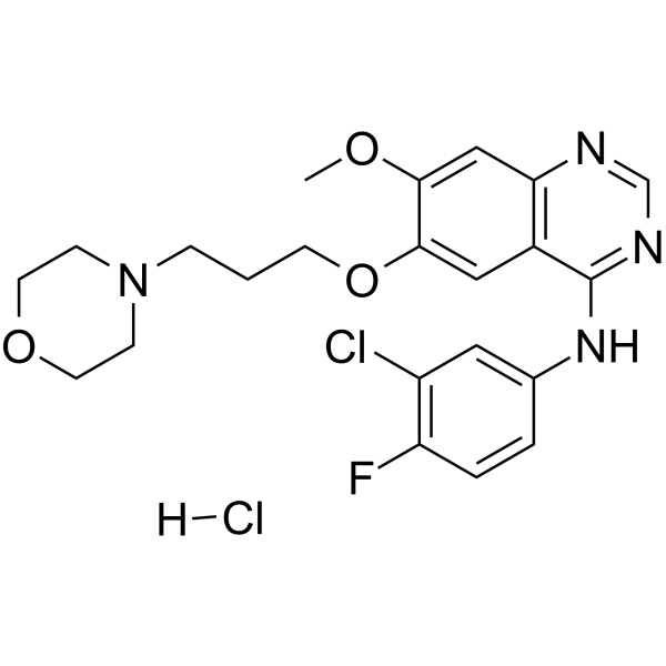
HY-50895A
Gefitinib hydrochlorideEGFR Inhibitor -
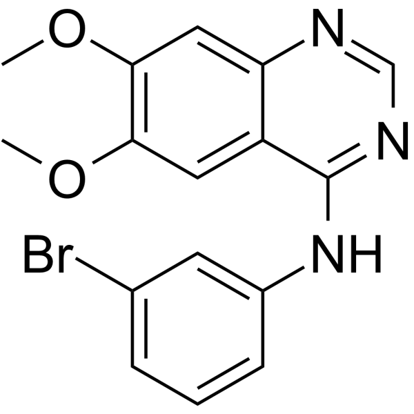
HY-14346
PD153035EGFR Inhibitor -
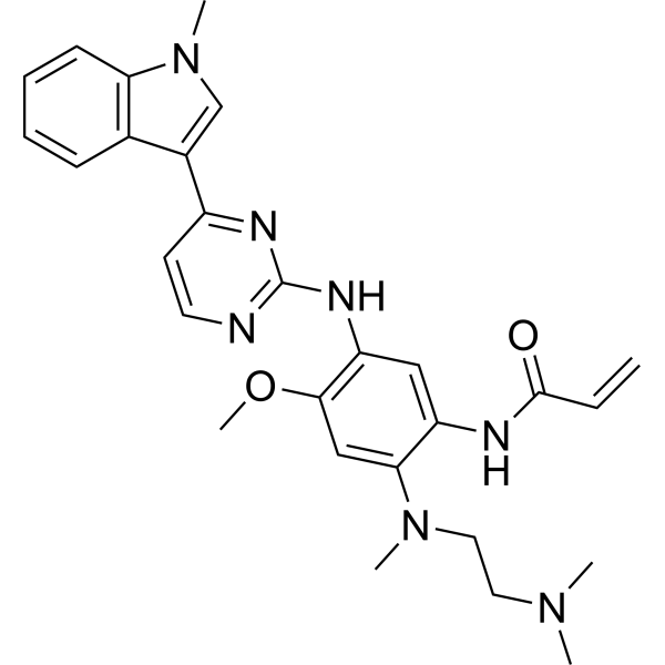
HY-15772
OsimertinibMutant-Selective EGFR Inhibitor -
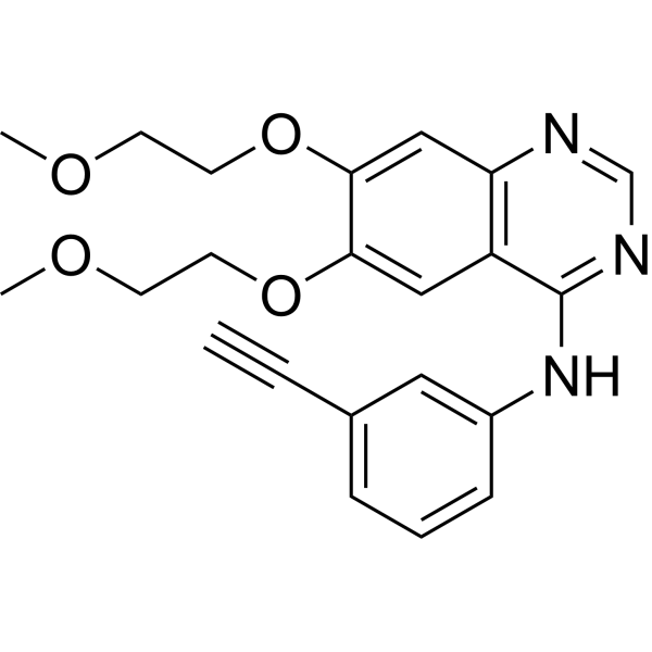
HY-50896
ErlotinibEGFR Tyrosine Kinase Inhibitor -
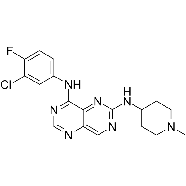
HY-10322
FalnidamolSelective EGFR Inhibitor -
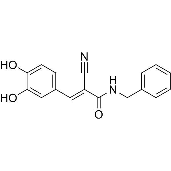
HY-12000
AG490EGFR Inhibitor -
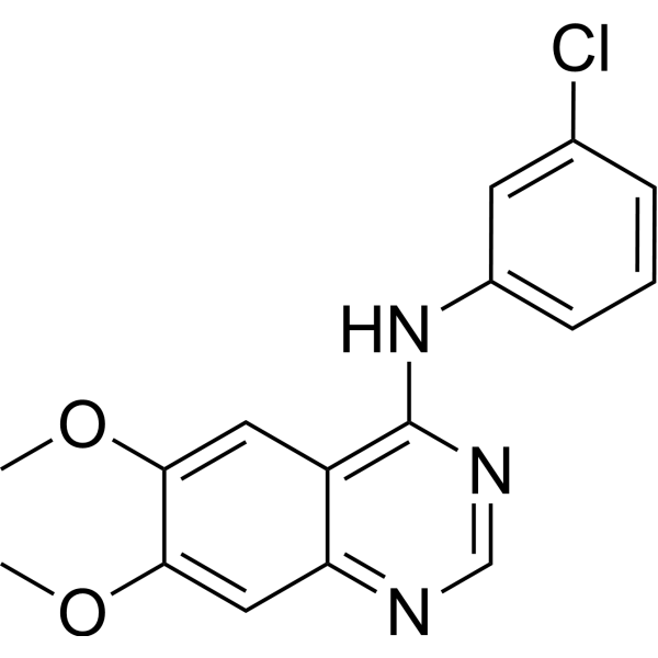
HY-13524
AG-1478EGFR Inhibitor -
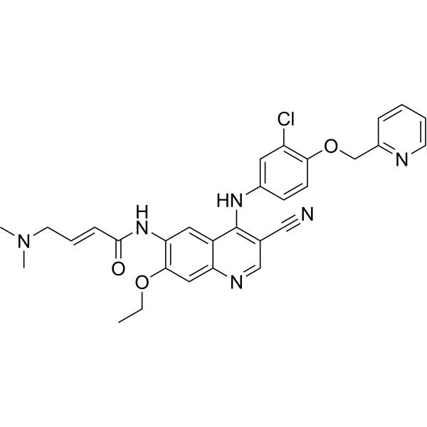
HY-32721
NeratinibHER2/EGFR Inhibitor
Publications Citing Use of MedChemExpress (MCE) Lavendustin A
MoreAlle EGFR Isoform-spezifische Produkte anzeigen
More

Biologische Aktivität
Beschreibung
IC50 & Target
IC50: 11 nM (EGFR-associated tyrosine kinase)[1]
Cellular Effect
|
Cell Line

|
Type | Value | Description | References |
|---|---|---|---|---|
| A-431 | IC50 |
4 ng/mL
Compound: 76
|
Inhibition of EGFR in human A431 cells
Inhibition of EGFR in human A431 cells
|
[PMID: 1479375] |
| A-431 | IC50 |
4 x 10⁻³ μg/mL
Compound: 76
|
Inhibition of EGFR in human A431 cells
Inhibition of EGFR in human A431 cells
|
[PMID: 1479375] |
| A-431 | IC50 |
26 μg/mL
Compound: 1
|
Cytotoxicity against human A431 cells
Cytotoxicity against human A431 cells
|
[PMID: 2614420] |
| HaCaT | IC50 |
>100 μM
Compound: 1
|
Inhibitory activity of HaCaT keratinocyte cell line
Inhibitory activity of HaCaT keratinocyte cell line
|
[PMID: 7990108] |
| L1210 | IC50 |
28 μg/mL
Compound: 1
|
Cytotoxicity against mouse L1210 cells
Cytotoxicity against mouse L1210 cells
|
[PMID: 2614420] |
| NIH3T3 | IC50 |
19.5 μg/mL
Compound: 1
|
Cytotoxicity against RSV transformed mouse NIH/3T3 cells
Cytotoxicity against RSV transformed mouse NIH/3T3 cells
|
[PMID: 2614420] |
| NIH3T3 | IC50 |
22 μg/mL
Compound: 1
|
Cytotoxicity against mouse NIH/3T3 cells
Cytotoxicity against mouse NIH/3T3 cells
|
[PMID: 2614420] |
| P388 | IC50 |
21.5 μg/mL
Compound: 1
|
Cytotoxicity against mouse P388 cells
Cytotoxicity against mouse P388 cells
|
[PMID: 2614420] |
In Vitro
Lavendustin A (5-50μM) dose-dependently inhibits progesterone production in cultures of ovarian dispersates[3].
MedChemExpress (MCE) has not independently confirmed the accuracy of these methods. They are for reference only.
In Vivo
MedChemExpress (MCE) has not independently confirmed the accuracy of these methods. They are for reference only.


Chemical Information
-
CAS. Nr. 125697-92-9
-
Appearance Solid
-
Molecular Weight 381.38
-
Formel C21H19NO6
-
Color Off-white to light brown
-
SMILES
O=C(O)C1=CC(N(CC2=CC(O)=CC=C2O)CC3=CC=CC=C3O)=CC=C1O
-
Synonyms
RG-14355
-
Structure Classification
-
Initial Source
Streptomyces griseolavendus
-
Versand
Room temperature in continental US; may vary elsewhere.
-
Speicherung
4°C, stored under nitrogen
* In solvent : -80°C, 6 months; -20°C, 1 month (stored under nitrogen)


Publications (1)
-
Journal Impact Factor
-
Most Recent
-
ISME J
Endophytic commensal bacteria capitalize on the AvrPto-FER pathway to enhance proliferation during early stages of pathogen invasion. [Abstract]2025 Jan 2;19(1):wraf145. PMID: 40632106


Lösungsmittel & Löslichkeit
In Vitro:
DMSO : 100 mg/mL (262.21 mM; Need ultrasonic; Hygroscopic DMSO has a significant impact on the solubility of product, please use newly opened DMSO)
Please refer to the solubility information to select the appropriate solvent. Once prepared, please aliquot and store the solution to prevent product inactivation from repeated freeze-thaw cycles.
Storage method and period of stock solution: -80°C, 6 months; -20°C, 1 month (stored under nitrogen). When stored at -80°C, please use it within 6 months. When stored at -20°C, please use it within 1 month.
Please refer to the solubility information to select the appropriate solvent. Once prepared, please aliquot and store the solution to prevent product inactivation from repeated freeze-thaw cycles.
Storage method and period of stock solution: -80°C, 6 months; -20°C, 1 month (stored under nitrogen). When stored at -80°C, please use it within 6 months. When stored at -20°C, please use it within 1 month.
Konzentration (Stammlösung) × Volumen (Stammlösung) = Konzentration (Ziellösung) × Volumen (Ziellösung)
In Vivo:
Select the appropriate dissolution method based on your experimental animal and administration route.
- For the following dissolution methods, please ensure to first prepare a clear stock solution using an In Vitro approach and then sequentially add co-solvents:
- To ensure reliable experimental results, the clarified stock solution can be appropriately stored based on storage conditions. As for the working solution for In Vivo experiments, it is recommended to prepare freshly and use it on the same day.
- The percentages shown for the solvents indicate their volumetric ratio in the final prepared solution. If precipitation or phase separation occurs during preparation, heat and/or sonication can be used to aid dissolution.
Add each solvent one by one: 10% DMSO 40% PEG300 5% Tween-80 45% Saline
Solubility: 2.5 mg/mL (6.56 mM); Clear solution; Need ultrasonic
This protocol yields a clear solution of 2.5 mg/mL.
Taking 1 mL working solution as an example, add 100 μL DMSO stock solution (25.0 mg/mL) to 400 μL PEG300, and mix evenly; then add 50 μL Tween-80 and mix evenly; then add 450 μL Saline to adjust the volume to 1 mL.
Preparation of Saline: Dissolve 0.9 g sodium chloride in ddH₂O and dilute to 100 mL to obtain a clear Saline solution.
Add each solvent one by one: 10% DMSO 90% (20% SBE-β-CD in Saline)
Solubility: 2.5 mg/mL (6.56 mM); Clear solution; Need ultrasonic
This protocol yields a clear solution of 2.5 mg/mL.
Taking 1 mL working solution as an example, add 100 μL DMSO stock solution (25.0 mg/mL) to 900 μL 20% SBE-β-CD in Saline, and mix evenly.
Preparation of 20% SBE-β-CD in Saline (4°C, storage for one week): 2 g SBE-β-CD powder is dissolved in 10 mL Saline, completely dissolve until clear.
In Vivo Dissolution Calculator
Please enter the basic information of animal experiments:
-
-
-
-
Recommended: Prepare an additional quantity of animals to account for potential losses during experiments.
Please enter your animal formula composition:
-
%DMSO +
Recommended: Keep the proportion of DMSO in working solution below 2% if your animal is weak.
-
%+
-
+%Tween-80 + +
-
%Saline +
The co-solvents required include: DMSO, . All of co-solvents are available by MedChemExpress (MCE). , Tween 80. All of co-solvents are available by MedChemExpress (MCE).
Working solution concentration: 0.22 mg/mL
Method for preparing stock solution: mg drug dissolved in μL DMSO. Stock solution concentration: mg/mL. * In solvent : -80°C, 6 months; -20°C, 1 month (stored under nitrogen)
1. Take μL DMSO stock solution;
2. Add μL .
μL , mix evenly;
3. Then add μL Tween 80, mix evenly;
4. Then add μL
Please ensure that the stock solution in the first step is dissolved to a clear state, and add co-solvents in sequence. You can use ultrasonic heating (ultrasonic cleaner, recommended frequency 20-40 kHz), vortexing, etc. to assist dissolution.


Reinheit & Dokumentation
-
Data Sheet (283 KB)
-
SDS (393 KB)
- English - EN (393 KB)
- Français - FR (393 KB)
- Deutsch - DE (393 KB)
- Norwegian - NO (393 KB)
- Español - ES (393 KB)
- Swedish - SV (393 KB)
- Italian - IT (393 KB)
- Korean - KR (393 KB)
- Portuguese - PT (393 KB)
-
Handling Instructions (2659 KB)
Verweise
[1]. Onoda T, et, al. Isolation of a novel tyrosine kinase inhibitor, lavendustin A, from Streptomyces griseolavendus. J Nat Prod. 1989 Nov-Dec;52(6):1252-7. [Content Brief]
[2]. Hu DE, et, al. Suppression of VEGF-induced angiogenesis by the protein tyrosine kinase inhibitor, lavendustin A. Br J Pharmacol. 1995 Jan;114(2):262-8. [Content Brief]
[3]. Whitehead SA, et, al. Protein tyrosine kinase activity of lavendustin A and the phytoestrogen genistein on progesterone synthesis in cultured rat ovarian cells. Fertil Steril. 2000 Mar;73(3):613-9. [Content Brief]


Complete Stock Solution Preparation Table
Please refer to the solubility information to select the appropriate solvent. Once prepared, please aliquot and store the solution to prevent product inactivation from repeated freeze-thaw cycles.
Storage method and period of stock solution: -80°C, 6 months; -20°C, 1 month (stored under nitrogen). When stored at -80°C, please use it within 6 months. When stored at -20°C, please use it within 1 month.
| Optional Solvent | Concentration Solvent Mass | 1 mg | 5 mg | 10 mg | 25 mg |
|---|---|---|---|---|---|
| DMSO | 1 mM | 2.6221 mL | 13.1103 mL | 26.2206 mL | 65.5514 mL |
| 5 mM | 0.5244 mL | 2.6221 mL | 5.2441 mL | 13.1103 mL | |
| 10 mM | 0.2622 mL | 1.3110 mL | 2.6221 mL | 6.5551 mL | |
| 15 mM | 0.1748 mL | 0.8740 mL | 1.7480 mL | 4.3701 mL | |
| 20 mM | 0.1311 mL | 0.6555 mL | 1.3110 mL | 3.2776 mL | |
| 25 mM | 0.1049 mL | 0.5244 mL | 1.0488 mL | 2.6221 mL | |
| 30 mM | 0.0874 mL | 0.4370 mL | 0.8740 mL | 2.1850 mL | |
| 40 mM | 0.0656 mL | 0.3278 mL | 0.6555 mL | 1.6388 mL | |
| 50 mM | 0.0524 mL | 0.2622 mL | 0.5244 mL | 1.3110 mL | |
| 60 mM | 0.0437 mL | 0.2185 mL | 0.4370 mL | 1.0925 mL | |
| 80 mM | 0.0328 mL | 0.1639 mL | 0.3278 mL | 0.8194 mL | |
| 100 mM | 0.0262 mL | 0.1311 mL | 0.2622 mL | 0.6555 mL |


Lavendustin A Related Classifications
HY-18963 Related Classifications
 Powered by Bioz